2,481
Views
14
CrossRef citations to date
0
Altmetric
Research Paper

Can plants serve as a vector for prions causing chronic wasting disease?

, , , , , , , , & show all
Pages 136-142 | Received 28 Nov 2013, Accepted 22 Jan 2014, Published online: 07 Feb 2014

Figures & data

Figure 1. Recombinant PrPC binds to the outside of wheat roots. Wheat plant roots were exposed to 50 µg/mL PrPC for 24 h then roots were either digested with (10 µg/mL) proteinase K (PK) for 5 min or left undigested. Control plants were exposed to the same solution lacking PrPC. Western Blotting on root protein extracts used the 8H4 mAb (1:10000). The last lane confirming antibody specificity is from the same blot with irrelevant lanes omitted. Results are representative of three independent replicates (n = 3). Lanes 1 and 2: control plants; Lanes 3 and 4: PrPC exposed plants; Lane 5: positive control (50 ng PrPC).
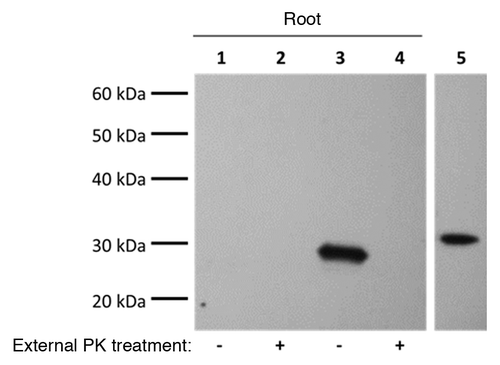
Figure 1. Recombinant PrPC binds to the outside of wheat roots. Wheat plant roots were exposed to 50 µg/mL PrPC for 24 h then roots were either digested with (10 µg/mL) proteinase K (PK) for 5 min or left undigested. Control plants were exposed to the same solution lacking PrPC. Western Blotting on root protein extracts used the 8H4 mAb (1:10000). The last lane confirming antibody specificity is from the same blot with irrelevant lanes omitted. Results are representative of three independent replicates (n = 3). Lanes 1 and 2: control plants; Lanes 3 and 4: PrPC exposed plants; Lane 5: positive control (50 ng PrPC).

Figure 2. Prion signal found in wheat roots exposed to CWD PrPTSE was protease sensitive but no prion signal in lower stem (Stem) extract was visible. CWD PrPTSE was purified from brain homogenate (BH) with Bio Rad-TeSeE® purification and re-suspended in phosphate buffered saline to a 1% solution (w/v) based on the initial BH solution. Wheat plant roots were exposed to the purified solution for 24 h. Normal BH processed with TeSeE® served as a negative control. Plant protein extracts were digested with proteinase K (PK) (10 µg/mL, 30 min, 37 °C) to determine PK-resistance of any proteins. Western blotting of plant protein extracts (plant total protein extraction kit) was done using P4 mAb (1:5000) and Prionics®-Check Western kit. Results are representative of three independent replicates (n = 3). Lanes 1, 3, 5, 7: plants exposed to normal BH processed with Bio Rad kit; Lanes 2, 4, 6, 8: plants exposed to CWD infected BH processed with Bio Rad kit; Lane 9: CWD infected BH (0.1%) processed with Bio Rad kit.
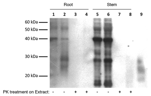
Figure 2. Prion signal found in wheat roots exposed to CWD PrPTSE was protease sensitive but no prion signal in lower stem (Stem) extract was visible. CWD PrPTSE was purified from brain homogenate (BH) with Bio Rad-TeSeE® purification and re-suspended in phosphate buffered saline to a 1% solution (w/v) based on the initial BH solution. Wheat plant roots were exposed to the purified solution for 24 h. Normal BH processed with TeSeE® served as a negative control. Plant protein extracts were digested with proteinase K (PK) (10 µg/mL, 30 min, 37 °C) to determine PK-resistance of any proteins. Western blotting of plant protein extracts (plant total protein extraction kit) was done using P4 mAb (1:5000) and Prionics®-Check Western kit. Results are representative of three independent replicates (n = 3). Lanes 1, 3, 5, 7: plants exposed to normal BH processed with Bio Rad kit; Lanes 2, 4, 6, 8: plants exposed to CWD infected BH processed with Bio Rad kit; Lane 9: CWD infected BH (0.1%) processed with Bio Rad kit.

Figure 3. Proteinase K (PK) digested CWD PrPTSE in a brain homogenate (BH) solution interacts with wheat roots but signal from lower stem (Stem) disappears when the stem is rinsed. (A) Wheat plant roots were exposed for 24 h to BH solutions either CWD positive (CWD BH) or CWD negative (Normal BH) and both digested with PK (50 µg/mL, 30 min, 37 °C) or not. Control plants were exposed to dH2O for 24 h. Only the roots were rinsed for 1 min in dH2O, with protein extracted from both the root and lower stem. Western blotting of plant extracts was done using P4 mAb (1:5000) and Prionics®-Check Western kit. Lanes 1 and 6: plants exposed to H2O; Lanes 2 and 7: plants exposed to Normal BH; Lanes 3 and 8: plants exposed to Normal BH digested with PK; Lanes 4 and 9: plants exposed to CWD BH; Lanes 5 and 10: plants exposed to CWD BH digested with PK; Lane 11: 0.1% Normal BH; Lane 12: 0.1% Normal BH digested with PK; Lane 13: 0.1% CWD BH; Lane 14: 0.1% CWD BH digested with PK. (B) Wheat plant roots were exposed to PK-digested (50 µg/mL, 30 min, 37 °C) CWD BH for 24 h then both the roots and stem were rinsed in dH2O for 1 min. Western blotting of extracted protein used P4 mAb (1:5000) and Prionics®-Check Western kit. Results are representative of three independent replicates (n = 3). Lanes 1 and 3: plants exposed to H2O; Lanes 2 and 4: plants exposed to CWD BH digested with PK; Lane 5: 0.1% CWD BH digested with PK.
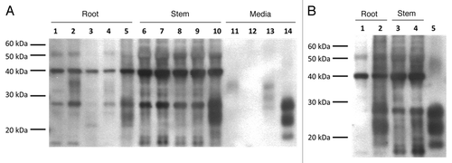
Figure 3. Proteinase K (PK) digested CWD PrPTSE in a brain homogenate (BH) solution interacts with wheat roots but signal from lower stem (Stem) disappears when the stem is rinsed. (A) Wheat plant roots were exposed for 24 h to BH solutions either CWD positive (CWD BH) or CWD negative (Normal BH) and both digested with PK (50 µg/mL, 30 min, 37 °C) or not. Control plants were exposed to dH2O for 24 h. Only the roots were rinsed for 1 min in dH2O, with protein extracted from both the root and lower stem. Western blotting of plant extracts was done using P4 mAb (1:5000) and Prionics®-Check Western kit. Lanes 1 and 6: plants exposed to H2O; Lanes 2 and 7: plants exposed to Normal BH; Lanes 3 and 8: plants exposed to Normal BH digested with PK; Lanes 4 and 9: plants exposed to CWD BH; Lanes 5 and 10: plants exposed to CWD BH digested with PK; Lane 11: 0.1% Normal BH; Lane 12: 0.1% Normal BH digested with PK; Lane 13: 0.1% CWD BH; Lane 14: 0.1% CWD BH digested with PK. (B) Wheat plant roots were exposed to PK-digested (50 µg/mL, 30 min, 37 °C) CWD BH for 24 h then both the roots and stem were rinsed in dH2O for 1 min. Western blotting of extracted protein used P4 mAb (1:5000) and Prionics®-Check Western kit. Results are representative of three independent replicates (n = 3). Lanes 1 and 3: plants exposed to H2O; Lanes 2 and 4: plants exposed to CWD BH digested with PK; Lane 5: 0.1% CWD BH digested with PK.

Figure 4. Proteinase K (PK) digested CWD PrPTSE interacts with wheat roots and remains slightly PK resistant after extraction while no CWD PrPTSE was detected in the lower stem (Stem). CWD positive brain homogenates (BH) were digested with PK (50 µg/mL, 30 min, 37 °C) prior to being exposed to wheat roots for 24 h. Wheat roots and stems were rinsed with dH2O for 1 min then protein was extracted with 1% SDS. Extracts were digested with PK (10µg/mL, 30 min, 37 °C) to determine any PK-resistant bands prior to blotting. Western blotting on protein extracts was done using P4 mAb (1:5000) and Prionics®-Check Western kit (Prionics). Images are from the same blot with irrelevant lanes omitted. Results are representative of three independent replicates (n = 3). Lanes 1, 4, 6: plants exposed to H2O; Lanes 2, 3, 5, 7: plants exposed to CWD BH digested with PK; Lanes 8 and 9: 0.1% CWD BH in presence of 1% SDS digested with PK.
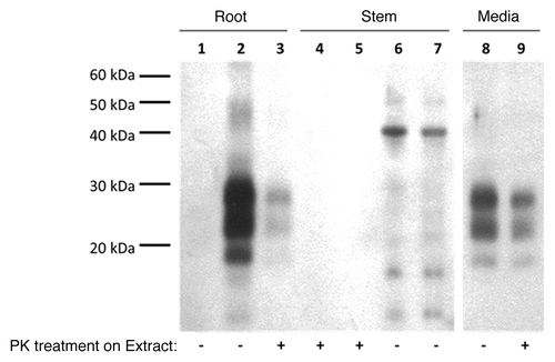
Figure 4. Proteinase K (PK) digested CWD PrPTSE interacts with wheat roots and remains slightly PK resistant after extraction while no CWD PrPTSE was detected in the lower stem (Stem). CWD positive brain homogenates (BH) were digested with PK (50 µg/mL, 30 min, 37 °C) prior to being exposed to wheat roots for 24 h. Wheat roots and stems were rinsed with dH2O for 1 min then protein was extracted with 1% SDS. Extracts were digested with PK (10µg/mL, 30 min, 37 °C) to determine any PK-resistant bands prior to blotting. Western blotting on protein extracts was done using P4 mAb (1:5000) and Prionics®-Check Western kit (Prionics). Images are from the same blot with irrelevant lanes omitted. Results are representative of three independent replicates (n = 3). Lanes 1, 4, 6: plants exposed to H2O; Lanes 2, 3, 5, 7: plants exposed to CWD BH digested with PK; Lanes 8 and 9: 0.1% CWD BH in presence of 1% SDS digested with PK.

Table 1. Analysis of plant protein extracts (1% SDS) from proteinase K-digested (PK) CWD BH treatment with Bio-Rad and IDEXX diagnostic tests

Supplemental material

Additional material

Download Zip (142.4 KB)

Reprints and Corporate Permissions

Please note: Selecting permissions does not provide access to the full text of the article, please see our help page How do I view content?

To request a reprint or corporate permissions for this article, please click on the relevant link below:

Academic Permissions

Please note: Selecting permissions does not provide access to the full text of the article, please see our help page How do I view content?

Obtain permissions instantly via Rightslink by clicking on the button below:

If you are unable to obtain permissions via Rightslink, please complete and submit this Permissions form. For more information, please visit our Permissions help page.