5,977
Views
82
CrossRef citations to date
0
Altmetric
REVIEW

Variant CJD

18 years of research and surveillance

, , , , , , & show all
Pages 286-295 | Received 17 Mar 2014, Accepted 14 May 2014, Published online: 01 Nov 2014

Figures & data

Figure 1. Vacuolation scoring in the mouse brain. Lesion profile comparison of vCJD and BSE following transmission to RIII mice. Data shows mean lesion profile ± standard error of the mean (n ≥ 6). G1-G9, gray matter scoring regions; (G1) dorsal medulla, (G2) cerebellar cortex, (G3) superior colliculus, (G4) hypothalamus, (G5) thalamus, (G6) hippocampus, (G7) septum, (G8) retrosplenial and adjacent motor cortex, (G9) cingulate and adjacent motor cortex.
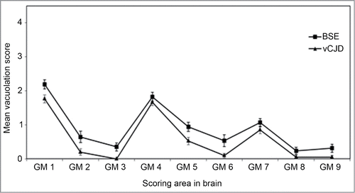
Figure 1. Vacuolation scoring in the mouse brain. Lesion profile comparison of vCJD and BSE following transmission to RIII mice. Data shows mean lesion profile ± standard error of the mean (n ≥ 6). G1-G9, gray matter scoring regions; (G1) dorsal medulla, (G2) cerebellar cortex, (G3) superior colliculus, (G4) hypothalamus, (G5) thalamus, (G6) hippocampus, (G7) septum, (G8) retrosplenial and adjacent motor cortex, (G9) cingulate and adjacent motor cortex.
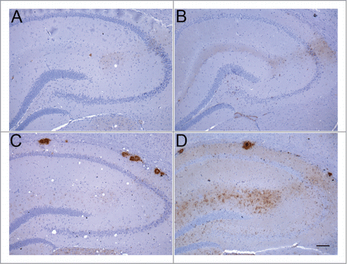
Figure 2. Abnormal PrP deposition in hippocampus of RIII and VM mice following inoculation with BSE or vCJD. (A) BSE in RIII mouse; (B) vCJD in RIII mouse; (C) BSE in VM mouse; (D) vCJD in VM mouse (bar, 100 μm; anti-PrP antibody: 6H4).

Figure 2. Abnormal PrP deposition in hippocampus of RIII and VM mice following inoculation with BSE or vCJD. (A) BSE in RIII mouse; (B) vCJD in RIII mouse; (C) BSE in VM mouse; (D) vCJD in VM mouse (bar, 100 μm; anti-PrP antibody: 6H4).

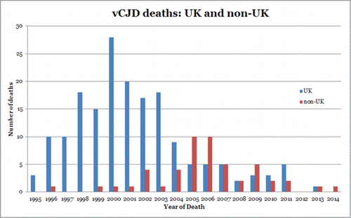
Figure 3. Reported incidence of vCJD deaths in the UK and in non-UK countries.

Figure 3. Reported incidence of vCJD deaths in the UK and in non-UK countries.

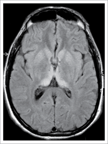
Figure 4. MRI brain scan in variant CJD. FLAIR axial section at the level of the basal ganglia showing bilateral symmetrical dorsomedial and pulvinar thalamic hyperintensity. Courtesy of Dr David Summers.

Figure 4. MRI brain scan in variant CJD. FLAIR axial section at the level of the basal ganglia showing bilateral symmetrical dorsomedial and pulvinar thalamic hyperintensity. Courtesy of Dr David Summers.