459
Views
15
CrossRef citations to date
0
Altmetric
Short Communication

Rapid, high-throughput detection of PrPSc by 96-well immunoassay

&
Pages 44-48 | Received 16 Jan 2009, Accepted 13 Mar 2009, Published online: 01 Jan 2009

Figures & data

Figure 1 96-well PrPSc immunoassay controls. (A) 96-well immunoassay and (B) quantification of brain homogenate from proteinase K digested uninfected (UN) or HY TME-agent infected (HY TME) hamsters with (+) and without (−) H2O2 in MeOH, primary antibody or secondary antibody to indicate the specificity of PrPSc immunodetection. Relative PrPSc intensity is based on well 16.
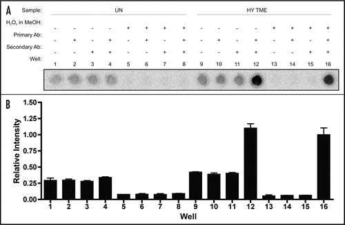
Figure 1 96-well PrPSc immunoassay controls. (A) 96-well immunoassay and (B) quantification of brain homogenate from proteinase K digested uninfected (UN) or HY TME-agent infected (HY TME) hamsters with (+) and without (−) H2O2 in MeOH, primary antibody or secondary antibody to indicate the specificity of PrPSc immunodetection. Relative PrPSc intensity is based on well 16.

Figure 2 Limits of PrP detection using western blot analysis. Western blot analysis of two-fold serial dilutions of brain homogenates treated with (+) and without (−) proteinase K (PK) from uninfected hamsters (UN, A) or hamsters infected with the HY TME (HY) or DY TME (DY) agents (B and C, respectively).
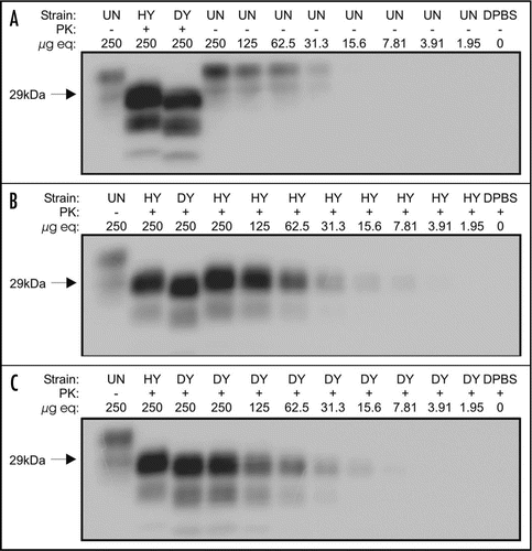
Figure 2 Limits of PrP detection using western blot analysis. Western blot analysis of two-fold serial dilutions of brain homogenates treated with (+) and without (−) proteinase K (PK) from uninfected hamsters (UN, A) or hamsters infected with the HY TME (HY) or DY TME (DY) agents (B and C, respectively).

Figure 3 Limits of PrP detection using 96-well immunoassay. 96 well immunoassay of two-fold serial dilutions of brain homogenate from uninfected (UN), HY TME agent (HY) or DY TME agent (DY) infected hamsters. Samples were either proteinase K (PK) digested (+) or were left untreated (−).
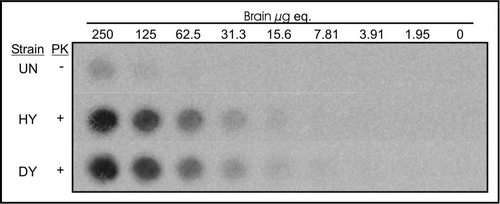
Figure 3 Limits of PrP detection using 96-well immunoassay. 96 well immunoassay of two-fold serial dilutions of brain homogenate from uninfected (UN), HY TME agent (HY) or DY TME agent (DY) infected hamsters. Samples were either proteinase K (PK) digested (+) or were left untreated (−).

Figure 4 Comparison of PrPSc detection limits by western blot and 96-well immunoassay. Quantification of PrPSc abundance of two-fold serial dilutions of uninfected (UN), HY TME-infected (HY) or DY TME-infected (DY) proteinase K digested brain material by either western blot (open bars) or 96-well immunoassay (solid bars). Represented data is the average and standard error of the mean of three independent experiments.
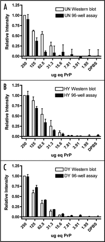
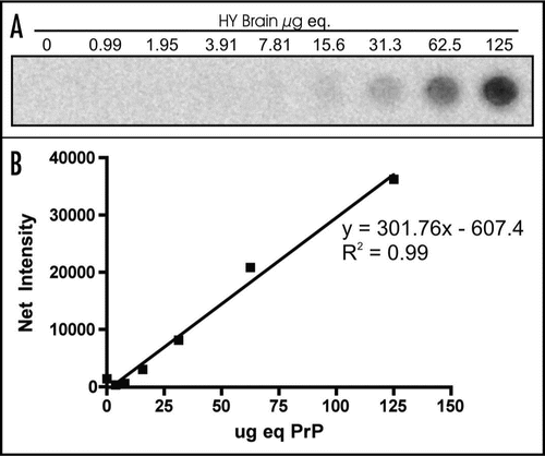
Figure 4 Comparison of PrPSc detection limits by western blot and 96-well immunoassay. Quantification of PrPSc abundance of two-fold serial dilutions of uninfected (UN), HY TME-infected (HY) or DY TME-infected (DY) proteinase K digested brain material by either western blot (open bars) or 96-well immunoassay (solid bars). Represented data is the average and standard error of the mean of three independent experiments.

Figure 5 Linear range of PrPSc detection by 96-well immunoassay. (A) 96-well immunoassay and (B) quantification of PrPSc from 2 fold serial dilutions of HY TME-agent infected brain homogenate digested with proteinase K.

Figure 5 Linear range of PrPSc detection by 96-well immunoassay. (A) 96-well immunoassay and (B) quantification of PrPSc from 2 fold serial dilutions of HY TME-agent infected brain homogenate digested with proteinase K.