514
Views
8
CrossRef citations to date
0
Altmetric
Commentary & View

Insights into prion protein function from atomistic simulations

&
Pages 13-19 | Received 11 Oct 2009, Accepted 16 Dec 2009, Published online: 01 Jan 2010

Figures & data

Figure 1 The binding site of the copper ion at high Cu concentration. Copper is shown in gold, oxygens are red, nitrogens blue, hydrogen white and carbons are cyan.
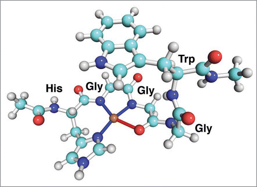
Figure 1 The binding site of the copper ion at high Cu concentration. Copper is shown in gold, oxygens are red, nitrogens blue, hydrogen white and carbons are cyan.

Figure 2 The structure of the low concentration copper binding site in the octarepeat domain of PrP.
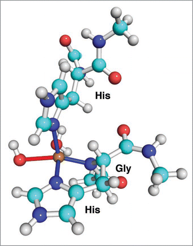
Figure 2 The structure of the low concentration copper binding site in the octarepeat domain of PrP.

Figure 3 The structure of the medium concentration copper binding mode in the octarepeat domain of PrP.
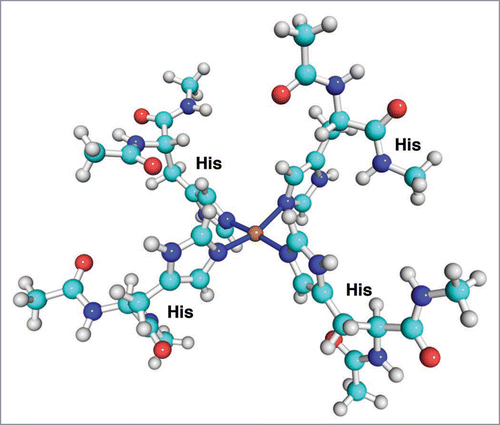
Figure 3 The structure of the medium concentration copper binding mode in the octarepeat domain of PrP.

Figure 4 Structures of full-length prion protein in various copper binding modes. (A) Free PrP, (B) Low concentration mode, one copper ion coordinated by His61, His69, His77 and His85, (C) medium concentration mode: two copper ions are attached to His61, His69 and His77, His85, respectively, (D) High concentration mode: four copper ions are attached, each ion is anchored by one of the following: His61, His69, His77 and His85. The copper atoms are shown as gold spheres. The octarepeat domain is colored blue, the non-octarepeat region of the N-terminal is colored grey. The α-helices and β-sheet in C-terminal are displayed in red and mangenta, respectively.
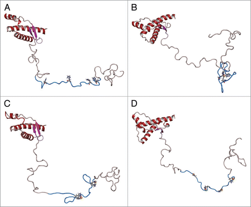
Figure 4 Structures of full-length prion protein in various copper binding modes. (A) Free PrP, (B) Low concentration mode, one copper ion coordinated by His61, His69, His77 and His85, (C) medium concentration mode: two copper ions are attached to His61, His69 and His77, His85, respectively, (D) High concentration mode: four copper ions are attached, each ion is anchored by one of the following: His61, His69, His77 and His85. The copper atoms are shown as gold spheres. The octarepeat domain is colored blue, the non-octarepeat region of the N-terminal is colored grey. The α-helices and β-sheet in C-terminal are displayed in red and mangenta, respectively.

Table 1 Energies of copper binding modes for octarepeat domain of PrP

Reprints and Corporate Permissions

Please note: Selecting permissions does not provide access to the full text of the article, please see our help page How do I view content?

To request a reprint or corporate permissions for this article, please click on the relevant link below:

Academic Permissions

Please note: Selecting permissions does not provide access to the full text of the article, please see our help page How do I view content?

Obtain permissions instantly via Rightslink by clicking on the button below:

If you are unable to obtain permissions via Rightslink, please complete and submit this Permissions form. For more information, please visit our Permissions help page.