Figures & data
Figure 1 PrPC levels in human CS F are attenuated following spinal cord injury. (A) Western analysis of PrPC in human CS F from control (lane1) and from spinal cord injured patient #19 (remaining lanes) at 12 h, 24 h, day 2, day 3, day 4, day 5 and day 6 following spinal cord injury. 40 µl of the indicated CS F samples were separated by SDS PAGE, the Western blot was probed with anti-PrPC 6H4 (Prionics) antibody and developed with SuperSignal® West Dura (Pierce). (B) Western analysis of representative human CS F from control, 12 h within spinal cord injury and 7 d after spinal cord injury (40 µl). Four patients are shown. Blots were probed with anti-PrPC 6H4 (Prionics) antibody or anti-PrPC 11C6 antibody (SPI-BIO).
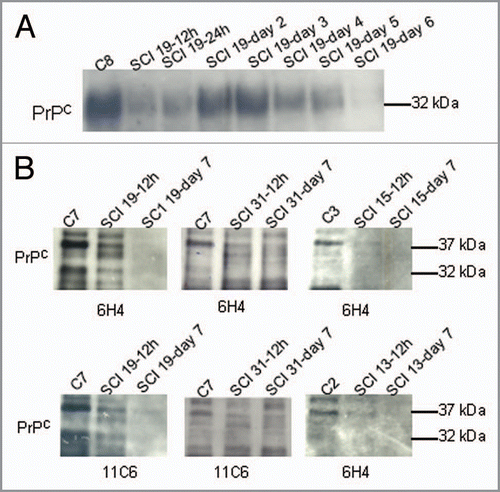
Figure 2 14-3-3 and IgG light chain levels in CS F after spinal cord injury. (A) Western analysis of 14-3-3 and IgG light chain after spinal cord injury. Three patients are shown. 14-3-3 is absent in all CS F samples. IgG light chains are present in varying amounts in all samples. 20 µg of rat brain supernatant and pellet fractions and 20 µg of PC12 cell homogenate (middle) are shown in the right lanes. (B) Western analysis of CS F (40 µl) after spinal cord injury for αA/αB crystallin and Hsp27.
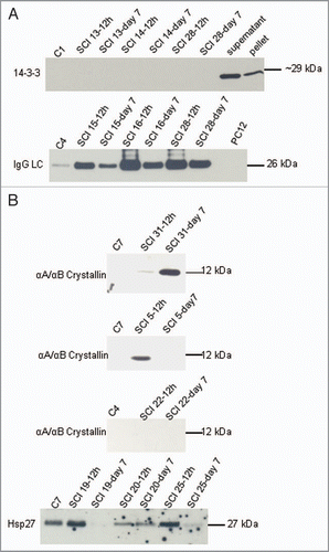
Figure 3 Molecular chaperones were not detected in CSF of control or spinal cord injured patients. Western analysis of Hsp40, Rdj-2, Hdj-2, Hsp110, Hsp90, Hsc70, Hsp70 and Hsp60 in CS F samples (40 µl) from control or spinal cord injured patients as indicated. 20 µg of rat brain supernatant and pellet fractions are shown in the right lanes.
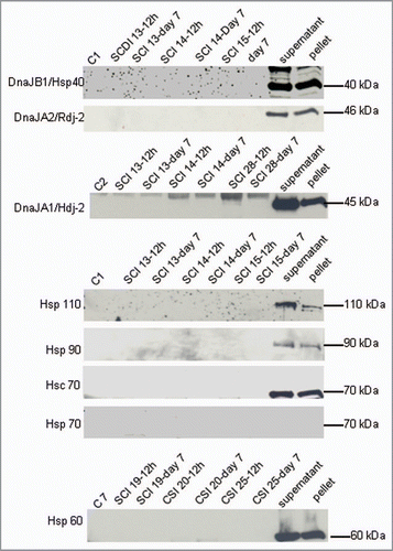
Table 1 Clinical characteristics of the patients included in the study