Figures & data
Figure 1 Model of the natural history of AD. Timeline from presumptive start of AD through patient diagnosis is plotted. The initiation of biological changes (stage I) marks the onset of disease and begins years to decades before any evidence is apparent (represented by dashed lines). At some point the first pathologic evidence of disease (stage II) begins and in theory can be detected with biomarkers such as CSF measurements of amyloid and tau or PET imaging with amyloid ligands. Subsequently, the first signs and symptoms of disease develop (stage III) followed by the patient seeking medical attention (stage IV) and finally a diagnosis is established (stage V). This timeline can be clustered into a presymptomatic phase (stages I–III) and a symptomatic phase (stages III–V). An alternative way to envision the disease spectrum is from the biological onset to the seeking of medical attention (stages I–IV) as the preclinical phase of disease with the clinical phase beginning with the initial clinical investigations into the cause of the patients' symptoms (stages IV and V). Stage III is the ideal time for dementia screening.
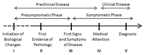
Figure 2 Changes in AD biomarkers by age and AD8 scores. AD biomarkers are plots as a function of age (x-axis) and AD8 scores. AD8 scores of 0 or 1 (no impairment) are depicted as filled circles while AD8 scores of 2 or greater (impairment) are depicted as open squares. Regression lines are plotted for the entire cohort (dashed black line) and for each subset (black for AD8 no impairment; gray for AD8 impairment). The top row (A–C) represents biomarker profiles for the entire cohort (n = 257) divided by their AD8 scores. With age, there are changes in biomarkers with decreasing CSF Aβ42 (A), increasing CSF Tau (B) and increased PiB-PET binding potential (C). The effect of age on CSF biomarkers is most marked in the AD8 no impairment group (black line) while changes in PiB binding is seen only in the AD8 impaired group (gray line). The bottom row (D–F) represents biomarker profiles for the individuals rated CDR 0 no dementia (n = 156), 25 of whom had AD8 scores in the impaired range. Similar age-related changes in CSF Aβ42 and PiB binding are seen with CSF Aβ42 having the greatest rate of decline in the AD8 no impairment group and PiB binding having the greatest rate of change in the AD8 impairment group. Increases in CSF Tau are seen as a function of age regardless of group.
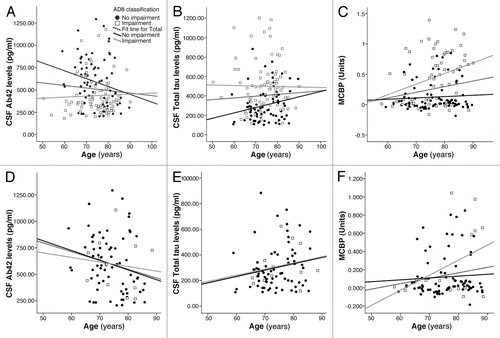
Table 1 Characteristics of nondemented CDR 0 individuals stratified by AD8 scores