Figures & data
Figure 1. Physical association of AtPP5 with MDH by FPLC and Co-IP analysis. After pre-incubating 20 µM AtPP5 with 10 µM MDH for 20 min at either (A) 22°C or (B) 42°C, the mixtures were separated by SEC (upper panels of A and B). Each fraction was analyzed on SDS-PAGE followed by silver staining (bottom panels of A and B). (C) In vitro co-IP of AtPP5 and MDH at 22°C and 42°C. The presence of MDH was detected on western blot using an anti-MDH antibody. The input lane was loaded with 2% of the reaction mixture. The arrow (→) indicates the IgG large subunit.
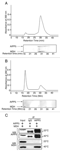
Figure 2. Interaction of AtPP5 and AtHsp90 by co-IP. In vivo co-IP of AtPP5 and AtHsp90. Co-IP of (lane 3) AtHsp90 with antiserum against AtPP5 and (lane 4) AtPP5 with antiserum against AtHsp90, followed by western blotting and detection with anti-AtHsp90 antibody and anti-AtPP5 antibody, respectively. The arrow (→) indicates the IgG large subunit.
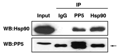
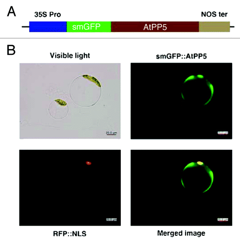
Figure 3. Subcellular localization of AtPP5 fused with GFP in Arabidopsis protoplasts. (A) Schematic diagram of plasmid construct for transforming plant cells for fluorescent confocal microscopy. Expression of the fused genes was driven by the CaMV 35S promoter (35S Pro) and terminated by the nopaline synthetase terminator (NOS ter). Arabidopsis protoplasts were transformed with the resultant constructs and fluorescent images were obtained 12 to 48 h after transformation. Green and red images are GFP and RFP fluorescence signals of smGFP::AtPP5 and RFP::NLS, respectively.