Figures & data
Figure 1. Morphology and structure of ovule in G. biloba during pollination stage. (A, B) Morphology of ovule before pollination. (C) PD secretion during pollination. (D) Morphology of ovule after pollination. (E) Anatomical structure of ovule. (F, G) Morphology of micropyle. Br, bract; I, integument; Le, leaf; Lh, long shoot; Mc, micropyle canal; Mi, micropyle; Nu, nucellus; O, ovule; Ob, ovule bracket; Pc, pollen chamber; PD, pollination drop; Sh, short shoot; S, stalk. Scale bars (A–D) = 1 cm, (E) = 1 mm, (F, G) = 200 μm.
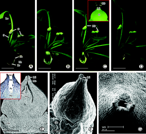
Figure 2. PDs secretion after different treatments. (A) Secretion of PD after removal of short shoot, leaves, ovular stalk, ovular collar, and micropyle. (B) Effects of sucrose and NaCl on PD secretion.
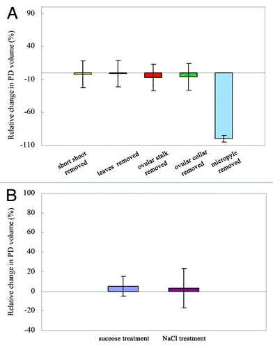
Figure 3. Structure of the nucellar apex and micropyle cells in G. biloba. (A) Ovule shape before pollination; (B) Nucellar cells before pollination; (C) Collapse of nucellar tip cells (arrow); (D) Nucellar cells below pollen chamber; (E) Morphology of micropyle canal; (F) Cells of micropyle; (G) Cells of micropyle canal. I, integument; Mc, micropyle canal; Nu, nucellus; Pc, pollen chamber; V, vacuole. Scale bars (A, C, E) = 100 μm; (B, D, F, G) = 50 μm.
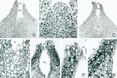
Figure 4. Ultrastructure of nucellar apex cells in G. biloba. (A, B) Nucellar apex cells; (C) Vacuome consisting of variously sized vacuoles; (D–G) Dense cytoplasm with abundant mitochondria, endoplasmic reticulum, protein bodies, and dictyosomes; (H) Vesicles near cell wall; (I) Plasmodesma connecting protoplasts of nucellar cells. Cw, cell wall; Di, dictyosome; Is, intercellular spaces; M, mitochondria; N, nucleus; Nu, nucellus; Pb, protein body; Pd, plasmodesma; Pl, plastid; Pm, plasma membrane; Rer, rough endoplasmic reticulum; V, vacuole; Ve, vesicle. Scale bars (A, B) = 5 μm; (C, D) = 2 μm; (E, F, G, I) = 1 μm; H = 0.5 μm.
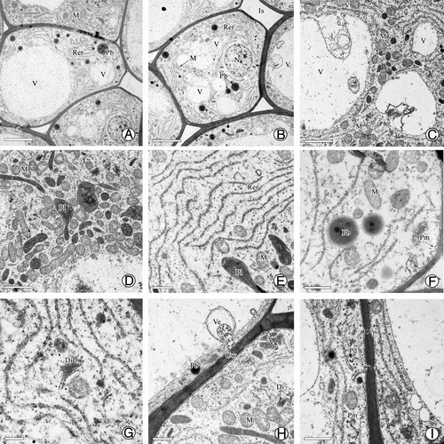
Figure 5. Characteristics of PD production in G. biloba. (A) Number of PDs on 50 ovules at different times; (B) Formation times of PDs and PD volume after removal and reformation (first time indicates when PD became detectable). (C) Number of PDs on 30 ovules at different relative humidity.
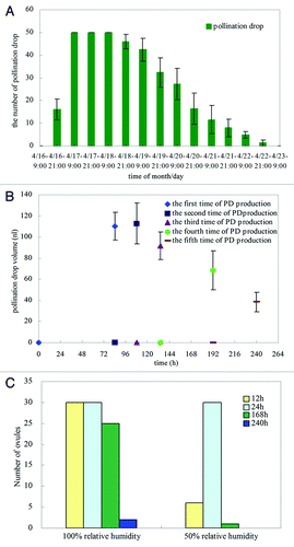
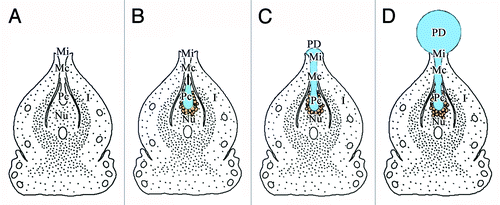
Figure 6. Pattern of PD production. (A) Formation of pollen chamber before PD production; (B) Collapse of nucellar apex cells during PD production; (C) Exudation of PD from pollen chamber to micropyle; (D) Constant production of PD by nucellar cells around pollen chamber, PD reaches maximum volume at pollination stage. I, integument; Mc, micropyle canal; Mi, micropyle; Nu, nucellus; Pc, pollen chamber; PD, pollination drop.