Figures & data
Figure 1. Non-supervised PCA representation of signals obtained from a non targeted analysis by HPLC-QTOFMS to monitor metabolomic changes during fungal invasion. (A) PCA1 vs PCA2 and (B) PCA1 vs PCA3. Three week old seedlings were sprayed inoculated with 10E3 spores.ml−1 of P. cucumerina and samples for analysis were collected 48hpi. Leaf material from 15 individual plants were pooled together for each treatment x genotype combination. Data points represent two technical replicates from three independent experiments injected randomly into the HPLC-QTOFMS. The signals corresponding to different treatments were compared using the non-parametric Kruskal-Wallis test, and only data with a P value lower than 0.01 between groups was used for subsequent processing.
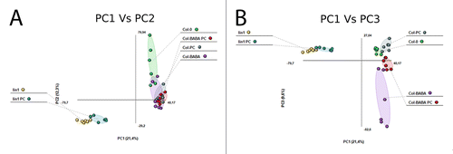
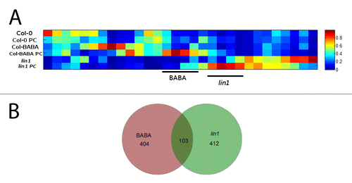
Figure 2. (A) Heat map analysis of the signals extracted from the Kruskal-Wallis test with a P value lower than 0.01 between groups performed with Marvis (Filter and Cluster packages) and (B) Venn diagrams of detected primed signals.