Figures & data
Figure 1 Zymogram of starch branching enzyme (SBE) activities. Barley leaves were incubated in sorbitol or sucrose with sense or antisense SBE ODNs followed by SBE zymogram analysis.Citation17 Antisense and corresponding sense ODNsCitation10 were constructed as follows. SBEI (NCBI Accession number AY304541), nt 15–32; SBEIIA (NCBI Accession number AF064560), nt 1–18; SBEIIB (NCBI Accession number AF064561, nt 116–133.
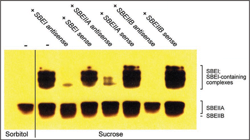
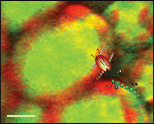
Figure 2 ODNs “piggyback” on transported sugar molecules. A pictorial representation of ODNs utilizing the sucrose translocator (SUT) to enter plant cells. The confocal microscope projection image shows uptake of fluorescently labeled ODNs (green) in barley leaves after 24 h incubation in 200 mM sucrose.Citation10 Autofluorescence from chloroplasts is shown in red. The superimposed cartoon shows the coupled symport of H+ (white dot), sucrose (blue) and ODN (green) through the SUT (red). Scale bar, 10 µm.