Figures & data
Figure 1 A model that describes a possible mechanism and sequence of events that lead to the [Ca2+]cyt transients and inhibition of root growth. (1) Al3+ interacts with Ca2+ channels in the plasma membrane of root cells in the root transition zone. The Ca2+channels open and external Ca2+ enters the cytosol. (2) [Ca2+]cyt rises producing the first peak of the biphasic [Ca2+]cyt signature. (3) Increased [Ca2+]cyt activates internal Ca2+ channels located in membranes of internal Ca2+ stores (e.g., tonoplast, ER, mitochondria or plastids) producing the second peak of the [Ca2+]cyt signature. (4) Al3+ permeates the PM through Ca2+- and non-selective cation channels. (5) Al3+ opens internal Ca2+ channels in the tonoplast, ER, mitochondria or plastids and as a result more Ca2+ is released into the cytosol. (6) The overall [Ca2+]cyt elevation stimulates mechanisms that inhibit root growth.
![Figure 1 A model that describes a possible mechanism and sequence of events that lead to the [Ca2+]cyt transients and inhibition of root growth. (1) Al3+ interacts with Ca2+ channels in the plasma membrane of root cells in the root transition zone. The Ca2+channels open and external Ca2+ enters the cytosol. (2) [Ca2+]cyt rises producing the first peak of the biphasic [Ca2+]cyt signature. (3) Increased [Ca2+]cyt activates internal Ca2+ channels located in membranes of internal Ca2+ stores (e.g., tonoplast, ER, mitochondria or plastids) producing the second peak of the [Ca2+]cyt signature. (4) Al3+ permeates the PM through Ca2+- and non-selective cation channels. (5) Al3+ opens internal Ca2+ channels in the tonoplast, ER, mitochondria or plastids and as a result more Ca2+ is released into the cytosol. (6) The overall [Ca2+]cyt elevation stimulates mechanisms that inhibit root growth.](/cms/asset/56441425-cfa0-4b25-a46c-1f9554b6ba93/kpsb_a_10911973_f0001.gif)
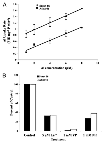
Figure 2 Al3+ uptake by PM vesicles isolated from 5 mm root tips of both the Al-sensitive cultivar Scout 66 and the Al-tolerant cultivar Atlas 66. (A) Rate of Al3+ uptake by PM vesicles incubated in increasing concentrations of Al3+. The PM vesicles from the Al sensitive cultivar Scout 66 were more permeable to Al3+ than those of the Al-tolerant cultivar Atlas 66. The values are means ± SD. Rates of Al3+ uptake are expressed in Fluorescence Intensity Units (FIU) mg−1 protein min−1. (B) Effect of Ca2+ channel blockers on the rate of Al3+ uptake by PM vesicles s percent of the control. All Ca2+ channel blockers tested inhibited the rate Al3+ uptake by the PM vesicles in both cultivars. The accumulation of Al3+ in the PM vesicles was monitored by measuring the fluorescence emitted by the Al-morin complex as described in the text. Both experiments were repeated three times in triplicate (n = 9). The PM vesicles were pooled from multiple independent membrane isolations in order to obtain enough membrane protein for the assays.
