Figures & data
Figure 1 Germination (A) and seedling development (B) in sunflower (Helianthus annuus). Achenes were sown in moist vermiculite and raised in darkness (D, days 0 to 2) or grown for 3 days in the dark and subsequently irradiated for 1 to 3 days with continuous white light (WL). The seedlings were kept in 99% relative humidity at 25°C.
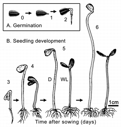
Figure 2 Bivariate plot of log10-transformed data for cellular respiration (R) versus fresh mass (Mf) during germination and seedling development. Sunflower seeds were raised in the dark or, after 3 days of plant growth, irradiated with continuous white light (WL). Lines are reduced major axis (RMA) regression curves (summary statistics provided in the text).
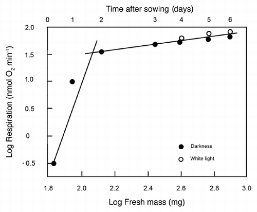
Figure 3 Representative scanning electron micrographs of the cotyledons (A) and epidermal cells (B) of 3-day-old sunflower seedlings that were grown in darkness. The peripheral cells (B) were photographed in the area indicated by the arrow (A). C, cotyledons; E, epidermal cells; S, open stomatum with open pore. Bars = 1 mm (A), 50 µm (B).
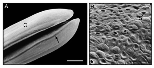
Figure 4 Ultrastructure of sub-epidermal cells in the region between the apical hook and the onset of the cotyledons in 3-day-old etiolated sunflower seedlings. The representative transmission electron micrograph shows the cytoplasmic region of three cells in the periphery of the organ that are separated by a thick wall. A, amyloplast; C, cytoplasm; L, lipid droplet (oleosome); M, mitochondrion; N, nucleus; S, starch; V, vacuole; W, cell wall. Bar, 2 µm.
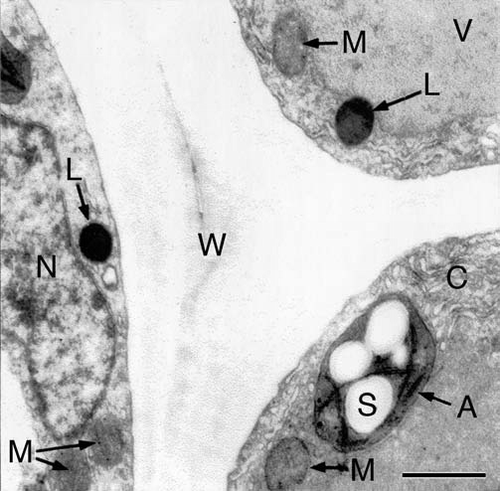
Figure 5 Bivariate plot of log10-transformed data for fresh mass (Mf) versus dry mass (Md) during germination and seedling development. Sunflower seeds were raised in the dark or, after 3 days of plant growth, irradiated with continuous white light (WL). The line is a reduced major axis (RMA) regression curve (summary statistics provided in the text).
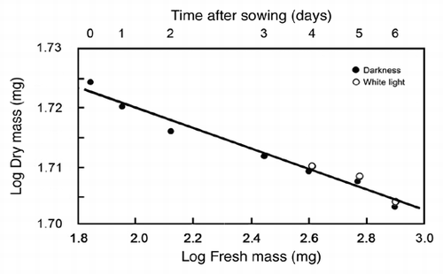
Table 1 List of abbreviations and definitions