843
Views
18
CrossRef citations to date
0
Altmetric
Addendum

The Nbeal2−/− mouse as a model for the gray platelet syndrome

, , , &
Article: e26561 | Received 26 Aug 2013, Accepted 20 Sep 2013, Published online: 26 Sep 2013

Figures & data

Figure 1. Platelet ultrastructure in Nbeal2−/− mice (A) and human GPS (B). Representative transmission electron microscopy (TEM) images of resting wild-type (+/+) and Nbeal2−/− (−/−) platelets and those of a control human subject as well as a characterized GPS patientCitation9,Citation15 with a homozygous L388P mutation. Platelets deficient in NBEAL2 both show lack of α-granules (#) and an increased number of vacuoles (arrows) while platelet size was increased and dense granule (*) content was unaltered. Bars = 0.5 µm.
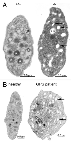
Figure 1. Platelet ultrastructure in Nbeal2−/− mice (A) and human GPS (B). Representative transmission electron microscopy (TEM) images of resting wild-type (+/+) and Nbeal2−/− (−/−) platelets and those of a control human subject as well as a characterized GPS patientCitation9,Citation15 with a homozygous L388P mutation. Platelets deficient in NBEAL2 both show lack of α-granules (#) and an increased number of vacuoles (arrows) while platelet size was increased and dense granule (*) content was unaltered. Bars = 0.5 µm.

Figure 2. Confocal microscopy of MKs cultured in vitro from CD34+ cells from the peripheral blood of a GPS patient with a homozygous L388P mutation in NBEAL2,Citation9 MK suspensions were incubated at day 14 of culture on polylysine-coated slides. Cells were fixed and permeabilized. Integrin αIIbβ3 and VWF were localized by a murine antibody (AP2) and VWF by a polyclonal antibody with bound IgG visualized using species-specific FITC (green) and Alexa-Fluor568-conjugated IgG. All technical details were as previously described.Citation24 In the upper panel, a round small MK shows abundant labeling for VWF; in the middle panel a MK shows VWF staining along the proplatelet extension, the VWF labeling is decreased compared to control and is absent from the proplatelet tip; the lower panel shows a very mature MK with a long proplatelet string with typical swellings along its length while the VWF labeling is minimal. This suggests that VWF is not maintained in the MK. Please note that the scale of this panel is reduced to allow the proplatelet to be fully seen.
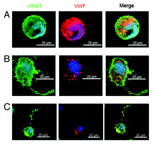
Figure 2. Confocal microscopy of MKs cultured in vitro from CD34+ cells from the peripheral blood of a GPS patient with a homozygous L388P mutation in NBEAL2,Citation9 MK suspensions were incubated at day 14 of culture on polylysine-coated slides. Cells were fixed and permeabilized. Integrin αIIbβ3 and VWF were localized by a murine antibody (AP2) and VWF by a polyclonal antibody with bound IgG visualized using species-specific FITC (green) and Alexa-Fluor568-conjugated IgG. All technical details were as previously described.Citation24 In the upper panel, a round small MK shows abundant labeling for VWF; in the middle panel a MK shows VWF staining along the proplatelet extension, the VWF labeling is decreased compared to control and is absent from the proplatelet tip; the lower panel shows a very mature MK with a long proplatelet string with typical swellings along its length while the VWF labeling is minimal. This suggests that VWF is not maintained in the MK. Please note that the scale of this panel is reduced to allow the proplatelet to be fully seen.