Figures & data
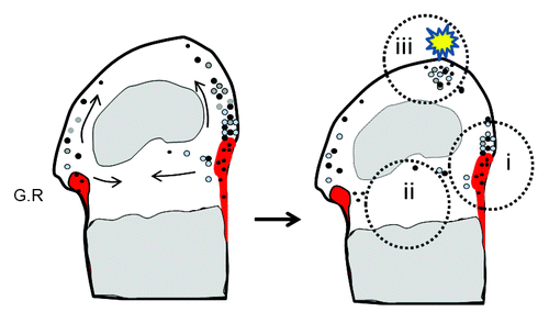
Figure 1. (A) PTPN11 mutations identified in MC patients. The locations of MC mutations in the corresponding SHP2 structure are indicated by red, blue and green colored lines, representing frameshift, nonsense and splice-site mutations, respectively. Predicted protein changes are indicated with arrows. Please see references Citation1 and Citation2 for original references. (B) Micro-CT and Faxitron images demonstrate the existence of osteochondromas and enchondromas (arrows) at hip, knee and ankles of 12-week-old mice lacking SHP2 in cathepsin K-expressing cells (CtskCre;Ptpn11fl/fl, Ctsk-KO) but not its littermate controls (CtskCre;Ptpn11fl/+, Ctsk-Control).
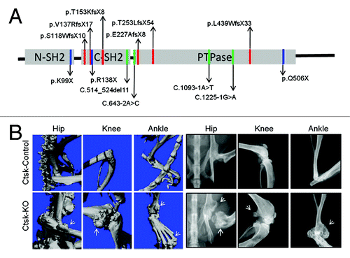
Figure 2. Location and potential functions of CCPs in epiphyseal cartilage Diagrams showing the location of perichondrial groove of Ranvier (G.R) (shaded in red) in the knee joint. CCPs and their differentiated progeny are denoted by dots of black and gray color, respectively. Note that CCPs reside primarily in the groove of Ranvier (i, black), but they can migrate toward, and live in, epiphyseal (ii) and articular cartilage (iii). In these alternative niches, they could either live quiescently (black) or undergo chondrocytic differentiation (gray) and replenish cartilage. We hypothesize that under physiological conditions, CCPs might be required for epiphyseal cartilage development and homeostasis. During cartilage injury (such as osteoarthritis and trauma) or disease conditions (mutations, etc.), these cells respond to pathogenic insults and start to expand to repair cartilage damage or cause tumorigenesis.