Figures & data
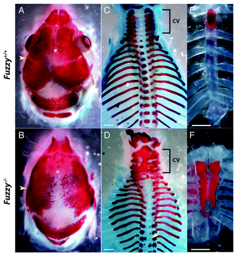
Figure 1. Skeletal preparations of wild-type and Fuz−/− embryos at E18.5. Alizarin red staining marks the bone. Alcian blue staining marks the cartilage. (A and B) Dorsal views of the skull. (A) Control. (B) Mutant mice display synostosis of the coronal suture (yellow arrowhead) as well as an open anterior fontanelle (yellow asterix). (C and D) Dorsal view of the axial skeleton. (C) Control. (D) In mutant animals, the cervical vertebra (cv, bracket) are fused. Ossification of the centrum in thoracic vertebra is lost or aberrant (yellow arrow). (E and F) Frontal views of the sternum. (E) Control. (F) In mutants, the sternum is shorter, hyperossified and cleft/bifid (black arrow).