8,588
Views
255
CrossRef citations to date
0
Altmetric
Review

Testosterone signaling and the regulation of spermatogenesis

Pages 116-120 | Received 15 Jun 2011, Accepted 16 Jun 2011, Published online: 30 Jun 2011

Figures & data

Figure 1 Testosterone signaling pathways. (1) The classical testosterone signaling pathway. Testosterone diffuses through the plasma membrane and binds with the AR. The AR undergoes an alteration in conformation allowing it to be released from heat shock proteins in the cytoplasm. AR then is able to translocate to the nucleus where it binds to specific DNA sequences called androgen response elements (AREs). AR binding to an ARE allows the recruitment of co-activator and co-repressor proteins that alter the expression of genes to alter cellular function. (2) The non-classical kinase activation pathway: testosterone interacts with the classical AR that then is able to recruit and activate Src, which causes the activation of the EGF receptor via an intracellular pathway. The EGF receptor then activates the MAP kinase cascade most likely through Ras resulting in the sequential activation of RAF and MEK and then ERK that activates p90Rsk-kinase, which is known to phosphorylate CREB on serine 133. As a result, CREB-regulated genes such as lactate dehydrogenase A (LDH-A) and early growth response 1 (Egr1) and CREB can be induced by testosterone.Citation42 (3) The non-classical Ca2+ influx pathway: Testosterone interacts with a receptor in the plasma membrane that has characteristics of a Gq coupled G-protein coupled receptor (GPCR). Phospholipase C (PLC) is activated to cleave PIP2 into IP3 and DAG. Lower concentrations of PIP2 inhibit K+ATP channels causing membrane depolarization and Ca2+ entry via L-type Ca2+ channels.
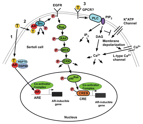
Figure 1 Testosterone signaling pathways. (1) The classical testosterone signaling pathway. Testosterone diffuses through the plasma membrane and binds with the AR. The AR undergoes an alteration in conformation allowing it to be released from heat shock proteins in the cytoplasm. AR then is able to translocate to the nucleus where it binds to specific DNA sequences called androgen response elements (AREs). AR binding to an ARE allows the recruitment of co-activator and co-repressor proteins that alter the expression of genes to alter cellular function. (2) The non-classical kinase activation pathway: testosterone interacts with the classical AR that then is able to recruit and activate Src, which causes the activation of the EGF receptor via an intracellular pathway. The EGF receptor then activates the MAP kinase cascade most likely through Ras resulting in the sequential activation of RAF and MEK and then ERK that activates p90Rsk-kinase, which is known to phosphorylate CREB on serine 133. As a result, CREB-regulated genes such as lactate dehydrogenase A (LDH-A) and early growth response 1 (Egr1) and CREB can be induced by testosterone.Citation42 (3) The non-classical Ca2+ influx pathway: Testosterone interacts with a receptor in the plasma membrane that has characteristics of a Gq coupled G-protein coupled receptor (GPCR). Phospholipase C (PLC) is activated to cleave PIP2 into IP3 and DAG. Lower concentrations of PIP2 inhibit K+ATP channels causing membrane depolarization and Ca2+ entry via L-type Ca2+ channels.

Figure 2 Deletion of exon 3 eliminates non-classical AR activity. 15P-1 Sertoli cells were infected with adenovirus constructs expressing wild type AR (AdhisAR) or exon 3-deleted AR (AdARΔexon 3). P-ER K, ER K and AR levels were determined by western blot after a 10 min stimulation in serum free media with vehicle (V) or 100 nM testosterone (T).
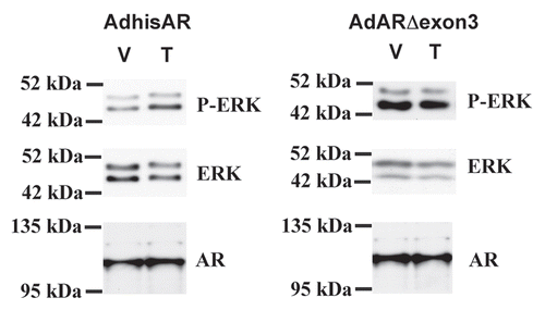
Figure 2 Deletion of exon 3 eliminates non-classical AR activity. 15P-1 Sertoli cells were infected with adenovirus constructs expressing wild type AR (AdhisAR) or exon 3-deleted AR (AdARΔexon 3). P-ER K, ER K and AR levels were determined by western blot after a 10 min stimulation in serum free media with vehicle (V) or 100 nM testosterone (T).