Figures & data
Table 1. Overview of studies addressing in vitro spermatogenesis.
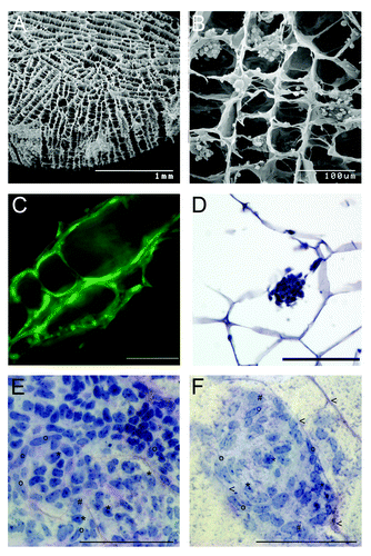
Figure 1. (A) Cytology of the mouse testis showing the relationship and the function of the interstitial, basal, intraepithelial, and adluminal compartment. (B) Schematic diagram of different approaches for male germ cell differentiation in vitro and their outcomes so far.
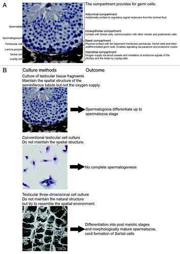
Figure 2. Artificial collagen sponges seeded with isolated testicular cells of mice (7 dpp). (A) Scanning electron microscope image of a collagen sponge prior to culture. (B) Scanning electron microscope image of a colonized collagen sponge. Isolated testicular cells (2 million) established aggregates in the structure of the sponge after one day of culture. (C) Testicular cells of eGFP mice on a collagen sponge after three days of culture. The cells have colonized the collagen scaffold along the given structure. Scale bar represents 100 µm. (D) Micrographs of a section of a paraffin embedded collagen sponge (hematoxylin staining) Scale bar represents 100 µm. (E, F) Histological micrographs for morphological identification of mouse testicular cell types after seven days of culture. Samples were embedded in resin (Technovit, Heraeaus Kulzer, GmbH, Wehrheim, Germany); stained with perjodic acid Schiff reagent; and cut to 3µm sections (protocol according to ref. Citation49). Scale bar represents 100 µm. (E) Cell clusters consist of a mixture of testicular cells. i.e peritubular cells (o), Sertoli cells (*) and some undifferentiated germ cells (#). (F) During colonization of the scaffolds, first signs of tubulogenic reassembly occur. The cells utilize the scaffold for structural reorganization when attaching to the collagen fibers (arrowheads).