Figures & data
Figure 1. Transduction of primary Sertoli cells using lentvirus vector. (A) Live reporter GFP fluorescence of HEK293T cells transduced at 5 × 10⁶ TU/ml (B) Flow cytometric analysis of control (no virus, black) and lentivirus transduced HEK293T cells (red). (C) Live GFP fluorescence of Sertoli cells transduced with 5 × 10⁶ TU/ml prior to and (D) after optimization. (E) Flow cytometric analysis of Sertoli cells prior to (red) and after (blue) optimization. (F) Expression of pSIH transfer vector normalized to β-actin in primary Sertoli cells 4 d (white) and 14 d (black) after transduction. (G) Experimental outline of optimized lentiviral vector transduction procedure in primary Sertoli cell culture. Scale bar = 50 μm.
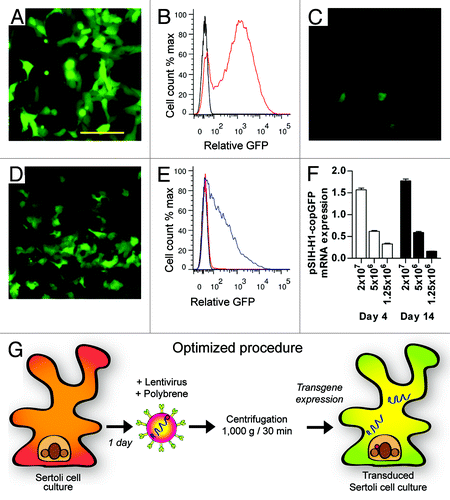
Table 1. Fold increase in genomic incorporation of lentiviral transfer vector by qPCR.
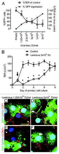
Figure 2. Effect of lentivirus administration on inter-Sertoli cell tight junction. (A) Transepithelial electrical resistance (%TER of control monolayers, white boxes) taken 48 h after Sertoli cell transduction with between 1.25 × 10⁶ and 2 × 10⁷ TU/ml. GFP reporter fluorescence was determined by flow cytometry (% GFP+ cells, flow cytometry, black circles). Increased lentivirus addition (and increased %GFP+ cells) is associated with loss of TER. TER data are mean, ± SD, n = 3, letters denote significant difference p < 0.01. (B) Daily TER of control (black boxes) and lentivirus transduced cells (5 x 10⁶ TU/ml, white boxes) over 8 d culture period. Black arrow indicates lentivirus addition 24 h after primary cell isolation. TER data are mean, ± SD, n = 3, * p < 0.01 between control and transduced cells. (C–F) Co-localization of endogenous reporter gene expression (GFP, green), Gata4 (blue) and tight junction protein (red); (C and D) Tjp1 and (E and F) Cldn11 with 1.25 × 10⁶ or 2 × 10⁷ TU/ml. White arrow indicates TJ co-localization between non-transduced cells, *indicates TJ localization between transduced (GFP) cells, and white arrowhead in (F) indicates reduced Cldn11 localization between adjacent transduced cells after high viral titer addition. Scale bar = 20 μm.