Figures & data
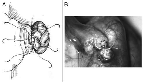
Figure 1. (A) Schematic representing re-approximation of vasal mucosa as part of a 4-layer closure using 10–0 nylon sutures with the microdot technique. (B) Intraoperative vasovasostomy repair – note the microdots spaced approximately 20 µm in diameter.
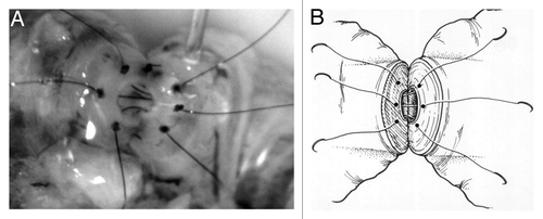
Figure 2. (A) Schematic demonstrating a vasoepididymostomy using a longitudinal intussuscepted technique with 10–0 sutures to intussuscept a longitudinally incised segment of the epididymal tubule into the lumen of the abdominal vasal remnant. The intussusception is then reinforced with multiple 9–0 and 8–0 sutures. (B) Intraoperative vasoepididymostomy using 10–0 sutures.