1,840
Views
75
CrossRef citations to date
0
Altmetric
Review

Dual role of immune cells in the testis

Protective or pathogenic for germ cells?

, , , , &
Article: e23870 | Received 17 Dec 2012, Accepted 02 Feb 2013, Published online: 01 Jan 2013

Figures & data

Figure 1. Schematic drawing illustrating the seminiferous epithelium and intestitium under normal (A) or inflammatory (B) conditions. (A) The seminiferous epithelium is composed of Sertoli cells and germ cells at different stages of development. The blood-testis barrier (BTB) is constituted by coexisting adherens, gap and tight junctions between adjacent Sertoli cells. As representative tight junction molecules, occludin and ZO-1 are shown. Macrophages close to Leydig cells and few immature dendritic cells (DCs), T cells and regulatory T cells (Treg) are present in the interstitium. (B) Spermatocytes and spermatids undergo apoptosis and sloughing in the lumen of the seminiferous tubule. Decreased occludin expression in Sertoli cell tight junctions associated to impairment of BTB is shown. An increased number of mature DCs, pro-inflammatory and intermediate type macrophages, CD4+ and CD8+ T cells and T regs are present in the interstitium. Pro-inflammatory cytokines (IL-6, TNF-α, FasL, IFN-γ, IL-17) and nitric oxide (NO) released by macrophages and T cells are involved in the induction of inflammation and germ cell apoptosis. Cytokines and other factors secreted by Sertoli, Leydig and peritubular cells are not illustrated.
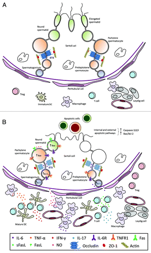
Figure 1. Schematic drawing illustrating the seminiferous epithelium and intestitium under normal (A) or inflammatory (B) conditions. (A) The seminiferous epithelium is composed of Sertoli cells and germ cells at different stages of development. The blood-testis barrier (BTB) is constituted by coexisting adherens, gap and tight junctions between adjacent Sertoli cells. As representative tight junction molecules, occludin and ZO-1 are shown. Macrophages close to Leydig cells and few immature dendritic cells (DCs), T cells and regulatory T cells (Treg) are present in the interstitium. (B) Spermatocytes and spermatids undergo apoptosis and sloughing in the lumen of the seminiferous tubule. Decreased occludin expression in Sertoli cell tight junctions associated to impairment of BTB is shown. An increased number of mature DCs, pro-inflammatory and intermediate type macrophages, CD4+ and CD8+ T cells and T regs are present in the interstitium. Pro-inflammatory cytokines (IL-6, TNF-α, FasL, IFN-γ, IL-17) and nitric oxide (NO) released by macrophages and T cells are involved in the induction of inflammation and germ cell apoptosis. Cytokines and other factors secreted by Sertoli, Leydig and peritubular cells are not illustrated.