Figures & data
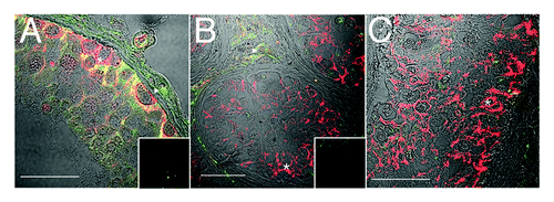
Figure 1. Evidence for Sertoli cell proliferation in men. Confocal immunofluorescence of proliferation markers in testis tissue from normal (A and E) and gonadotropin suppressed men [2 wk, B; 12 wk, C and D (enlarged portion of C) and F and G (enlarged portion of panel F)], in testis with carcinoma in situ (H) and in a tubule adjacent to a seminoma (I). Sections were probed with a combination of either GATA4 (green, in the nuclei of Sertoli cells) within the seminiferous epithelium (asterisks), and PCNA antibodies (red, staining cells with proliferation capacity - triangles) (A–D, H–I), or GATA4 (red) and Ki67 (green, staining proliferating cells) (E–G). Colocalization of GATA4 and PCNA or GATA4 and Ki67 in Sertoli cell nuclei (indicating a proliferative state, arrows) is shown by a yellow color. includes a transmitted light channel to illustrate histological detail. Inserts are negative controls. (Bar = 50 µm).
![Figure 1. Evidence for Sertoli cell proliferation in men. Confocal immunofluorescence of proliferation markers in testis tissue from normal (A and E) and gonadotropin suppressed men [2 wk, B; 12 wk, C and D (enlarged portion of C) and F and G (enlarged portion of panel F)], in testis with carcinoma in situ (H) and in a tubule adjacent to a seminoma (I). Sections were probed with a combination of either GATA4 (green, in the nuclei of Sertoli cells) within the seminiferous epithelium (asterisks), and PCNA antibodies (red, staining cells with proliferation capacity - triangles) (A–D, H–I), or GATA4 (red) and Ki67 (green, staining proliferating cells) (E–G). Colocalization of GATA4 and PCNA or GATA4 and Ki67 in Sertoli cell nuclei (indicating a proliferative state, arrows) is shown by a yellow color. Figure 1E–G includes a transmitted light channel to illustrate histological detail. Inserts are negative controls. (Bar = 50 µm).](/cms/asset/f1250c13-b788-4dd0-a368-45143294601a/kspe_a_10924014_f0001.gif)
Figure 2. Downregulation of androgen receptor immunoreactivity in PCNA-positive Sertoli cells. Confocal immunofluorescence of human testis sections from normal (A) or gonadotropin-suppressed men (B, 2 wk; C–I, 12 wk). Sections were probed for GATA-4 [green, Sertoli cells within the seminiferous epithelium, asterisks), androgen receptor (blue, Sertoli (asterisks), Leydig and peritubular cells], and PCNA (red, labeling predominantly germ cells, triangles). Colocalization of GATA-4 and PCNA in Sertoli cells indicated by yellow (arrows). G–I are individual channels of the merged image in F, highlighting a GATA4 and PCNA-positive, AR-negative Sertoli cell (arrow) and a GATA4 and AR-positive, PCNA-negative Sertoli cell (asterisk). Inserts are controls where primary antibody was substituted for non-specific antibody of the same isotype. (Bar = 50 µm).
![Figure 2. Downregulation of androgen receptor immunoreactivity in PCNA-positive Sertoli cells. Confocal immunofluorescence of human testis sections from normal (A) or gonadotropin-suppressed men (B, 2 wk; C–I, 12 wk). Sections were probed for GATA-4 [green, Sertoli cells within the seminiferous epithelium, asterisks), androgen receptor (blue, Sertoli (asterisks), Leydig and peritubular cells], and PCNA (red, labeling predominantly germ cells, triangles). Colocalization of GATA-4 and PCNA in Sertoli cells indicated by yellow (arrows). G–I are individual channels of the merged image in F, highlighting a GATA4 and PCNA-positive, AR-negative Sertoli cell (arrow) and a GATA4 and AR-positive, PCNA-negative Sertoli cell (asterisk). Inserts are controls where primary antibody was substituted for non-specific antibody of the same isotype. (Bar = 50 µm).](/cms/asset/f3962110-2fd0-4567-9770-f30553e45fb7/kspe_a_10924014_f0002.gif)
Figure 3. Claudin-11 and Connexin 43 are disorganized in testicular dysgenesis. Confocal immunofluorescence of normal (A) and carcinoma in situ (B and C) human testis sections detecting claudin-11 (red, tight junctions, asterisks) and connexin 43 (green, gap junctions, open squares and triangles). Co-localization appears in yellow. Inserts are negative controls. (Bar = 50 µm).
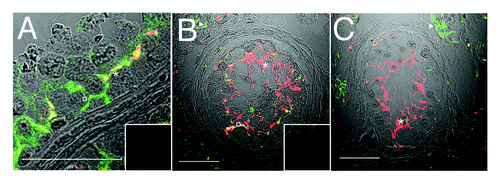
Figure 4. JAM-A localization is disorganized in carcinoma in situ and becomes highly expressed in malignant seminoma. Confocal immunofluorescence of normal human testis (A), carcinoma in situ (B and C) and seminoma sections (D) detecting JAM-A (red, asterisks) and connexin 43 (green, gap junctions, squares). Inserts are negative controls. (Bar = 50 µm).
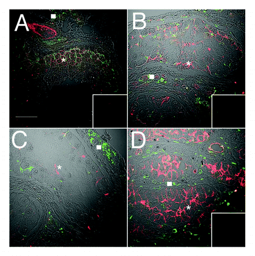
Figure 5. ZO-1 localization is disorganized in carcinoma in situ, in the presence of normal connexin 43 reactivity. Confocal immunofluorescence of normal human testis (A) and carcinoma in situ (B) detecting ZO-1 (tight and associated junctions, red, asterisks) and connexin 43 (gap junctions, green, triangles). Inserts are negative controls. (Bar = 50 µm).