Figures & data
Figure 1 (A) Crystal-Structure of Thermotoga maritima NusA and solution NMR-structures of the two AR-domains of E. coli NusA. The NusA-NTD of T. maritima (green) is linked via a connecting helix (grey) to the central RNA-binding domains S1 (red), KH1 (blue) and KH2 (aquamarine) of NusA (PDB-ID: 1HH2).Citation4 Additionally shown are the solution NMR-structures of AR1 (brown; PDB-ID: 1WCL) and AR2 (yellow; PDB-ID: 1WCH) domains,Citation6 which so far could only been identified within E. coli. (B) Crystal-Structure of the E. coli NusB:NusE complex. NusE (green) and NusB (purple) form a tight complex (PDB-ID: 3D3B).Citation17 The single sphere within NusE denotes Ser46, which replaces the ribosome binding loop 46–67 in the crystallized construct (see details in reference Citation17). (C) Solution NMR-Structure of NusG (NusG-NTD; PDB-ID: 2K06; NusG-CTD; PDB-ID: 2JVV).Citation24
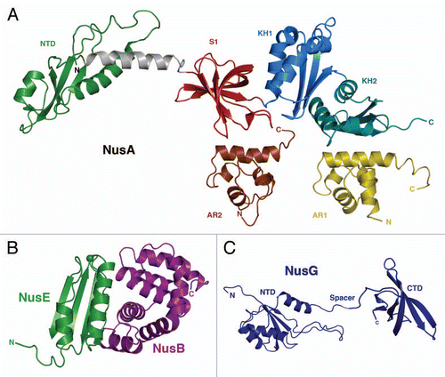
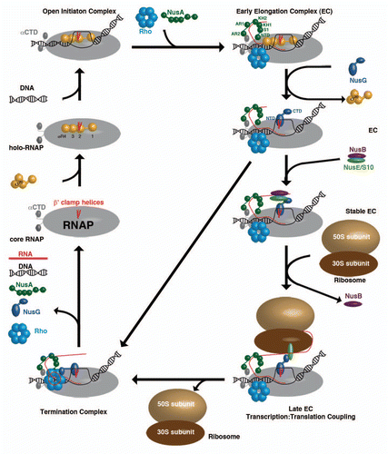
Figure 2 The different ECs within a transcription cycle consisting of RNAP (grey), β′ clamp helices (red), σ70 (yellow), DNA (black), Rho-hexamer (light-blue), NusA (dark-green), RNA (red), NusG (dark-blue), NusB (purple), NusE (light-green), 50S ribosomal subunit (sand) and 30S ribosomal subunit (brown).