Figures & data
Figure 1. The MS2 tethering strategy to decipher the potential contribution of the nuclear retention signal (NRS) in SRSF2 to transcription activation. (A) Schematic illustration of the MS2 tethering strategy. Three copies of the MS2 binding sites were cloned in the first exon of the reporter. The mutant version deleted the stem-loop from the MS2 binding site. (B) Dual-luciferase assay and RT-qPCR analysis upon co-transfection of the GFP-MS2-SRSF2 fusion protein with the luciferase reporters. (C) Dosage-dependent activation of the wild type luciferase reporter at the enzymatic and RNA levels by the GFP-MS2-SRSF2 fusion with or without the NRS (simply labeled as SRSF2 or SRSF2∆NRS). (D) Dual-luciferase assay (left panel) and RT-qPCR (right panel) upon expressing the wild type GFP-MS2-SRSF1 protein or the protein further fused with the NRS from SRSF2.
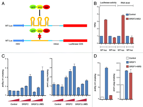
Figure 2. Dissecting the sequence requirement for SRSF2 to act as a transcription activator. (A) Illustration of SRSF2 domains and sub-domains. (B and C) Dual-luciferase and RT-qPCR assays upon expression of different SRSF2 fragments fused to GFP-MS2.
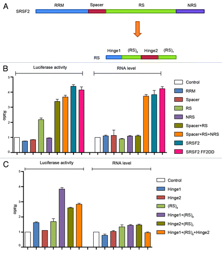
Figure 3. Comparison among SR and hnRNP proteins. (A and D) western blot analysis of MS2-GFP fused to individual SR (panel A) and hnRNP (panel D) proteins detected by anti-GFP antibody. GAPDH protein was probed as the loading control. (B and E) Dual luciferase assays for SR (panel B) and hnRNP (panel E) proteins on wild type (red) and mutant (blue) luciferase reporters. (C and F) RT-qPCR analysis of luciferase mRNA from cells transfected with individual SR (panel C) and hnRNP (panel F) proteins.
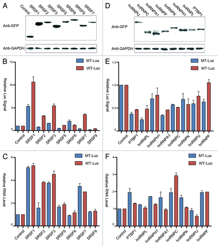
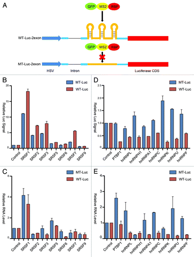
Figure 4. Position-dependent effects of SR and hnRNP proteins in transcriptional and posttranscriptional control. (A) Schematic diagram of the constructs with and without carrying the MS2 binding sites in the second exon of the luciferase reporter. (B and D) Dual luciferase assays for the response to SR (panel B) and hnRNP (panel D) proteins tethered to the second exon of wild type (red) and mutant (blue) luciferase reporters. (C and E) RT-qPCR analysis of reporter RNA in cells transfected with individual SR (panel C) and hnRNP (panel E) proteins fused to GFP-MS2.