
Abstract
Pine wilt disease caused by the pine wood nematode (PWN), Bursaphelenchus xylophilus, has been epidemic and has had disastrous impacts on pine forests and forest ecosystems in eastern Asia. Many pine species in this area are susceptible to this disease. Pinus thunbergii is particularly susceptible. In Japan, tree breeders have selected surviving trees from severely damaged forests as resistant candidates, and have finally established several resistant varieties of P. thunbergii. However, this breeding procedure requires much time and effort due to the lack of physiological and phenotypical information about resistance. To investigate the resistance mechanisms of selected P. thunbergii, we compared histochemical responses, tissue damage expansion, and PWN distribution in resistant and susceptible clones of P. thunbergii after PWN inoculation. The results suggested that the mechanisms of resistance are as follows: damage expansion in the cortex, cambium, and xylem axial resin canals are retarded in resistant trees soon after inoculation, probably due to the induction of wall protein-based defenses. Suppression of PWN reproduction was particularly caused by inhibition of damage expansion in the cambium. The slow expansion of damage in each tissue provides time for the host to complete the biosynthesis of lignin in the walls of cells that surround the damaged regions. This lignification of cell walls is assumed to effectively inhibit the migration and reproduction of the PWNs. The mechanism of initial damage retardation is presumed to be a key for resistance.
Introduction
Pine wilt disease caused by the pine wood nematode (PWN), Bursaphelenchus xylophilus (Kiyohara and Tokushige Citation1971), has brought disastrous damage to pine forests in Japan, China (Zhao Citation2008), and Korea (Shin Citation2008), and is a potential threat to pine forests in Europe (Mota et al. Citation1999; Mota and Vieira Citation2008). In Japan, PWNs are vectored from dead to healthy pine trees by pine sawyer beetles, primarily Monochamus alternatus (Mamiya and Enda Citation1972), and invade young pine branches through wounds made by feeding beetles. PWNs initially enter cortical resin canals and subsequently migrate through the resin canals of the cortex and xylem (Mamiya Citation1985; Myers Citation1986; Ishida et al. Citation1993; Ichihara et al. Citation2000a; Son et al. Citation2010). During migration, they feed on the living cells of the host and reproduce. Various physiological changes are simultaneously induced in the host trees by PWN infection: denaturation, and necrosis of the parenchyma cells (Nobuchi et al. Citation1984; Mamiya Citation1985; Kuroda et al. Citation1988; Fukuda et al. Citation1992; Ishida et al. Citation1993; Ichihara et al. Citation2000a); pathogenesis- and wall-related gene expression (Nose and Shiraishi Citation2011; Hirao et al. Citation2012); increase of acidic (Fukuda et al. Citation1992; Hara et al. Citation2006) and peroxidized (Yamada Citation1987) lipids; ethylene production (Mori and Inoue Citation1986; Fukuda et al. Citation1994); and cavitation and embolism in the tracheids (Ikeda and Suzaki Citation1984; Kuroda et al. Citation1988; Fukuda et al. Citation1992; Umebayashi et al. Citation2011). Dysfunction of water conduction eventually causes the death of host trees.
The PWN is believed to have originated in North America (de Guiran and Bruguier Citation1989) and the damage caused by it was first reported in 1905 (Yano Citation1913). Unfortunately, Japanese pine species such as P. thunbergii and P. densiflora are highly susceptible to this invasive nematode. Researchers and tree breeders started the first resistance breeding program in western Japan in 1978 as a cooperative effort between the national forest tree breeding institute and 14 prefectural research organizations (Toda Citation2004). Surviving pine trees were selected from severely damaged stands as resistant candidates, and, based on the results of two PWN inoculation tests, a moderate number of resistant individuals were finally selected. The resistance of the selected pine trees was ranked from grade 1 (low) to 5 (high) in the order of the survival rate of openly pollinated progenies that were inoculated with PWNs.
The breeding projects have been advanced through the great effort of many tree breeders in order to develop resistant pine varieties. However, few studies have reported the resistance mechanisms of the selected resistant pine trees against PWNs. Kawaguchi (Citation2006) indicated that the cortical resin canals of resistant P. thunbergii had a smaller diameter than those of non-selected P. thunbergii and that this characteristic might restrict the migration of PWNs. Kuroda (Citation2004) indicated that the disordered connection of cortical and xylem resin canals at branch joints retarded PWN migration in resistant P. densiflora. Nose and Shiraishi (Citation2011) and Hirao et al. (Citation2012) reported differential expression of several defense genes in resistant and susceptible P. thunbergii after PWN inoculation. Although some characteristics that might be related to PWN resistance have been proposed by researchers, details of resistance mechanisms are not fully understood.
In this study, we compared histological responses and tissue damage expansion in resistant and susceptible P. thunbergii that were inoculated with the PWNs in order to determine the tissues and processes that directly inhibit the migration and reproduction of PWNs. In addition, while most previous studies prepared samples for light microscopy by subjecting them to fixation and dehydration procedures, we prepared samples for light microscopy without employing such procedures in order to avoid the dissolution of chemicals, including resin.
Materials and methods
Plant materials and nematode inoculation
A resistant tree of the variety ‘Namikata 73’, the most highly resistant variety (grade 5) of P. thunbergii selected as part of a breeding project (Toda Citation2004), was planted in the Forestry and Forest Products Research Institute, Forest Tree Breeding Center (FFPRI-FTBC) in Ibaraki, Japan. A susceptible tree of the variety ‘Mizunami 1’, selected as a plus-tree for growth traits, was also planted in the FFPRI-FTBC. Both clones used in the present study were grafts obtained from the original trees at the FFPRI-FTBC in 2006. The grafted saplings were cultivated for 3.5 years.
Inoculation with a highly virulent isolate (Ka-4) of PWN was conducted on July 1, 2009. The tips (2 cm) of six susceptible clones (tree numbers: S1–S6) and six resistant clones (R1–R6) were cut off, and 10,000 nematodes that had been suspended in 100 μL sterile water were injected into each cut edge (Fig. ). Non-inoculated samples were created by injecting sterile water (without nematodes) into the cut edges of the main stem of an additional three susceptible clones and three resistant clones. Stem segments at 1–3 and 15–17 cm below the stem apex were collected from two inoculated trees and one non-inoculated tree at 3, 10, and 20 days after inoculation (dai). Two-centimeter-long segments were frozen immediately in liquid nitrogen, and stored at −20 °C. Stem segments 3–5 cm below the stem apex were also collected from the inoculated trees. PWNs were extracted from these segments using the Baermann funnel method and counted.
Fig. 1 Scheme of nematode inoculation and stem segment sampling. Pine wood nematodes were inoculated into the cut edge of the current shoot of the main stem. Stem segments 1–3 and 15–17 cm below the inoculation point were used for light microscopic observations and a segment 3–5 cm below the inoculation point was used for nematode extraction using the Baermann funnel method 3, 10, and 20 days after inoculations
Tissue sectioning and staining
Several cross sections were obtained from the middle part of each 2-cm-long segment (Fig. ). The frozen segments were embedded in frozen glue and sectioned using a cryostat CM1500S (Leica Microsystems, Japan). Using a multi-purpose cryosection preparation kit (Leica Microsystems), 10 μm-thick sections were bonded to an adhesive clear plastic film (Kawamoto Citation2003). The use of a cryosection preparation kit enabled sectioned nematodes and pine tissues to be fixed in position throughout the staining procedure. Sections were stained with toluidine blue O to observe cell walls and phenolic compounds (O'Brien et al. Citation1964; Ramalingam and Ravindranath Citation1970), fast blue B to detect phenolics (Heintz and Blaich Citation1990), phloroglucinol-HCl to detect lignin (Jensen Citation1962), or fluorescein isothiocyanate-conjugated wheat germ agglutinin (F-WGA) to detect nematodes by green fluorescence under blue excitation light (Komatsu et al. Citation2008; Son et al. Citation2010). Suberin was detected by autofluorescence under UV light using the sections stained with phloroglucinol-HCl (Biggs Citation1984). Protein cross-linking was visualized using 30-μm-thick sections. The sections were submerged in 1 % sodium dodecyl sulfate (SDS) for 24 h at 80 °C, stained with 0.1 % Coomassie blue in 40 % ethanol/10 % acetic acid, and rinsed with 40 % ethanol/10 % acetic acid solution (Mellersh et al. Citation2002). Nematode sections were shed from the pine sections during this staining procedure. All stained sections were mounted with 30 % glycerol and examined under a light microscope (Nikon E400) equipped with an epifluorescence attachment (Nikon Y-FL).
Evaluation of tissue damage
Damaged and undamaged cortical and xylem axial resin canals in the sections were counted under a light microscope. The areas of healthy and discolored bark on the cross-cut surface of the segments were measured using digital photographs and the graphics software package ImageJ (http://rsbweb.nih.gov/ij/). The circular length of cambium and cambial cavities were measured using digital photographs of the sections under a light microscope and the graphics software package ImageJ. Damage in each tissue was evaluated as the proportion of damaged cortical and xylem axial resin canals, the area of discolored bark, and the circular length of cambial cavities.
Results
Damage expansion in cortical resin canals
At 3 dai, almost all cortical resin canals were damaged by PWNs in segments 2 and 16 cm below the inoculation point in both resistant and susceptible P. thunbergii (Fig. ). However, few PWNs were observed in these canals (Fig. ). Several epithelial cells of the cortical resin canals collapsed due to PWN attack and living epithelial cells were swollen (Fig. a). At 10 and 20 dai, PWNs had destroyed most of the epithelial cells in the cortical resin canals in segments of both resistant and susceptible trees 2 and 16 cm below the inoculation point. Occlusion of the cortical resin canals in these sections by swollen epithelial cells was occasionally observed.
Fig. 2 Percentages of individual tissues damaged by pine wood nematodes in resistant (a) and susceptible (b) P. thunbergii. Data were calculated as the proportion of damaged cortical and xylem axial resin canals, the area of discolored bark, and the circular length of cambial cavities. R1–R6 and S1–S6 indicate individual resistant and susceptible trees, respectively
Fig. 3 Number of pine wood nematodes distributed in individual tissues in cross-sections of resistant (a) and susceptible (b) P. thunbergii. For tissues containing more than 50 nematodes, the numbers of nematodes are shown in parentheses on the column. R1–R6 and S1–S6 indicate individual resistant and susceptible trees, respectively
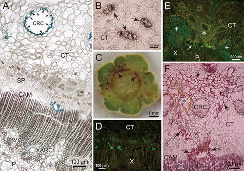
Fig. 4 Cross-sections of resistant P. thunbergii stems inoculated with pine wood nematodes. a A section obtained 2 cm below the inoculation point at 3 days after inoculation (dai) that was stained with Coomassie blue following SDS treatment. Blue color indicates accumulation of protein cross-linking. Arrows and arrow heads indicate swelling epithelial cells and epithelial cells collapsed by nematodes, respectively. b A non-stained section taken from 16 cm below the inoculation point at 10 dai. Arrows indicate cortical resin canals occluded by gel-like resin. c Cut surface of the stem of R3, 2 cm below the inoculation point at 10 dai. Discoloration of bark occurred beside damaged cambium (arrow). d Epifluorescent image of fluorescein isothiocyanate-conjugated wheat germ agglutinin (F-WGA)-stained section obtained 2 cm below the inoculation point of R3 at 10 dai. Round and rectangular green fluorescence indicates the presence of nematodes. Many nematodes were distributed in cambial cavities. e Epifluorescent image of F-WGA-stained section obtained 2 cm below the inoculation point of R6 at 20 dai. Nematodes are distributed in discolored bark tissue (asterisk), cambial cavity (arrow), and pith, but not in healthy bark tissue (plus). f A section obtained 2 cm below the inoculation point of R6 at 20 dai that was stained with phloroglucinol-HCl. Red color indicates lignin accumulation. Lignification induced by nematodes (arrow) occurred in the walls surrounding a cambial cavity and discolored bark tissue (asterisk). CAM cambium, CRC cortical resin canal, CT cortex, P pith, SP secondary phloem, X xylem, XARC xylem axial resin canal
Many cortical resin canals accumulated some amount of liquid resin. However, at 10 and 20 dai, the resin was weakly solidified, gel-like, and occluded canals of the resistant pine trees 16 cm below the inoculation point (Fig. b). Such physical metamorphosis of resin was not observed in the living cortex of the susceptible trees.
Damage expansion in cortex, cambium, and xylem, and distribution of PWNs
The damage in the xylem and cambium was more advanced 2 cm below the inoculation point in one of the two susceptible trees examined at 3 dai (i.e., S1) than in the other susceptible tree and two resistant trees. The epithelial cells were destroyed in 72 % of the axial resin canals in the xylem and ∼7 % of the circumference of the cambium (Figs. , a). In this sample, there were more PWNs in the cavities of the cambium, the damaged axial resin canals, and the intercellular space of the cortex and pith than in other stem sections (Figs. , a). In addition, more PWNs were extracted from the segment 3–5 cm below the inoculation point in S1 than from the corresponding segments in the other susceptible (S2) and two resistant (R1 and R2) trees (Fig. ). In segments 2 and 16 cm below the inoculation point from S2, R1, and R2 at 3 dai, there was little or no damage to any tissue, except the cortical resin canals (Fig. ).
Fig. 5 Cross-sections of susceptible P. thunbergii stems inoculated with pine wood nematodes. a A section obtained 2 cm below the inoculation point of S1 at 3 days after inoculation (dai) that was stained with toluidine blue. Arrows indicate the section of nematodes. b A section obtained 2 cm below the inoculation point at 10 dai that was stained with Coomassie blue following SDS treatment. Blue color indicates the accumulation of protein cross-linking. c Epifluorescent image of a section obtained 16 cm below the inoculation point at 20 dai that was stained with fluorescein isothiocyanate-conjugated wheat germ agglutinin. Round and rectangular green fluorescence indicates the presence of nematodes. Many nematodes were distributed in cambial cavities, cortex, and xylem parenchyma. d A section obtained 2 cm below the inoculation point at 10 dai that was stained with phloroglucinol-HCl. Red color indicates lignin accumulation. Arrows and an arrowhead indicate the lignification of cortical walls and a xylem axial resin canal, respectively. The surrounding of cambial cavities (asterisk) were browned but little lignified. CAM cambium, CRC cortical resin canal, CT cortex, P pith, SP secondary phloem, X xylem, XARC xylem axial resin canal
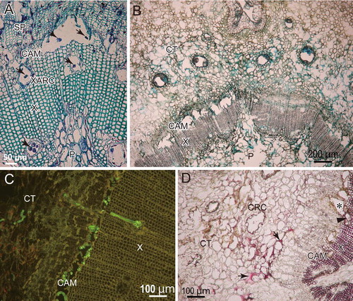
Fig. 6 Number of pine wood nematodes extracted from P. thunbergii stem segments 3–5 cm below the inoculation site. R1–R6 and S1–S6 indicate individual resistant and susceptible trees, respectively
At 10 dai, almost all epithelial cells of the xylem axial resin canals were destroyed and most parts of the bark were discolored 2 cm below the inoculation point in both susceptible trees (S3 and S4). In these trees, ∼55 % of the circumference of the cambium was destroyed (Fig. ) and there were large and continuous cavities (Fig. b). The PWNs were distributed in the cortical resin canals, the cortex, the cavities of the cambial zone, the ray parenchyma, the axial resin canals in the xylem, and the pith (Fig. ). The large number of PWNs suggested by sections of the region 2 cm below the inoculation point in S3 and S4 was supported by PWN extraction from segments 3–5 cm below the inoculation point (Fig. ). Resinosis was partly observed in the cortex, the cavities of the cambium, the xylem axial resin canals, and the pith in sections 2 cm below the inoculation point of the susceptible trees. At the site 16 cm below the inoculation point, 10–20 % of the xylem axial resin canals were destroyed, while 0–3 % of the cambium was damaged (Fig. ). In one of two resistant trees at 10 dai (R3), the percentage of the cambial destruction at 2 cm below the inoculation point was similar to that of the susceptible trees, but the destruction of the axial resin canals in the xylem and the discoloration of the bark were not as severe (Fig. ). The discoloration of the bark was limited to areas surrounding the destroyed cambium (Fig. c). There were a greater number of PWNs in the cavities of the cambium than in the cortex and the xylem axial resin canals (Figs. , d). In addition, while the total number of the PWNs in the section 2 cm below the inoculation point in R3 was similar to that in susceptible trees (Fig. ), relatively few PWNs were extracted from the segment 3–5 cm below the inoculation point (Fig. ). In the other resistant tree (R4), the damage caused by PWNs was limited at 10 dai (Fig. ). No destruction was observed in the cambium, cortex, or xylem 16 cm below the inoculation point in either resistant tree.
At 20 dai, the tissues 2 cm below the inoculation point in susceptible trees (S5 and S6) were completely dead and dried. However, for individual tissues, the percentage that had been destroyed was similar to that observed at 10 dai (Fig. ). There were fewer PWNs in the dried sections of susceptible trees 20 dai than in the corresponding sections of susceptible trees at 10 dai (Fig. ). Similarly, relatively few PWNs were extracted from segments 3–5 cm below the inoculation point (Fig. ). In contrast, a very large number of PWNs were distributed 16 cm below the inoculation point of these trees. Several hundred PWNs were observed in the cavities of the cambium (Figs. , c), ∼95 % of which was destroyed (Fig. ). Though many PWNs were observed in the cambium, cortex, axial resin canals, and pith, few PWNs were observed in the cortical resin canals (Fig. ). In one resistant tree at 20 dai (R5), most parts of the tissues in the section 2 cm below the inoculation point were more damaged than in the other resistant tree (R6) (Fig. ) and the bark was partially deformed because of tissue death. There were more PWNs in the segment 3–5 cm below the inoculation point in R5 than in the corresponding segment in R6 (Fig. ). In the section 2 cm below the inoculation point of R6, the cambial destruction and the bark discoloration were restricted to a narrow area (Figs. , e, f). Many PWNs were distributed in the cambial cavities and the pith, and some were found in the discolored region of the cortex and the destroyed axial resin canals (Fig. e). The number of PWNs and percentage tissue damaged by PWNs gradually increased with time in the sections 16 cm below the inoculation points in resistant trees, but were at a much lower level than in susceptible trees.
Histochemical responses
At 3 dai, the inner surface of the damaged cortical resin canals had accumulated protein cross-linking, particularly in collapsed cells (Fig. a), and were slightly lignified in both susceptible and resistant trees. The accumulation of protein cross-linking was also observed in the walls of the parenchyma cells that surrounded the destroyed cambium and axial resin canals (Fig. a) and in part of apparently intact axial resin canals, cortex, and pith. The accumulation of protein cross-linking was widely observed in those sections in which many PWNs were distributed (i.e., 2 cm below the inoculation point of S1).
At 10 dai, in both susceptible and resistant trees, protein cross-linking accumulated around the destroyed cells of the cambium and the resin canals in the xylem and cortex, and in the discolored cortex and pith (Fig. b). Because the distribution of protein cross-linking coincided with the regions damaged by PWNs, protein cross-linking was evident in more of the tissue in susceptible tree sections than in resistant tree sections. In both susceptible and resistant trees, lignification of the inner walls of the cortical resin canals was more advanced than that at 3 dai. There was little evidence of lignification around the cambial cavities (Fig. d), while the walls of the destroyed axial resin canals, cortex, and pith were partly lignified in many sections (Fig. d). The accumulation of phenolic compounds was observed in parenchyma cells of the xylem and phloem that were near heavily damaged cambium.
In the resistant tree in which tissue damage was restricted at 2 cm below the inoculation point until 20 dai (R6), the cell walls surrounding the cambial cavities and the discolored cortex were highly lignified (Fig. f) and the PWNs were distributed in the compartmentalized areas (Fig. e). In other sections with extensive damage at 20 dai, the lignified cell walls in the cortex were scattered and no compartmentalization due to lignification was observed.
Suberization of the cell walls and wound periderm formation were not observed in any samples in this study.
Discussion
This study revealed the differences between susceptible and resistant P. thunbergii in damage expansion after inoculation with a highly virulent isolate of B. xylophilus. Overall, the damage expansion of the cortex, cambium, and xylem parenchyma tissues, including the axial resin canals, was slower in resistant pine samples than in susceptible samples. The distribution of PWNs was concentrated in the cambial cavities of some resistant pine trees and in the segment that was 16 cm below the inoculation point of susceptible samples at 20 dai, suggesting that PWNs primarily reproduce in the cambium by feeding on cambial cells. Furthermore, the concentrated distribution of PWNs in the cambial cavities of resistant pine trees implied that PWNs could not easily move from the cambium to the bark and xylem. The retarded damage expansion in the cortex and the xylem is assumed to result from this difficulty of horizontal migration because the areas in which PWNs were found closely coincided with the damaged regions of cortex and xylem in this and previous studies (Ichihara et al. Citation2000a). In contrast to resistant pine trees, a moderate number of PWNs were distributed among all tissues of the susceptible pine trees. The distribution of PWNs was accompanied by tissue destruction, and PWNs soon migrated to, and reproduced in, the lower part of the stem. The limitation of PWN migration in resistant samples probably affected the early distribution of the PWNs inoculated into resistant trees, i.e., the distribution at 3 dai. When PWNs are inoculated into the cross-cut surface of stems, they migrate vertically through both cortical and xylem axial resin canals (Son et al. Citation2010). PWNs migrate horizontally from the cortical resin canals to the cortex, and from the xylem axial resin canals to ray parenchyma tissues (Ichihara et al. Citation2000a; Son et al. Citation2010). In this study, PWNs easily migrated via the cortical resin canals in both resistant and susceptible trees, as indicated by the damage to the epithelial cells even at the site 16 cm from the inoculation point in both varieties at 3 dai. Nevertheless, the PWN populations in the cortex, cambium, and xylem parenchyma were smaller in the resistant trees than in susceptible trees, probably due to the difficulty of horizontal migration in resistant trees.
The accumulation of protein cross-linking was the quickest histochemical response in cell walls damaged and stimulated by PWN attack. The cross-linkage of proteins results from the oxidation of proteins by reactive oxygen species in the absence of O2 (Berlett and Stadtman Citation1997; Dean et al. Citation1997). It is reported that protein cross-linking in plant cell walls occurs in response to infections with bacteria (Brown et al. Citation1998), fungi (Thordal-Christensen et al. Citation1997; Mellersh et al. Citation2002), and parasitic plants (Pérez-de-Luque et al. Citation2006), and it is assumed that the cross-linkage of wall proteins, such as extensins, (hydroxy)proline-rich proteins, and glycine-rich proteins, contribute to the strength of cell walls (Bradley et al. Citation1992; Showalter Citation1993; Brisson et al. Citation1994). Hirao et al. (Citation2012) reported that the expression of putative hydroxyproline-rich glycoprotein and extensin genes was higher in ‘Namikata 73’ at 14 dai than in susceptible trees at 7 dai. In the present study, resistant trees at 20 dai and susceptible trees at 10 dai showed similar damage and protein cross-linking in the tissues. These observations imply the possibility that proteins related to wall strength are more highly expressed in individual walls of resistant pine trees than in those of susceptible pine trees, and that the expression of these proteins is beneficial to the restriction of nematode migration and cell destruction.
Another resistance mechanism observed in this study was the occlusion of the cortical resin canals by gel-like resin, which was formed up to 10 dai in resistant samples. The hardening of cortical resin may help to prevent the vertical migration of PWNs. It is reported that interruption of cortical resin canals restricts vertical migration of PWNs (Ishida et al. Citation1993; Ichihara et al. Citation2000b; Kuroda Citation2004; Son et al. Citation2010) and that this interruption may be related to resistance (Kuroda Citation2004). In addition, the inner walls of the cortical resin canals started to lignify 3 dai in both resistant and susceptible samples. Lignification of the cortical resin canals may prevent the transfer of PWNs between the cortical resin canals and the cortex.
The retarded expansion of cell destruction as a result of resistance mechanisms provides trees with sufficient time to accumulate chemical metabolites, such as lignin, in the surrounding tissues. Lignin synthesis and lignification are both more time-consuming and more powerful defenses than protein-based defenses (Kusumoto Citation2005). In the resistant sample in which cambial destruction was limited until 20 dai, the tissues surrounding the destroyed cambium were highly lignified. The hardness of the lignified cell walls surrounding the cambial cavities presumably restricted the feeding of the PWNs and resulted in the suppression of PWN reproduction. Limiting the size of the PWN populations helps to limit the expansion of tissue damage.
Many previous studies that compared different resistant pine species or inoculation with virulent PWNs to inoculation with avirulent PWNs have indicated that the migration and reproduction of PWNs are important factors that determine the death or survival of pine trees (Tamura and Dropkin Citation1984; Kuroda et al. Citation1991; Yamada and Ito Citation1993; Ichihara et al. Citation2000a). The present study demonstrated that the resistant variety of the susceptible pine species, P. thunbergii, retarded the initial migration of inoculated PWNs by blockages in the cortex, cambium, and xylem, which probably depended on constitutive and/or induced protein-based defenses. Exclusion of PWNs from the cambium is particularly important for suppression of PWN reproduction (Sasaki et al. Citation1984; Fukuda Citation1997). If PWNs easily overcome cambial defenses and reproduce, they can spread throughout the tree and mass-attack living tissues before time-consuming defenses, such as lignification, can be deployed. The damage expansion in xylem eventually results in embolism of tracheids and tree death (Ikeda and Kiyohara Citation1995). Therefore, it is presumed that the restriction of tissue destruction soon after PWN invasion is a key factor of resistance against pine wilt disease in P. thunbergii.
Acknowledgment
This work was supported by JSPS Grants-in-Aid for Scientific Research (No. 20380085, 23248024).
Notes
Open Access This article is distributed under the terms of the Creative Commons Attribution License which permits any use, distribution, and reproduction in any medium, provided the original author(s) and the source are credited.
References
- BerlettBSStadtmanERProtein oxidation in aging, disease, and oxidative stressJ Biol Chem1997272203132031610.1074/jbc.272.33.20313
- BiggsARIntracellular suberin: occurrence and detection in tree barkIAWA Bull NS1984524324810.1163/22941932-90000899
- BradleyDJKjellbornPLambCJElicitor- and wound-induced oxidative cross-linking of a proline-rich plant cell wall protein: a novel, rapid defense responseCell199270213010.1016/0092-8674(92)90530-P
- BrissonLFTenhakenRLambCFunction of oxidative cross-linking of cell wall structural proteins in plant disease resistancePlant Cell1994617031712160556
- BrownITrethowanJKerryMMansfieldJBolwellGPLocalization of components of the oxidative cross-linking of glycoproteins and of callose synthesis in papillae formed during the interaction between non-pathogenic strains of Xanthomonas campestris and French bean mesophyll cellsPlant J19981533334310.1046/j.1365-313X.1998.00215.x
- GuiranGBruguierNHybridization and phylogeny of the pine wood nematode (Bursaphelenchus spp.)Nematologica19893532133010.1163/002825989X00421
- DeanRTFuSStockerRDaviesMJBiochemistry and pathology of radical-mediated protein oxidationBiochem J19973241181218394
- FukudaKPhysiological process of the symptom development and resistance mechanism in pine wilt diseaseJ For Res1997217118110.1007/BF02348216
- FukudaKHogetsuTSuzukiKCavitation and cytological changes in xylem of pine seedlings inoculated with virulent and avirulent isolates of Bursaphelenchus xylophilus and B. mucronatusJ Jpn For Soc199274289299
- FukudaKHogetsuTSuzukiKEthylene production during the symptom development of pine wilt disease caused by Bursaphelenchus xylophilusEur J For Pathol19942419320210.1111/j.1439-0329.1994.tb00985.x
- HaraNTakeuchiYFutaiKCytological changes in ray parenchyma cells of seedlings of three pine species infected with the pine wilt diseaseJpn J Nematol200636233310.3725/jjn.36.23
- HeintzCBlaichRUltrastructural and histochemical studies on interactions between Vitis vinifera L. and Uncinula necator (Schw.) BurrNew Phytol199011510711710.1111/j.1469-8137.1990.tb00928.x
- HiraoTFukatsuEWatanabeACharacterization of resistance to pine wood nematode infection in Pinus thunbergii using suppression subtractive hybridizationBMC Plant Biol20121213339826810.1186/1471-2229-12-13
- IchiharaYFukudaKSuzukiKEarly symptom development and histological changes associated with migration of Bursaphelenchus xylophilus in seedling tissues of Pinus thunbergiiPlant Dis20008467568010.1094/PDIS.2000.84.6.675
- IchiharaYFukudaKSuzukiKThe effect of periderm formation in the cortex of Pinus thunbergii on early invasion by the pinewood nematodeFor Pathol20003014114810.1046/j.1439-0329.2000.00199.x
- IkedaTKiyoharaTWater relations, xylem embolism and histological features of Pinus thunbergii inoculated with virulent or avirulent pine wood nematode, Bursaphelenchus xylophilusJ Exp Bot19954644144910.1093/jxb/46.4.441
- IkedaTSuzakiTInfluence of pine-wood nematodes on hydraulic conductivity and water status in Pinus thunbergiiJ Jpn For Soc198466412420
- IshidaKHogetsuTFukudaKSuzukiKCortical responses in Japanese black pine to attack by the pine-wood nematodeCan J Bot1993711399140510.1139/b93-168
- JensenWABotanical histochemistry1962San FranciscoFreedom
- KawaguchiERelationship between the anatomical characteristics of cortical resin canals and migration of Bursaphelenchus xylophilus in stem cuttings of Pinus thunbergii seedlingsJ Jpn For Soc20068824024410.4005/jjfs.88.240
- KawamotoTUse of a new adhesive film for the preparation of multi-purpose fresh-frozen sections from hard tissues, whole-animals, insects and plantsArch Histol Cytol20036612314310.1679/aohc.66.123
- KiyoharaTTokushigeYInoculation experiments of a nematode, Bursaphelenchus sp., onto pine treesJ Jpn For Soc197153210218
- KomatsuMSonJMatsushitaNHogetsuTFluorescein-labeled wheat germ agglutinin stains the pine wood nematode, Bursaphelenchus xylophilusJ For Res20081313213610.1007/s10310-008-0065-9
- KurodaKInhibiting factors of symptom development in several Japanese red pine (Pinus densiflora) families selected as resistant to pine wiltJ For Res2004921722410.1007/s10310-004-0076-0
- KurodaKYamadaTMineoKEffects of cavitation on the development of pine wilt disease caused by Bursaphelenchus xylophilusAnn Phytopathol Soc Jpn19885460661510.3186/jjphytopath.54.606
- KurodaKYamadaTItoSBursaphelenchus xylophilus induced pine wilt: factors associated with resistanceEur J For Pathol19912143043810.1111/j.1439-0329.1991.tb00780.x
- KusumotoDConcentrations of lignin and wall-bound ferulic acid after wounding in the phloem of Chamaecyparis obtusaTrees20051945145610.1007/s00468-004-0404-1
- MamiyaYInitial pathological changes and disease development in pine trees induced by the pine wood nematode, Bursaphelenchus xylophilusAnn Phytopathol Soc Jpn19855154655510.3186/jjphytopath.51.546
- MamiyaYEndaNTransmission of Bursaphelenchus lignicolus (Nematoda: Aphelenchoididae) by Monochamus alternatus (Coleoptera: Cerambycidae)Nematologica19721815916210.1163/187529272X00395
- MellershDGFouldsIVHigginsVJHeathMCH2O2 plays different roles in determining penetration failure in three diverse plant-fungal interactionsPlant J20022925726810.1046/j.0960-7412.2001.01215.x
- MoriTInoueTPine-wood nematode-induced ethylene production in pine stems and cellulase as an inducerJ Jpn For Soc1986684350
- MotaMMVieiraPCZhaoBGFutaiKSutherlandJRTakeuchiYPine wilt disease in PortugalPine wilt disease2008TokyoSpringer3338
- MotaMMBraaschHBravoMAPenasACBurgermeisterWMetgeKSousaEFirst report of Bursaphelenchus xylophilus in Portugal and in EuropeNematology1999172773410.1163/156854199508757
- MyersRFCambium destruction in conifers caused by pinewood nematodesJ Nematol1986183984022618560
- NobuchiTTominagaTFutaiKHaradaHCytological study of pathological changes in Japanese black pine (Pinus thunbergii) seedlings after inoculation with pine-wood nematode (Bursaphelenchus xylophilus)Bull Kyoto Univ For198456224233
- NoseMShiraishiSComparison of the gene expression profiles of resistant and non-resistant Japanese black pine inoculated with pine wood nematode using a modified Long SAGE techniqueFor Pathol20114114315510.1111/j.1439-0329.2010.00646.x
- O'BrienTPFederNMcCullyMEPolychromatic staining of plant cell walls by toluidine blue OProtoplasma19645936837310.1007/BF01248568
- Pérez-de-LuqueAGonzález-VerdejoCILozanoMDDitaMACuberoJIGonzález-MelendiPRisueñoMCRubialesDProtein cross-linking, peroxidase and β-1,3-endoglucanase involved in resistance of pea against Orobanche crenataJ Exp Bot2006571461146910.1093/jxb/erj127
- RamalingamKRavindranathMHHistochemical significance of green metachromasia to toluidine blueHistochemie197024322327
- SasakiSOdaniKNishiyamaYHayashiYDevelopment and recovery of pine wilt disease studied by tracing ascending sap flow marked with water soluble stainsJ Jpn For Soc198466141148
- ShinSCZhaoBGFutaiKSutherlandJRTakeuchiYPine wilt disease in KoreaPine wilt disease2008TokyoSpringer2632
- ShowalterAMStructure and function of plant cell wall proteinsPlant Cell19935923160246
- SonJAKomatsuMNatsushitaNHogetsuTMigration of pine wood nematodes in the tissues of Pinus thunbergiiJ For Res20101518619310.1007/s10310-009-0171-3
- TamuraHDropkinVResistance of pine trees to pine wilt caused by the nematode, Bursaphelenchus xylophilusJ Jpn For Soc198466306312
- Thordal-ChristensenHZhangZWeiYCollingeDBSubcellular localization of H2O2 in plants. H2O2 accumulation in papillae and hypersensitive response during the barley-powdery mildew interactionPlant J1997111187119410.1046/j.1365-313X.1997.11061187.x
- TodaTStudies on the breeding for resistance to the pine wilt disease in Pinus densiflora and P. thunbergiiBull For Tree Breed Cen20042083217
- UmebayashiTFukudaKHaishiTSotookaRZuhairSOtsukiKThe developmental process of xylem embolisms in pine wilt disease monitored by multipoint imaging using compact magnetic resonance imagingPlant Physiol2011156943951317728810.1104/pp.110.170282
- YamadaTLipid peroxidation during the development of pine wilt diseaseAnn Phytopathol Soc Japn19875352353010.3186/jjphytopath.53.523
- YamadaTItoSHistological observations on the responses of pine species, Pinus strobus and P. taeda, resistant to Bursaphelenchus xylophilus infectionAnn Phytopathol Soc Jpn19935965966510.3186/jjphytopath.59.659
- YanoMInvestigations on the cause of pine mortality in Nagasaki prefectureSanrin-Koho19134 (suppl)114
- ZhaoBGZhaoBGFutaiKSutherlandJRTakeuchiYPine wilt disease in ChinaPine wilt disease2008TokyoSpringer1825