Abstract
The aim of this study was to compare modified Limberg flap procedure with excision and primary closure in the treatment of uncomplicated pilonidal disease.
Methods: This study was conducted on 120 patients with uncomplicated sacrococcygeal pilonidal disease that were randomly allocated into two groups: group I underwent excision and primary closure; group II underwent modified Limberg flap procedure. The duration of operation, postoperative pain, length of hospital stay, duration of incapacity for work, postoperative complications and postoperative recurrence were recorded.
Results: Duration of operation was longer in group II than in group 1 (P < 0.001). However, postoperative pain was less (P < 0.001), duration of hospital stay shorter (P < 0.001), time to resumption of work shorter (P < 0.001) and postoperative complications were significantly fewer in group II. During follow-up period of 21.5 ± 6.82 (months) for group I and 22 ± 7.64 (months) for group II, single case of recurrence 1.67% was detected in patients in group II versus four patients (6.67% patients) in group I (P = 0.032).
Conclusion: Wide excision with a modified Limberg transposition flap reconstruction is a very effective operative procedure for uncomplicated pilonidal sinus, associated with a low complication rate, short hospitalization and disability, and a low recurrence rate.
1 Introduction
Pilonidal sinus is known to be a simple condition that refers to a tract or cavity that contains loose hair and is associated with repeated infection and abscess formation. Its incidences are higher in male than in female and increase with obesity and hairy skin.Citation1 It is more common in people aged 15–30 years after puberty due to the effect of sex hormones on pilosebaceous glands and change in healthy body hair growth.Citation2
Although different surgical approaches have been used to manage sacrococcygeal pilonidal sinus, none of these approaches eliminate the postoperative morbidity including delayed wound healing, discomfort and high rate of recurrence which range between 1% and 43% in different studies.Citation3
The surgical wound may be left to heal by open healing (secondary intention). Advocates of this technique state that reduced wound tension facilitates trouble free healing without recurrence if all sinus tracts are fully excised. Alternatively, the wound may be closed to heal by primary closure (primary intention). Methods can be broadly categorized as midline closure techniques (with the wound lying within the natal cleft) or other techniques (where the wounds placed out with the midline). Advocates of primary closure perceive benefits of faster tissue healing.Citation4,Citation5
Excision and primary closure involve excision of the entire sinus with closure of the wound. This procedure has the advantage of avoiding wound packing. One problem is that the incision tends to be situated in a deep midline cleft where there is tension and also the propensity to accumulate hair.Citation4
Skin flaps have been described to cover a sacral defect after wide excision; this keeps the scar off the midline and flattens the natal cleft. The techniques available include the cleft closure, advancement flap (Karydakis procedure), local advancement flap (V-Y advancement flap), and rotational flap (Limberg flap, modified Limberg flap, gluteus maximus myocutaneous flap).Citation5,Citation6
However, there have been few clinical studies to compare the rhomboid fasciocutaneous transposition flap procedure (modified Limberg flap) with other conventional procedures in the treatment of uncomplicated pilonidal disease. The recurrence rate in the modified Limberg flap group was lower than the recurrence rate in the other flap techniques. The modified Limberg flap technique provides a more efficient flattening of the natal cleft, including the most inferior part that is inclined to invert towards the anal region, lateralization of the inferior apex of the classic Limberg flap decrease recurrences which could occur in the inferior midline.Citation6,Citation7 The aim of this study was to perform a randomized clinical trial to compare the modified Limberg flap procedure with excision and primary closure in the treatment of uncomplicated pilonidal disease.
2 Methods
This study was conducted in Colorectal Unit, Surgery Department at the Alexandria University Hospital during the period from February 2008 to March 2011. One hundred and twenty patients who were treated for uncomplicated pilonidal disease were eligible for the study. The patients who presented with acute pilonidal abscesses were excluded from this study. Recurrent or complex pilonidal sinuses were also excluded from this study.
Informed consent was obtained from all patients included in the study which was approved by the local ethics committee. All patients were subjected to history taking, clinical examination, and laboratory test. Randomization was achieved through a computer-generated schedule and the results were sealed into envelopes. The envelopes were drawn and opened by a nurse in the operating room.The patients were then randomized into two groups: group I underwent excision and primary closure; group II underwent modified Limberg flap procedure.
All patients were operated under general anaesthesia. Patients were placed in prone Jack-knife position with two adhesive straps in each glutted region to pull them laterally to allow better visualization of the natal cleft, then shaving off the hairs around the sinus and cleaning the area with povidone iodine. Antibiotic prophylaxis of 1.2 g amoxicillin–clavulinic acid was given intravenously at the time of induction and continued for 48 h. Then this was changed to oral form for five days.
2.1 Group I (excision and primary closure procedure)
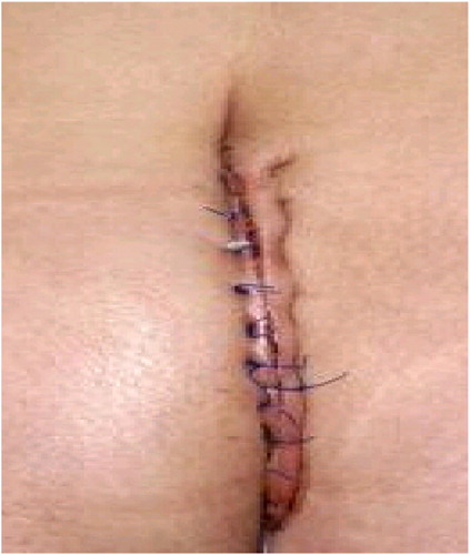
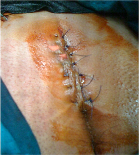
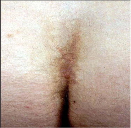
The procedure begins by excising the wound, with the sinuses removed en bloc with a vertical elliptical specimen of overlying skin to the level of the sacrococcygeal fascia. After placing deep approximating 0 polyglactin sutures (VicrylTM; Ethicon, New Jersey, USA), the skin was approximated with 3/0 polyglactin interrupted subcutaneous sutures (VicrylTM; Ethicon, New Jersey, USA) and the skin edges were closed with 2/0 polypropylene interrupted mattress sutures (Propilen; Dogsan, Trabzon, Turkey). At the end of the procedure, a suction drain was inserted from a separate incision ( and ).
2.2 Group II (modified Limberg flap)
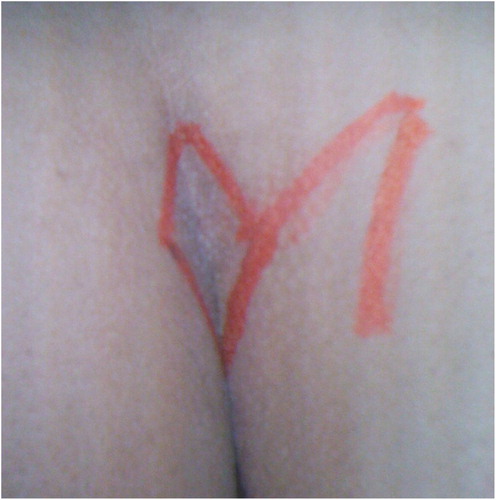
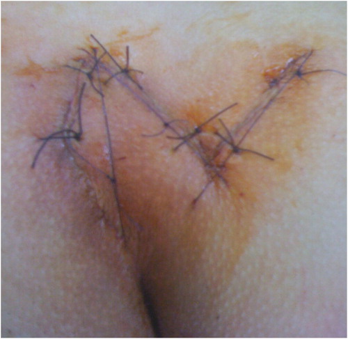
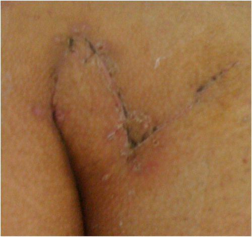
The lesion was excised with a rhomboid shaped incision with each side equal in length ( and ) with lateralization of the inferior apex. The depth of the rhomboid excision was extended to the gluteal fascia. The rhomboid flap was then rotated from the gluteal fascia to the excised area without tension. Subcutaneous tissue sutured with interrupted 3/0 vicryl (VicrylTM; Ethicon, New Jersey, USA) and the skin was sutured separately with interrupted proline 2/0 sutures (Propilen; Dogsan, Trabzon, Turkey) (). At the end of the procedure, a suction drain was inserted. Methylene blue was not used to identify the tracks in either group. A single dose of antibiotic prophylaxis was used immediately before incision.

Patients were discharged when clinically free after the operation, removal of the drain 4–5 days after operation and all patients were advised to visit the outpatient clinic every week for one month and then every 3 months for at least 12 months during the follow-up period. Stitches were removed 14 days postoperatively ( and ). All patients were recommended to walk freely but not to exercise until removal of stitches. All patients were advised to shave the area well around the operative site at least monthly.
The duration of operation, postoperative pain, length of hospital stay, duration of incapacity for work, postoperative complications (infection, flap oedema, wound dehiscence), and postoperative recurrence were recorded. Duration of operation was defined as the length of time between the first incision and placement of the last suture. Postoperative pain was assessed according to a visual analogue scale (VAS) from 0 (no pain) to 10 (worst pain imaginable) on the first postoperative day. Duration of incapacity for work was defined as the time from the date of surgery to the date on which the patient returned to normal activities including employment and leisure activities. Infection was considered as leakage of purulent secretion through the surgical wound and not only peri-incisional hyperaemia.
2.3 Statistical analysis
The statistical analysis of data was done by using excel programme and SPSS program statistical package for social science version 10. The description of the data was done in the form of mean ± SD for quantitative data, frequency and proportion for qualitative data. The analysis of the data was done to test statistical significant difference between groups. For quantitative data Student's t-test was used to compare between two groups. Chi square test, and Fisher's exact test was used for qualitative data.
N.B.: P is significant if ⩽0.05 at confidence interval 95%.
3 Results
This study was conducted on 120 patients with uncomplicated sacrococcygeal pilonidal disease that were randomly allocated by closed envelop technique into two groups, group I (60 patients) the mean age was 27 ± 9.4 years (46 men, and 14 women) who underwent excision and primary closure procedure and group II (60 patients) the mean age was 26 ± 8.6 years (45 men, and 15 women) that underwent modified Limberg flap procedure. There was no significant difference between both groups regarding age, sex, preoperative symptoms, and period of follow-up. Intermitted discharge and pain were the most common symptoms. Follow-up (months) was 21.5 ± 6.82 for group I and 22 ± 7.64 for group II ().
Table 1 Demographic and pre-treatment symptoms of patients with uncomplicated sacrococcygeal pilonidal disease.
Operative data showed that the operative time (minute) for group I was 40.6 ± 6.8 which was significantly less than that for group II 55.2 ± 7.6. However, hospital stay, pain score, period off work, and healing period were significantly higher in group I than for group II ().
Table 2 Outcome after surgical treatment.
Postoperative follow-up showed that incidence of complications was significantly higher among patients of group I as wound infection occurs in six patients 10%, flap oedema in eight patients 13.33%, wound dehiscence in four patients 6.67% and recurrence in four patients 6.67% ().
Table 3 Postoperative complications.
4 Discussion
The varied surgical techniques proposed for the eradication of pilonidal sinuses are evidences of the lack of a completely satisfactory method of management of this surgical problem. Most of the procedures have some merits and are based on seemingly sound surgical principles, but regardless of the methods employed, there is recurrence in a significant proportion of cases.
Complete excision of the sinus is widely practiced, but controversy remains about what to do with the wound after excision.Citation5 Excision and packing, excision and primary closure, marsupialization, and flap techniques are surgical procedures that have been developed for treatment of pilonidal sinus.Citation8 Despite the controversy about the best surgical technique for the treatment of pilonidal sinus, an ideal operation should minimize financial cost, allow patients to return earlier to work, be simple to perform, not require a prolonged hospital stay, inflict minimal pain, and have a low disease recurrence rate.Citation9
Wide excision and healing by secondary intention enjoys considerable popularity, the advantage of this method is that all inflamed tissues are removed and the chance of recurrence is low, but need long hospitalization and daily dressing, moreover time to healing is long and may exceed one year in the most unfortunate patient.Citation10 Compared with open packing and marsupialization, excision and primary closure are known to provide quicker healing and quicker return to work. Most patients return to work in 3–4 weeksCitation11 but recurrence rates of 7–42% have been reported following excision and primary closure,Citation12,Citation13 while a number of studies have reported a recurrence rate of 0–3% after rhomboid excision and Limberg flap repair outweigh the disadvantages related to an unfavourable cosmetic appearance following rhomboid excision and Limberg flap closure.Citation14–Citation16,Citation6
The higher morbidity of surgical techniques was naturally reflected by hospital stay and time off work. In our study, the hospital stay for patients treated with modified Limberg flap was 2.1 ± 1.2 days, which was shorter than 5.5 days as reported by Rossi et al.Citation17 for Limberg flap and 5.7 days as reported by Singh et al.Citation18 for adipo-fasciocutaneous flap. In our study, the mean hospital stay was 3.8 ± 1.6 in primary closure group, and the difference between the groups was significant. Time off work was significantly shorter for patients treated with modified Limberg flap as compared with patients treated with primary closure (16.2 ± 2.1 versus 21.6 ± 3.5 days), and this shorter duration in the flaps group was similar to that reported by Abu Galala et al.Citation19 and Eryilmaz et al.Citation15, while Gilani et al.Citation20 found that the mean time to return to work after primary closure was 25.5 days, while Lieto et al.Citation21 found that the mean time to return to work was 7 days in patients with rhomboid flap while Muzi et al.Citation22 found that there was no significant difference found in time to return to work and wound disruption.
In the current study postoperative pain scores (VAS scores) were significantly lower in group II than in group I (2.1 ± 1.2 versus 5.2 ± 1.4), presumably because less wound tension was created with the Limberg flap procedure, similar results were achieved by MahdyCitation23 and Akca et al.Citation24 Healing period was significantly shorter in group II than in group I (14.1 ± 2.9 days versus 19.1 ± 3.7 days) this is also achieved by Eryilmaz et al.,Citation15 Akca et al.,Citation24 and Muzi et al.Citation22
In the current study the rate of infection in group II was 3.33% which is significantly lower than group I with six cases of infection ie 10%, similar results were reported in literature with an infection rate range of 1.5–6% with Limberg flap,Citation15,Citation24,Citation25 and infection rate around 10% with excision and primary closureCitation24,Citation26 which could be explained by tension on the suture line and the formation of a serosanguineous collection in the subcutaneous layer leads to infection and breakdown of the wound with excision and primary closure.
Preventing recurrence is a major concern in the surgical treatment of pilonidal sinus. Postoperative complications and recurrences, like the original sinus, develop in the midline and as the natal cleft becomes deeper, an anaerobic medium is created, resulting in an increased anaerobic bacterial content.Citation27 Furthermore, the vacuum effect created between the heavy buttocks sucks the anaerobic bacteria, hair, and debris into the subcutaneous fat tissue.Citation28 We believe that our successful results after flap reconstructions stem from the fact that the deep midline is eliminated. When the midline is lateralized or flattened, recurrences are less likely to occur than after primary closure. In the current series recurrence observed in only one patient 1.67% with modified Limberg flap procedure which was significantly lower than recurrence with excision and primary closure which was observed in four patients ie 6.67%. Other authors have reported recurrence rates of 0–3% with Limberg flap repair.Citation14–Citation16,Citation6 FossCitation29 reported on a collective series of 1129 patients treated with excision and closure of their pilonidal disease. Failure of primary healing was 16% and recurrence rate was 16%.
In conclusion, the results of this series provided further evidence that wide excision with a modified Limberg transposition flap reconstruction is a very effective operative procedure for uncomplicated pilonidal sinus, associated with a low complication rate, short hospitalization and disability, and a low recurrence rate.
Notes
Peer review under responsibility of Alexandria University Faculty of Medicine.
Available online 3 December 2011
Related Research Data
References
- G.E.KarydakisEasy and successful treatment of pilonidal sinus after explanation of its causative processAust N Z J Surg621992385389
- H.C.LeeY.H.HoC.F.SeowK.W.EuD.NyamPilonidal disease in Singapore: clinical features and managementAust N Z J Surg702000196198
- A.S.AzabM.S.KamalF.El BassyoniThe rationale of using the rhomboid fasciocutaneous transposition flap for the radical cure of pilonidal sinusJ Dermatol Surg Oncol12198612951299
- K.SondenaaI.NesvikE.AndersenSoreide JA: recurrent pilonidal sinus after excision with closed or open treatment: final result of a randomised trialEur J Surg16231996237240
- H.SpivakV.L.BrooksM.NussbaumFriedman I: treatment of chronic pilonidal diseaseDis Colon Rectum3910199611361139
- B.B.MentesS.LeventogluA.CihanE.TatliciogluM.AkinM.OguzModified Limberg transposition flap for sacrococcygeal pilonidal sinusSurg Today342004419423
- R.RyilmazM.SahinO.AlimogluF.DasiranSurgical treatment of sacrococcygeal pilonidal sinus with the Limberg transposition flapSurgery1342003745749
- J.ArmstrongP.BarciaPilonidal sinus diseaseArch Surg1291994914918
- O.F.AkinciA.CoskunA.UzunkoySimple and effective surgical treatment of pilonidal sinusDis Colon Rectum432000701702
- I.J.McCallumP.M.KingJ.BruceHealing by primary closure versus open healing after surgery for pilonidal sinus: systematic review and meta-analysisBMJ3362008868871
- H.S.KhairaJ.H.BrownExcision and primary closure of pilonidal sinusAnn R Coll Surg Engl771995242244
- K.SondenaaI.NesvikE.AndersenO.NatasJ.A.SoreideBacteriology and complications of chronic pilonidal sinus treated with excision and primary sutureInt J Colorectal Dis101995161166
- I.IesalnieksA.FurstM.RentschK.W.JauchPrimary midline closure after excision of a pilonidal sinus is associated with a high recurrence rateChirurg742003461468
- A.S.AzabM.S.KamalF.el BassyoniThe rationale of using the rhomboid fasciocutaneous transposition flap for the radical cure of pilonidal sinusJ Dermatol Surg Oncol12198612951299
- R.EryilmazM.SahinO.AlimogluF.DasiranSurgical treatment of sacrococcygeal pilonidal sinus with the Limberg transposition flapSurgery1342003745749
- M.KapanS.KapanS.PekmezciV.DurgunSacrococcygeal pilonidal sinus disease with Limberg flap repairTech Coloproctol620022732
- P.RossiF.RusooP.GentileschiThe pilonidal sinus: its surgical management, our experience and a review of the literatureG Chir141993120123
- S.RaghubirM.PavithranAdipo-fascio-cutaneous flaps in the treatment of pilonidal sinus: experience with 50 casesAsian J Surg282005198201
- K.H.Abu GalalaI.M.SalamK.R.Abu SamaanTreatment of pilonidal sinus by primary closure with a transposed rhomboid flap compared with deep suturing: a prospective randomized clinical trialEur J Surg1651999468472
- S.N.GilaniH.FurlongK.ReichardtA.O.NasrG.TheophilouT.N.WalshExcision and primary closure of pilonidal sinus disease: worthwhile option with an acceptable recurrence rateIr J Med Sci18012011173176
- E.LietoP.CastellanoM.PintoA.ZamboliC.PignatekkiG.GaliziaDufourmentel rhomboid flap in the radical treatment of primary and recurrent sacrococcigeal pilonidal diseaseDis Colon Rectum53201010611068
- M.G.MuziG.MilitoF.CadedduC.NigroF.AndreoliF.AmabileRandomized comparison of Limberg flap versus modified primary closure for the treatment of pilonidal diseaseAm J Surg2002010914
- T.MahdySurgical treatment of the pilonidal disease: primary closure or flap reconstruction after excisionDis Colon Rectum5112200818161822 [Epub 2008 October 21]
- T.AkcaT.ColakB.UstunsoyA.KanikS.AydinRandomized clinical trial comparing primary closure with the Limberg flap in the treatment of primary sacrococcigeal pilonidal diseaseBr J Surg9220051081
- K.TopgulE.OzdemirK.KilicH.GokbayirZ.FerahkoseLong-term results of Limberg flap procedure for treatment of pilonidal sinus: a report of 200 casesDis Colon Rectum46200315451548
- A.TocchiG.MazzoniM.BononiV.FornasariM.MicciniA.DrumoOutcome of chronic pilonidal disease treatment after ambulatory plain midline excision and primary sutureAm J Surg19620082833
- J.MarksK.G.HardingL.E.HughesC.D.RibeiroPilonidal sinus excision—healing by open granulationBr J Surg721985637640
- J.U.BascomPilonidal sinusCurr Pract Surg61994175180
- M.V.FossPilonidal sinus: excision and closureProc R Soc Med631970752758