
Abstract
Introduction
Knowledge of the course and anatomical relations of the marginal mandibular branch of the facial nerve (MMBFN) in the head and neck is important to avoid its injury during surgery.
Aim
The aim of this work was to study the course and relations of the (MMBFN) to the lower border of the mandible and parotid gland.
Material and methods
The material of this work included thirty halves of formalin preserved head and neck specimens obtained from the Dissecting Room of Anatomy Department, Faculty of Medicine, Alexandria University.
Results
Results showed that the (MMBFN) arises as a single branch, two branches, and three branches in 36.7%, 43.3% and 20% of specimens, respectively. In 83.3% of cases, one of the main or secondary branches of the marginal mandibular nerve crosses superficial (lateral) to the facial vessels. There are communications either between the main or the secondary branches of the marginal mandibular nerve itself in 53.6% of specimens and with the buccal branch of the facial nerve in 40%, also with the anterior branch of the great auricular nerve in 3.3%, and with the transverse cervical nerve in 3.3% of specimens. The relationship of the nerve to the lower border of the mandible at a point midway between the angle of the mandible and symphysis menti is variable; it is either totally above it in most of the specimens 80%, or below it in 10% or at it in the remaining 10% of the specimens. The branches that lie above the lower border of the mandible are always deep into the superficial layer of the parotid fascia, while those branches that lie below the lower border of the mandible are intrafascially. The termination of the nerve is deep into the muscles of the ipsilateral lower lip in all specimens.
Abbreviation:
1 Introduction
All muscles of facial expression derive their nerve supply from the facial nerve.Citation1 Concerning the extracranial course of the facial nerve, it exits through the stylomastoid foramen and immediately gives off branches to the auricular muscles, the posterior belly of digastric and stylohyoid muscles. Then the nerve courses anteriorly till the posterior edge of the parotid gland, it splits into upper and lower divisions. Within the parotid gland, there are many individual variations in the branching pattern of the facial nerve. As a rule, the upper division of the facial nerve gives off temporal, zygomatic and buccal branches, whereas the lower division gives off marginal mandibular and cervical branches. From these five major branches of the facial nerve, the marginal mandibular branch of the facial nerve (MMBFN) supplies all the muscles of the lower lip.Citation2,Citation3
The most frequent cause of affection of this nerve is iatrogenic injury during operations in the mandibular or parotid regions. It may be injured in parotidectomy, submandibular gland excision, carotid endarterectomy, and liposuction surgery or during the deep dissection of the neck. The high incidence of (MMBFN) injury is due to lack of an accurate description of the course of this nerve in most of the known textbooks of anatomy.Citation4,Citation5
Injury to the (MMBFN) results in a significant cosmetic deformity. The deformity is caused by interruption of nerve fibers to the depressor anguli oris and the depressor labii inferioris that results in flattening and inversion of the ipsilateral half of the lower lip and the inability to move the lip downwards and laterally. Elevation of the affected lower lip is evident when the patient smiles and the asymmetry is the most obvious when the patient cries ‘the asymmetric crying faces’.Citation6
Knowledge of the course and anatomical relations of the (MMBFN) in the upper neck is important to avoid such injury. The choice of the surgical approach is very relevant in parotid surgery because of the extreme anatomical variability of the parotid area and the functional importance of the branches of the facial nerve.Citation7
2 Aim
The aim of this work was to study the course and relations of the (MMBFN) to the lower border of the mandible and parotid gland.
3 Materials and methods
The material of this work included thirty halves of formalin preserved cadaveric head and neck specimens. These specimens were obtained from the Dissecting Room of the Anatomy Department, Faculty of Medicine, Alexandria University. A submandibular incision was done extending antero-inferiorly from the tragus of the auricle down to the symphysis menti, running below the lower border of the mandible. After reflection of skin flap and fascia upwards and downwards, the platysma muscle was separated from the underlying structures by blunt dissection. Exposure of the parotid duct was done as a landmark. Facial artery and the (anterior) facial vein were identified near the antero-inferior angle of the masseter muscle. The mode of origin, termination, course and number of branches of the (MMBFN) was noted. The distance from a point midway between the angle of mandible and symphysis menti was measured in millimeters using a dividal and a ruler. Also its relations to the lower border of the mandible, facial artery and (anterior) facial vein and other surrounding structures were recorded. Its peripheral anastomoses with other branches of the facial nerve and with other nerves in this region were also reported.Citation8
4 Results
By dissection of thirty halves head and neck specimens, the (MMBFN) was described under the following headings:
4.1 Number of branches
4.1.1 Marginal mandibular nerve represented by one branch
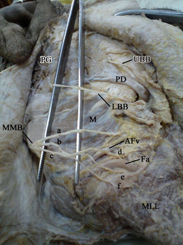
In 11 out of 30 specimens (36.7%) of this study, the (MMBFN) was represented by one branch. In these cases, the nerve emerged from the lower pole of the parotid gland ().
4.1.2 Marginal mandibular nerve represented by two branches
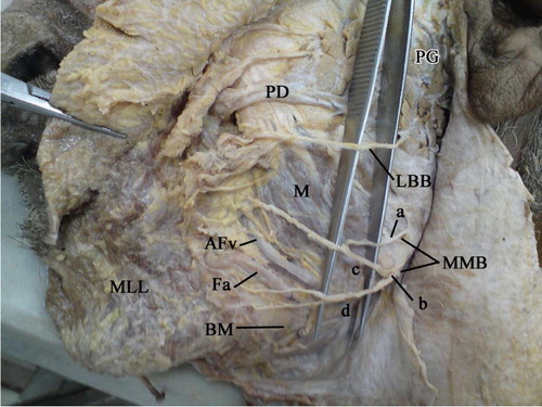
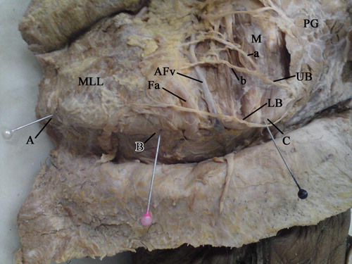
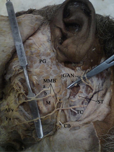
In 13 out of the 30 specimens (43.3%), the (MMBFN) was represented by two branches. In these specimens, the upper main branch emerged from the lower part of the anterior border of the parotid gland in 5 specimens (38.5%), while in the remaining 8 specimens (61.5%); the lower main branch arise from the lower pole of the parotid gland ().
4.1.3 Marginal mandibular nerve represented by three branches
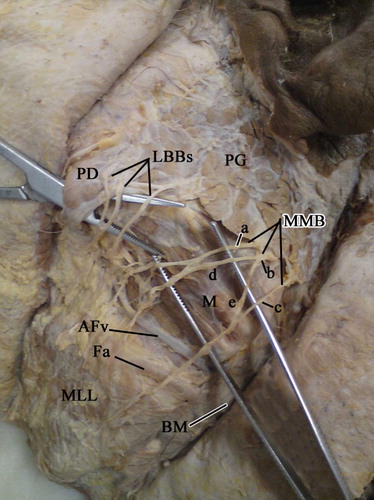
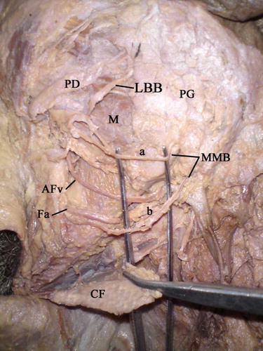
In the remaining 6 specimens (20%), the (MMBFN) was represented by three branches. In these specimens, the upper and middle main branches emerged from the lower part of the anterior border of the parotid gland in 4 out of 6 specimens (66.7%) while in the remaining 2 specimens (33.3%), the lower main branch emerged from the lower pole of the parotid gland ( and ).
4.2 Relation of these branches to the facial vessels
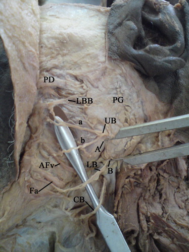
In 28 out of 30 specimens (93%), either one of the main or secondary branches of the (MMBFN) crossed superficial (lateral) to the facial vessels (, and –)
In the remaining 2 specimens (7%), one of the secondary branches of the (MMBFN) passed deep (medial) into the facial artery ( and ).
4.3 Communications of the marginal mandibular nerve
In 16 out of 30 specimens (53.6%), there were communications between the main and the secondary branches of the (MMBFN) (–).
In 12 out of 30 specimens (40%), the main or the secondary branches of the (MMBFN) communicated with the buccal branch of the facial nerve ( and ).
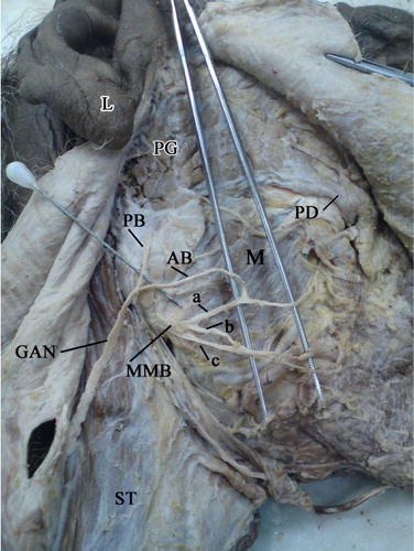
In 1 out of 30 specimens (3.3%), one of the main branches of the (MMBFN) communicated with the anterior branch of the great auricular nerve ().
In the remaining specimen (3.3%), one of the main branches of the (MMBFN) communicated with the transverse cervical nerve ().
4.4 Relation of MMBFN to the lower border of the mandible at a point midway between the angle of the mandible and symphysis menti
In 24 out of 30 specimens (80%), the (MMBFN) or its secondary branches lied above this point at a distance ranged from 1.1 to 1.7 cm with a mean distance of 1.3 (SD 0.12) (, , and ).
In 3 out of 30 specimens (10%), the lower main branch of the (MMBFN) lied at the same level of the lower border of the mandible ().
In the remaining 3 specimens (10%), the lower main branch of the (MMBFN) lied below this point (midway between angle of mandible and symphysis menti) at a distance ranged from 1.5 to 1.7 cm with a mean distance of 1.6 (SD 0.1).
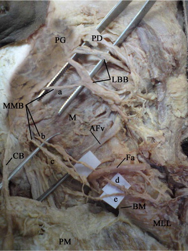
Then it crossed the base of the mandible and continued upwards to reach the muscles of the lower lip ( and ).
4.5 Relation of MMBFN to the deep fascia
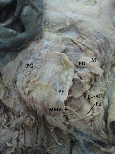
In the specimens where the branches of the (MMBFN) lied above the lower border of the mandible, they passed deep into the superficial layer of the parotid fascia (). However, all the branches of the nerve that passed below the lower border of the mandible ran intrafascially in the investing layer of deep cervical fascia (). The termination of this nerve was deep into the muscles of the lower lip in all specimens.
5 Discussion
Injury to the marginal mandibular branch of the facial nerve results in a significant cosmetic deformity due to paralysis of the muscles of the lower lip that is difficult to correct. Precise knowledge of the anatomical relationship of the nerve with its surroundings will allow safe identification and preservation of this important structure.Citation5
In the present study, by dissection of thirty halves head and neck specimens, the (MMBFN) was represented by one branch in (36.7%), two branches in (43.3%) and three branches in the remaining (20%) of the specimens.
Dingman and GrabbCitation7 found that the (MMBFN) was represented by a single branch in (20%), 2 branches in (67%), 3 branches in (9%) and 4 branches in (4%) of their specimens. The same study was done by Al-HayaniCitation5 on the (MMBFN); he found that this nerve arise as a single branch in (32%), 2 branches in (40%) and three branches in (28%) of cases which were nearly similar to the findings of the present study.
The above results showed that the (MMBFN) was represented by more than one branch that had highest incidence in all studies (especially the two branches pattern). This limits the extent of paralysis in case of injury of one of the branches of the (MMBFN).
In the present study, in (93%) of specimens, either one of the main or the secondary branches of the (MMBFN) crossed superficial (lateral) to the facial vessels while in (7%) of the cases one of the secondary branches of the (MMBFN) passed deep (medial) into the facial artery.
Singh et al.Citation8 Batra et al.Citation9 Charles et al.Citation10 Barron et al.Citation11 and StarkCitation12 found that the (MMBFN) passed superficial into the facial vessels in 100% of the cases which was nearly similar to the findings of the present study although in a few cases of the present study there was one of the secondary branches passed deep into the artery.
Thus, the facial artery can be used as an important landmark in the course of the nerve. The pulsations of the facial artery can be readily palpated by the surgeon at the antero-inferior angle of the masseter muscle. This landmark is an important guide in locating the (MMBFN) during surgical procedures.
The present study revealed that some of the branches of the (MMBFN) passed medial into facial artery. So care must be taken during manipulations deep to this artery.
Regarding the communications between the (MMBFN) with other nerves, the present study revealed that in 53.6% of specimens there were communications between either the main or the secondary branches of the (MMBFN) while in (40%) either the main or the secondary branches of the (MMBFN) communicated with the buccal branch of the facial nerve, while in (3.3%) there was a communication with the anterior branch of the great auricular nerve, and in another (3.3%) of specimens, there was a communication with the transverse cervical nerve.
Brennan et al.Citation13 added a case report where he found a communication between the anterior branch of the great auricular nerve and the (MMBFN).
One embryological explanation could be that as the (MMBFN) passed into the neck; it is joined by the cervical nerve during its development. Whether this finding is a rare developmental anomaly or the cervical plexus sometimes provides sensory communication with the facial nerve branches requires further electromyographic cadaveric studies.
Kim et al.Citation14 found that in (40%) of specimens the (MMBFN) communicated with the buccal or with the cervical branch of the facial nerve while in the present study this communication was found with the buccal branch of the facial nerve only in (3.3%) of the specimens.
Also, Domet et al.Citation15 found a communication between (MMBFN) with the transverse cervical nerve and with the cervical branch of facial nerve which was similar to the findings of the present study regarding the communication with the transverse cervical nerve only where it was found in (3.3%) of specimens but there was no communication between this nerve and the cervical branch of the facial nerve in this study.
From the above communications, a clinical intraoperative stimulation of the nerves of the cervical plexus that communicate with a branch of the facial nerve may result in a certain motor activity (such as depression of the lower lip).Citation15
In the present study, in (80%) of the specimens, the (MMBFN) or its secondary branches passed above a point midway between the angle of the mandible and symphysis menti at a distance ranged from 1.1 to 1.7 cm with a mean distance of 1.3 cm (SD 0.12), In another 10% of specimens the lower main branch of (MMBFN) passed at the same level of the lower border of the mandible while in the remaining (10%), the lower main branch of (MMBFN) ran below the mentioned point at a distance ranged from 1.5 to 1.7 cm with a mean distance of 1.6 (SD 0.1).
Batra et al.,Citation9 found that in (52%) of specimens, the (MMBFN) ran along the angle and the lower border of the mandible while it was found below the inferior border of the mandible in (32%) of cases. When this nerve became below the angle and the body of the mandible, its maximum distance was found to be 1.6 and 1.4 cm, respectively.
However, Freilinger et al.Citation16 and another study done by Corriea and ZaniCitation17 reported that the nerve crossed the lower border of the mandible halfway between its angle and its mental protuberance.
ChatterjeeCitation18 found that an average distance of the loop of the (MMBFN) was 1.2 cm below the inferior border of the mandible.
From the above results, in order to avoid damage to the (MMBFN) the submandibular incision should be planned at a distance not less than 1.6 cm below a point midway between the angle of the mandible and symphysis menti. The surgeons should put into consideration that the extension of the head during surgery, laxity and atrophy of the skin due to aging may cause slight downward displacement of the position of this nerve.
In the present study, all the branches of the (MMBFN) that ran above the lower border of the mandible was deep into the superficial layer of the parotid fascia while all the branches of the nerve below the mandible passed intrafascially in the investing layer of the deep cervical fascia.
SternCitation19 found that the (MMBFN) passed through the platysma muscle anterior to the facial artery. Liebman et al.Citation20 in addition to Greyling and Meiring,Citation21 Kirici et al.Citation22 and Karapinar et al.Citation23 found that the nerve passed superficially as it travels to supply the effector muscles.
From the above results, skin flaps should be carefully elevated in a plane immediately deep into the platysma and superficial in the investing layer of the deep cervical fascia.
6 Conclusion
The branching pattern of (MMBFN) by more than one branch limits the extent of paralysis in case of injury of one of its branches. The facial artery can be used as an important landmark in locating the (MMBFN) during surgical procedures. In order to avoid injury to the marginal mandibular branch of the facial nerve, the submandibular incision should be planned at a distance not less than 1.6 cm below a point midway between angle of the mandible and symphysis menti. Skin flaps should be carefully elevated in a plane immediately deep into the platysma and superficial in the investing layer of the deep cervical fascia.
Conflict of interest
None declared.
Notes
Peer review under responsibility of Alexandria University Faculty of Medicine.
Available online 24 January 2014
References
- D.EgasseFacial nerve: particular aspects of the cervicofacial anatomyAnn Dermatol Venereol12820015962
- D.C.A.RodriguesJ.C.AndreoD.F.L.MenezesP.T.ChinellatoG.M.RosajúniorAnatomy of the facial nerve and its implication in the surgical proceduresInt J Morphol272009183186
- C.SaylamH.UcerlerM.OrhanA.UckanC.OzekLocalization of the marginal mandibular branch of the facial nerveJ Craniofac Surg1812007137
- A.AssadianC.SenekowitschN.PfaffelmeyerO.AssadianH.PtakovskyG.HagmüllerIncidence of cranial nerve injuries after carotid eversion endarterectomy with a transverse skin incision under regional anaesthesiaEur J Vasc Endovasc Surg282004421424
- A.Al-HayaniAnatomical localization of the marginal mandibular branch of the facial nerveFolia Morphol662007307313
- R.W.NasonA.BinahmedM.G.TorchiaJ.ThliversisClinical observations of the anatomy and function of the marginal mandibular nerveInt J Oral Maxillofac Surg362007712715
- R.O.DingmanW.C.GrabbSurgical anatomy of the mandibular ramus of the facial nerve based on the dissection of 100 facial halvesPlast Reconstr Surg291962266272
- A.P.SinghA.MahajanK.Karunesh GuptaMarginal mandibular branch of the facial nerve: an anatomical studyIndian J Plast Surg43120106064
- A.P.BatraA.MahajanK.GuptaMarginal mandibular branch of the facial nerve: An anatomical studyIndian J Plast Surg4320106064
- R.CharlesS.RodneyRob CharlesOperative surgery1983ButterworthsLondon524525
- J.N.BarronM.N.SaadManual of operative plastic and reconstructive surgeryEdinburgh1980Churchill LivingstoneUnited Kingdom437 vol. 1
- R.B.StarkSecondary surgery of the lipsPlastic Surgery of the Head and Neck1987Churchill LivingstoneNew York810822 Vol. 2
- P.A.BrennanR.WebbF.KemidiJ.SprattS.StandringGreat auricular communication with the marginal mandibular nerve, a previously unreported anatomical variantBr J Oral Maxillofac Surg462008492493
- H.J.KimK.S.KohC.S.OhK.S.HuJ.W.KangI.H.ChungEmerging patterns of the cervical cutaneous nerves in AsiansInt J Oral Maxillofac Surg3120025356
- M.A.DometN.P.ConnorD.M.HeiseyG.K.HartiqAnastomoses between the cervical branch of the facial nerve and the transverse cervical cutaneous nerveAm J Otolaryngol262005168171
- G.FreilingerH.GuberW.HappakU.PechmanSurgical anatomy of the mimic muscle system and the facial nerve: importance for reconstructive and aesthetic surgeryPlast Reconstr Surg801987686690
- D.W.CorrieaR.ZaniSurgical anatomy of the facial nerve as related to ancillary operations in rhytidoplastyPlast Reconstr Surg521973549560
- A.ChatterjeeMarginal mandibular nerve – Interpolation from cadaver anatomy may be followedIndian J Plast Surg January432010140143
- S.J.SternPrecise localization of the marginal mandibular branch of facial nerve during nerve dissectionHead Neck Plast Surg141992328331
- E.P.LiebmanR.C.WebsterJ.K.GanlT.GiffinThe marginal mandibular branch of facial nerve in rhytidectomy and liposuction surgeryArch Otolaryngol Head Neck Surg1141988179181
- L.M.GreylingJ.H.MeiringMorphological study on the convergence of the facial muscles at the angle of the mouthActa Anatomica1431992127129
- Y.KiriciC.KilicS.DeveliMarginal mandibular branch of the facial nerve in human fetusesSaudi Med J322011459462
- U.KarapinarThe course of the marginal mandibular branch of the facial nerve in adult cadavers. An anatomic studySaudi Med J342013364368