
Abstract
Background
Acute appendicitis is a disease of the young presenting in children and early adolescents although no age group is exempt. It is the most common cause of acute surgical abdomen worldwide. This clinicopathological study aims to determine the various lesions of the surgically removed appendix in our centre and if any, changing trend in this lesion in our environment.
Method
A retrospective study was undertaken to review the histopathology reports of all appendicectomy specimens submitted to the Department of Pathology of the Asokoro District Hospital, Abuja; Nigeria from November 2009 to October 2012 Patient’s biodata, clinical signs and symptoms were extracted from the request form.
Result
A total of 293 appendices were received during the 3-year study period constituting about 10% of total specimens. The male to female ratio was 1:1.3 with a mean age of 26.33 ± 11.39 years. Acute appendicitis was found in 81.2% of our cases while other lesions constitute 14.7% and negative appendectomy occurred in 4.1% of the cases. No mortality was recorded.
Conclusion
The findings in this study compared favourably with those of our environment and in the developed world although acute appendicitis in this study showed slight female preponderance.
1 Introduction
The diagnosis of acute appendicitis is most commonly a clinical one; many patients present with a typical history and examination findings. The cause of acute appendicitis is unknown but is probably multifactorial; luminal obstruction and dietary and familial factors have all been suggested.Citation1–Citation3 Appendicectomy is the treatment of choice and is increasingly done as a laparoscopic procedure.Citation2 Acute appendicitis is a disease of the young presenting in children and early adolescents although no age group is exempt. It is the most common cause of acute surgical abdomen worldwide and its incidence varies with geographical location.Citation3
In the United States, a crude estimate of the incidence of acute appendicitis is 11 cases per 10,000 population.Citation4 Studies have shown acute appendicitis to be more common in the whites than non whites.Citation5 Appendicitis is the most common abdominal emergency and accounts for more than 40,000 hospital admissions in England every year.Citation6
In Nigeria, incidence of acute appendicitis is relatively low with varying reports of average annual frequencies ranging from 22.1 to 49.8 new cases but in other African countries, annual frequencies are relatively higher ranging from 22.9 to 129 new cases per 100,000 persons.Citation7–Citation12 Clinicopathological studies of appendiceal lesions on the African continent are relatively few and this study was conducted to determine the various lesions of surgically removed appendix in our centre and compare our findings with other studies elsewhere.
2 Materials and methods
A retrospective study was undertaken to review the histopathology reports of all appendicectomy specimens submitted at the Department of Pathology of the Asokoro District Hospital, Abuja; the capital city of Nigeria from November 2009 to November 2012. The number of cases/year for the 3 years was 93, 95 and 105, respectively. Our laboratory is a referral centre for 12 other government district hospitals and private hospitals in the Federal Capital Territory and its environs. The Federal Capital Territory is an urban community with a population of 1,405,201 people (2006 census). Patient’s biodata, clinical signs and symptoms were extracted from the laboratory request form. Routine haematoxylin and eosin (H&E) staining and where necessary histochemical studies were carried out. Alcian blue/Periodic Acid Schiff stain was done for a case of suspected mucocoele. The data were analysed in terms of frequency, age and sex distribution, nature of clinical signs and symptoms as well as histological characteristics of pathologic lesions (normal, acute appendicitis with or without peritonitis, lymphoid hyperplasia, eosinophilic appendicitis and schistosomal appendicitis) using the SPSS version 17. The data for these patients were presented in tables and figures.
3 Result
A total of 293 appendices were received in our laboratory during the 3-year study period constituting about 10% of total specimens. The annual incidence is 8.4 per 100,000 population. There were 128 males and 165 females constituting a male to female ratio of 1:1.3. The age range of patients in this study is 3–57 years with a mean of 26.33 ± 11.39 years.
3.1 Age and sex distribution
The age and sex distribution of the patients is shown in . The peak age of occurrence in this study is 20–29 years closely followed by 10–19 year age group both constituting over 60% of the cases. The least number of cases (13 patients) were seen in the age group 50–59 years constituting 4.4%.
Table 1 Distribution of cases according to age groups and sex.
3.2 Clinical presentation
The most common form of presentation by our patients was right iliac abdominal pain (95%) which later became generalised in 21.8% of cases. The other symptoms include fever (81%), vomiting (56%), and loss of appetite (48%). The mean duration of symptoms was 3.05 days.
Twelve patients had perforation at surgery and 2 out of these presented primarily in shock.
3.3 Histological diagnosis
The distribution of histological diagnosis is as shown in . Twelve cases (4.1%) were found to be normal. Uncomplicated acute appendicitis was seen in 174 patients constituting 59.4% and acute appendicitis with peritonitis constitutes 21.8%. Submucosal fibrosis, schistosomiasis, lymphoid hyperplasia and subacute appendicitis constitute 5.1%, 2.4%, 3.4% and 1.7%, respectively. Others include eosinophilic appendicitis (0.7%) and mucocoele was seen in one patient (0.3%). The histology of acute appendicitis, acute appendicitis with lymphoid hyperplasia and schistosomal appendicitis is shown in –. shows the distribution of the histological diagnosis according to age groups. Acute appendicitis with peritonitis () occurs more in males than females with a ratio of approximately 2:1. The peak age of occurrence is 20–29 years, constituting 31.3% and closely followed by age groups 10–19 years and 30–39 years constituting 23.9% each. The distribution of patients with histological diagnosis of acute appendicitis and acute appendicitis with peritonitis is shown in and , respectively. Acute appendicitis occurs more in females (63.8%) than males (36.2%) while acute appendicitis with peritonitis occurs more in males (65.7%) than females (34.3%). The peak age of occurrence for both is the 3rd decade of life.
Figure 1 This is a section of the appendix showing transmural infiltration by acute inflammatory cells and dilated and congested vessels (H&E ×200).

Figure 2 This is a section of the appendix showing lymphoid nodules within the lamina propria that also extend into the submucosa (H&E ×40).
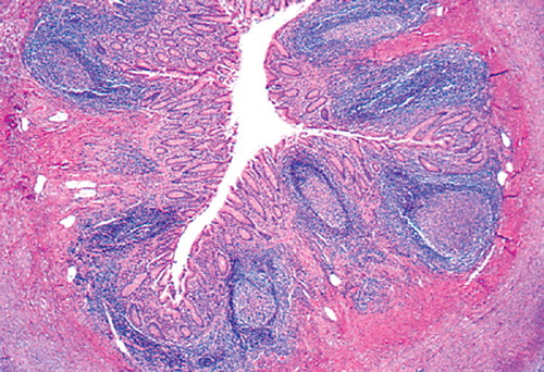
Figure 3 Section of appendix showing schistosomal appendicitis (H&E ×100).
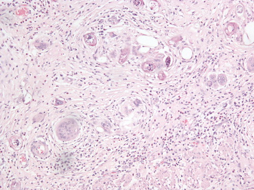
Table 2 Distribution of cases according to histological diagnosis.
Table 3 Distribution of the histological diagnosis according to age.
Table 4 Age and sex distribution of patients with histological diagnosis of acute appendicitis.
Table 5 Age and sex distribution of patients with histological diagnosis of acute appendicitis with peritonitis.
4 Discussion
Acute appendicitis is a very common disease and appendix is a common specimen received in the histopathology laboratory worldwide. The incidence varies widely from region to region with higher incidence among the whites than blacks. Appendix is one of the most common specimens received in our laboratory during the study period. This is exemplified by the fact that it constituted about 10% of the total histological specimens received in our hospital during the 3 year period. The annual incidence in our study is 8.4 per 100,000 which is about twice that reported in suburban population of Sagamu, Southwest, Nigeria.Citation13 The incidence of acute appendicitis is increasing in our community probably due to urbanisation as Abuja is the capital of Nigeria which is occupied by the affluent in the society. The diet is westernised and consists of low fibre. It is believed that high fibre diet which increases the bowel motion reduces the incidence of acute appendicitis. Therefore in regions (Africa, Southeast Asia) where high fibre diet is the staple food, the incidence of acute appendicitis is less.Citation14
The male to female ratio of 1:1.3 in this study compares favourably with the studies by Ali et al.Citation15 in Maiduguri and Blair et al.Citation16 in Canada where the male to female ratio was 1:1.2 but contrasts most reports within and outside the country in which males predominate.Citation7,Citation8,Citation13
The peak age of occurrence in our centre is the 3rd decade of life. Most of the reports showed peak age of occurrence in the 2nd and 3rd decades of life. Our finding compares with that of Fashina et al.Citation17 in Lagos, Alatise et al.Citation18 in Ile-Ife while Ohene-Yeboa et al.Citation10 in Ghana and Ali et al.Citation15 in Maiduguri, Nigeria reported peak age in the 4th decade of life. Some studies especially in the developed world reported peak age in the 2nd decade of life.Citation19,Citation20 Humes and SimpsonCitation2 in a clinical review of acute appendicitis in England reported peak age incidence of occurrence as 10–20 years and male to female ratio of 1.4:1 although they acknowledged variations in incidence across geographical zones as no age is exempt from acute appendicitis. The frequency of occurrence of acute appendicitis decreases with increasing age. This is similar to most studies from within and outside our country.Citation8–Citation10,Citation19
The diagnosis of acute appendicitis is mainly through history and physical examination. The findings in this study concur well with what was obtained in other studies. The mean duration of symptoms in our study was 3.05 days which compares favourably with other reports locally and internationally.Citation9,Citation10 Thirty-six patients (12.1%) had perforation at surgery and 2 of these presented in shock. This is probably due to late presentation at the emergency room. Fear of surgical operation and relatively high cost of treatment are the main reasons for late presentation in our environment.Citation13 The 2 patients who presented in shock were adequately resuscitated and had appendectomy. The average hospital stay post operation was 6.5 days with a range of 5–14 days. This is comparable with other studies.Citation9,Citation10 No mortality was recorded in the patients studied.
This study showed a wide range of histological diagnosis. The negative appendicectomy rate of 4.1% is low compared with most studies reviewed which reported a range of 8.6–35.8%.Citation8,Citation9,Citation17,Citation21–Citation23 This low rate may not be unconnected with the growing clinical acumen of our surgeons coupled with radiological investigations. The abdominal pains in these patients were mainly due to pelvic inflammatory diseases and urinary tract infection.
Acute appendicitis constituted 82.3% in this study which is comparable with the study of Abudu et al.Citation13 in Sagamu, Nigeria. Ojo et al.Citation8 and Abdulkareem et al.Citation9 reported slightly lower rates of 69.9% and 70.3%, respectively. Most studies from outside Nigeria reported a range of 45.7–82.5% which compared favourably with this study.Citation10,Citation20
Lymphoid hyperplasia constituted 3.4% which compares with the study conducted in the UK by Singhal and Jadhav, 2007 and contrasts that of Abdulkareem (Lagos, Nigeria) and Al-Fujairah (United Arab Emirates) which reported higher frequency of 19% and 25%, respectively. About 70% of these patients are in the 2nd and 3rd decades of life which compares with most studies. The presence of reactive lymphoid hyperplasia in appendix in varying degrees lends credence to the theory that obstruction of the lumen by lymphoid tissue aggregation is important in the pathogenesis of acute appendicitis especially in the 2nd and 3rd decades of life.
Submucosal fibrosis was seen in 5.1% of the cases with peak age in the 2nd decade of life. This compares with the finding of Andreou et al.Citation24 that fibrosis and faecoliths were more common in the older individuals at autopsy than in the younger surgically resected group. Submucosal fibrosis is characterised by deposition of fibrous tissue in the submucosa due to repeated acute or chronic inflammation which does not resolve completely in most cases. This fibrous tissue deposition causes narrowing and obstruction of the lumen and sometimes superimposed infection with resultant acute appendicitis. Submucosal fibrosis could also be seen in conjunction with an infective aetiology or following interval appendicectomy.Citation8,Citation25–Citation27
Schistosomal appendicitis constituted 2.4% of the total patients studied. All these cases were associated with varying degrees of fibrosis in the submucosa. This lends credence to the fact that schistosomal appendicitis is associated with inflammation, repair, deposition of fibro-connective tissue and subsequent obliteration of the appendiceal lumen which will result in the typical presentation of acute abdomen.Citation16,Citation27
The cases of subacute appendicitis seen in this study were from patients who had interval appendicectomy. These patients also had varying degrees of submucosal fibrosis.Citation27
5 Conclusion
The finding in our study compared favourably with those of our environment and in the developed world although we found a slight female preponderance. The incidence in our environment is increasing especially in a very urban area like our study has shown. Acute appendicitis is one of the most common surgical emergencies in our environment therefore prompt and adequate intervention will go a long way in reducing its morbidity and mortality.
Conflict of interest
None declared.
Notes
Peer review under responsibility of Alexandria University Faculty of Medicine.
Available online 17 September 2014
References
- J.DanielE.MontgomeryInflammatory disorders of the appendixR.D.OdzeJ.R.GoldblumSurgical pathology of the GI tract, liver, biliary tract and pancreas2nd ed.2009Saunders Elsevier Inc.395408
- D.J.HumesJ.SimpsonAcute appendicitisBMJ3332006530534
- R.PieperL.KagerU.TidefeldtObstruction of appendix vermiformis causing acute appendicitis. An experimental study in the rabbitActa Chir Scand148119826372
- P.M.BaoPresence or absence of gas in the appendix: additional criteria to rule out or confirm acute appendicitis–evaluation with ultrasoundRadiology2172000599600
- C.S.GraffeoF.L.CounselmanAppendicitis in textbook of GI emergencies, part IIEmerg Med Clin North Am141996
- Hospital episode statistics: primary diagnosis summary. Available from: http://www.hesonline.nhs.uk/Ease/%20servlet/ContentServer?siteID%20sta:summary.=1937%26categoryID=202 [accessed 28 Aug 2006].
- A.S.OguntolaM.L.AdeotiT.A.OyemoladeAppendicitis: trend in incidence, age, sex and seasonal variations in South Western NigeriaAnn Afr Med942010213217
- O.S.OjoS.C.UdehW.O.OdesanmiReview of the histopathological findings in removed appendices for acute appendicitis in NigeriansJ R Coll Surg Edinb3641991245248
- F.B.AbdulkareemD.I.AwelimoborSurgical pathology of the appendix in a tropical teaching HospitalNiger Med Pract55320093236
- M.Ohene-YeboahB.TogbeAn audit of appendicitis and appendectomy in Kumasi, GhanaWest Afr J Med2522006138143
- S.T.EdinoA.Z.MohammedO.OchichaM.AnumahAppendicitis in Kano, Nigeria: a 5 year review of pattern, morbidity and mortalityAnn Afr Med J3120043841
- S.K.ChavdaS.HassanG.A.MagohaAppendicitis at Kenyatta National Hospital, NairobiEast Afr Med J82102005526530
- E.K.AbuduT.Y.OyebadeyoA.O.TadeN.A.AwololaSurgical pathologic review of appendectomy at a sub urban tropical tertiary hospital in AfricaJ Med Med Sci262011932938
- J.BlackAcute appendicitis in Japanese soldiers in Burma: support for the “fibre” theoryGut5122002297
- N.AliS.AliyuAppendicitis and its surgical management experience at the University of Maiduguri Teaching Hospital NigeriaNiger J Med2122012223226
- P.BlairS.P.BugisS.J.TurnerM.M.MacLeodReview of the pathologic diagnoses of 2216 appendectomy specimenAm J Surg16551993618620
- I.B.FashinaA.A.AdesanyaO.A.AtoyebiO.O.OsinowoC.J.AtimomoAcute appendicitis in Lagos: a review of 250 casesNiger Postgrad Med J1642009268273
- O.I.AlatiseT.OgunweideAcute appendicitis: incidence and management in NigeriaIFEMED J14120086670
- D.G.AddissN.ShafferB.S.FowlerR.V.TauxeThe epidemiology of appendicitis and appendectomy in the United StatesAm J Epidemiol1321990910925
- M.Al-OmranM.MamdaniR.S.McLeodEpidemiologic features of acute appendicitis in Ontario, CanadaCan J Surg4642003263268
- M.N.MuthupheiP.MorwamocheThe surgical pathology of the appendix in South African blacksCent Afr J Med4411998911
- I.ChamisaA clinicopathological review of 324 appendices removed for acute appendicitis in Durban, South Africa: a retrospective analysisAnn R Coll Surg Engl9182009688692
- T.E.MadibaA.A.HaffejeeD.L.MbeteH.ChaithramJ.JohnAppendicitis among African patients at King Edward VIII Hospital, Durban, South Africa: a reviewEast Afr Med J75219988184
- P.AndreouS.BlainC.E.Du BoulayA histopathological study of the appendix at autopsy and after surgical resectionHistopathology1751990427431
- A.W.MeshikhesC.J.ChandrashekarQ.Al-DaolahO.Al-SaifA.S.Al-JoaibS.S.Al-HabibSchistosomal appendicitis in the Eastern Province of Saudi Arabia: a clinicopathological studyAnn Saudi Med19119991214
- L.W.LampsAppendicitis and infections of the appendixSemin Diagn Pathol21220048687
- G.GuoJ.K.GreensonHistopathology of interval (delayed) appendectomy specimens: strong association with granulomatous and xanthogranulomatous appendicitisAm J Surg Pathol278200311471151