
Abstract
A 16 year old patient had ingested two sewing needles about 4.5 cm long accidentally that eventually resided in extra intestinal tract position, and being asymptomatic, she ignored the problem for a long period about 2 years. She only consulted the hospital after having symptoms. The needles were located by X-ray imaging and retrieved with the aid of C-arm fluoroscope at a laparotomy. Her postoperative period was uneventful.
Keywords:
1 Introduction
Foreign body (FB) ingestion is a common problem among children and psychologically deranged, inebriated people and rare among psychologically healthy people which occurs usually by accident. It is one of the most common causes of death in babies under 12 months of age. The patient usually presents with mild symptoms or as an emergency situations. Many foreign bodies had swallowed and need immediate operation while others remain dormant. This case was peculiar because of the presence of two needles which were dormant in her body for a long time (two years).
2 Case report
A 16-year-old female presented after swallowing a sewing needle 2 years ago. At that time she consulted a health center, took X-rays and was advised to wait for spontaneous passage of the needles. The patients was asymptomatic in the following period, so she ignored the problem. Three months ago she began to feel pricking sensations after meals in the region of the epigastrium, so she consulted a private hospital on 2014.
On examination she was a thin healthy young girl with no abnormal physical signs. Abdominal examination revealed soft abdomen with only mild tenderness in the epigastrium.
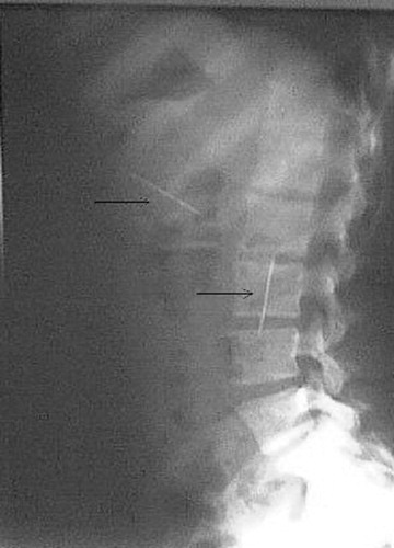
Plain X-ray of the abdomen showed two longitudinal metallic foreign bodies, identical in shape about 4.5 cm in length, in the right paravertebral region one at the level of L4 and the other at the level of T11–T12 ( and ).
Fig. 1 Plain X-ray of the abdomen showing the needles at the epigastrium and right flank.
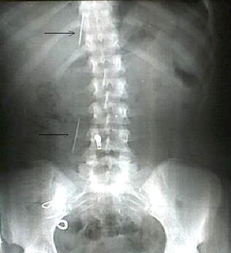
Fig. 2 Plain X-ray lateral showing both needles.
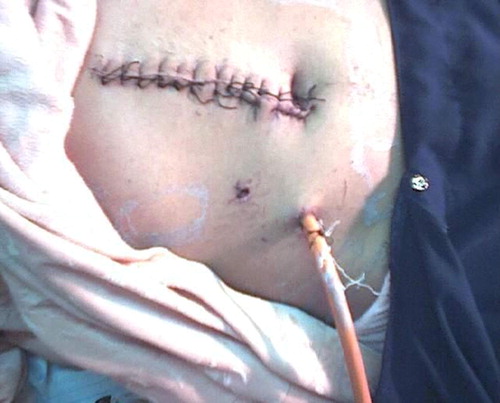
Upper gastrointestinal endoscopy showed empty stomach and duodenum with abnormal findings. Laparotomy was done for her with the aid of C-arm fluoroscope (OCE 2006 GE USA). Laparoscopy was not used initially because it was not available in our hospital at that time. It revealed a firm mass in the retrocolic space of the ascending colon bout 6 × 3 cm in size, inside which was a sewing needle which indicates the rarity of the case because the needle has passed two constrictions namely the pylorus and ileocecal valve to eventually travers the colonic wall posteriorly. There was a small scar at the lesser curvature of the stomach facing the eye of the second needle, which was embedded in the left lobe of the liver. Retrieval of both needles was done (), and the patient passed her postoperative period uneventfully (). After her discharge from hospital, she was referred to psychiatric department for assessment of her psychiatric status.
Fig. 3 Size of the needles.
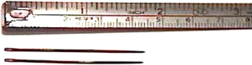
Fig. 4 Post operative condition of the patient.
3 Discussion
Foreign body ingestion is an international problem among children starting from fishbone impaction which is more commonly observed in countries where fish is the main dietarysnackCitation1 to large object that lead to esophageal perforation.Citation2 Some foreign bodies need urgent removal like button batteriesCitation3 while others remains dormant for years like in this case. At times FB ingestion is intentional and at others it is accidental.Citation4 In our patient the foreign bodies were long sharp ones (sewing needles).
Foreign body ingestion can be diagnosed by historyCitation5 taken from the patient or his or her parents, and approved by plain X ray, of course if the object is radio opaque, which was the case in our patient. A hand held metal detector is a simple, non invasive device that localize metallic foreign bodies in the GIT,Citation5 but this was not available to us. Laparoscopy is very useful tool in performing surgery foreign body removal but unfortunately it was not available.
Sometimes objects remain in the GIT for many years.Citation6 Henderson and Gaston reported nine incidences of perforation in 800 cases of foreign body ingestion at Boston City hospital, perforation occurred without signs of peritonitis; they observed two asymptomatic patients with straight pins lodged in their spleens.Citation6 Sharp pointed objects such as sewing needles may penetrate the bowel wall. If abdominal pain, tenderness, fever or leukocytosis occur, immediate surgical removal of the offending object is indicated. Abscess or foreign body granuloma (type IV hypersensitivity reaction – delayed type of cell mediated immunity) formation are the usual outcome without surgical therapy.Citation7 In our case one of the needles evidently had perforated the ascending colon and lodged in a foreign body granuloma in the retrocolic area while the other perforated the stomach and was lodged in the left lobe of the liver. Abel et al. reported a similar case of a pin in the left lobe of the liver, but the patient was much younger (11 months old).Citation8
If a pin stays in one place on abdominal roentgenogram for 5 days or more, It has most likely penetrated the wall or is lodged in the appendix or a Meckel'sdeverticulum.Citation9 Some believe that such objects should be surgically removed even if there are no symptomsCitation5, others believe that sharp objects should be retrieved from the stomach because 15–35% will cause intestinal perforation.Citation6 In our patient on observing the site of the needles it showed that it has not changed its place for the last 3 months, and knowing that these foreign bodies are long and sharp sewing needles so surgery was indicated.
The lessons learnt from our case lies in that:
| • | The period since first ingesting the needles was long i.e. two years, which makes our case peculiar. | ||||
| • | The amazing ability of the human body to segregate foreign bodies and isolate them from harming the rest of the body. | ||||
| • | Perfect health of the patient in spite of perforation of the bowel by the needles and being lodged outside the wall of the gut. | ||||
| • | Laparotomy was aided by the C arm fluoroscope because attempted removal at laparotomy can be very difficult as the object may be much more difficult to find than might be expected.Citation10 | ||||
| • | Laparoscopy if available is an excellent tool to remove foreign bodies because of its minimally invasive characteristic. | ||||
Ingested foreign bodies are common encountered in medical practice. The majority can be managed with endoscopic removal like fish bones.Citation11 Migration of foreign bodies into other parts of the body may necessitate using more invasive surgical procedures for removal mussel shell,Citation12 coins,Citation13 nasal splint after septoplasty,Citation14 and partial denturesCitation15 and needles.
In conclusion, when a patient come with foreign body ingestion to causality, rapid and accurate diagnostic confirmation is necessary. After diagnosis, the patients should be followed up clinically and radiologically, if any symptoms occur e.g. peritoneal irritation or foreign body was constant in position for a long period then prompt removal is mandatory to prevent complications like perforation and foreign body granuloma. Lateral chest radiography and CT scan must be performed for a correct diagnosis and initiating treatment is necessary to prevent complications and to lower mortality risk. Every case must be referred to psychological department for assessment because many cases are found to be psychologically deranged.
Conflict of interest
None.
Funding
None.
Notes
Peer review under responsibility of Alexandria University Faculty of Medicine.
Available online 9 June 2017
References
- C.W.LimM.H.ParkH.J.DoFactors associated with removal of impacted fishbone in children, suspected ingestionPediatr Gastroenterol Hepatol Nutr192016168174
- N.J.PetersJ.K.MahajanM.BawaA.ChabbraR.GargK.L.RaoEsophageal perforations due to foreign body impaction in childrenJ Pediatr Surg8201512601263
- J.H.LeeJ.H.LeeJ.O.ShimJ.H.LeeB.L.EunK.H.YooForeign body ingestion in children: should button batteries in the stomach be urgently removed?Pediatr Gastroenterol Hepatol Nutr1920162028
- K.R.MurshidG.E.KhairyLaproscopic removal of a foreign body from the intestineJ R Coll Surg Edinb4321998 Apr109111
- M.H.BeersR.BerlowThe Merck manual of diagnosis and therapy17th ed. (Centennial ed.)2591999Merck research LaboratoriesWest Point
- F.F.HendersonE.A.GastonIngested foreign body in the gastrointestinal tractArch Surg36193866
- B.Mark EversM.CourtneyJames C.TownsendThompsonJr.Small intestineSeymourSchwartzG. TomShiresFrank C.SpencerJohn M.DalyJosef E.FisherAubrey C.GallowayPrinciples of Surgery7th ed.vol. 21999Mc Graw-Hill Book CompanyNew York1251
- R.M.AbelJ.E.FischerH.H.HendersonPenetration of the alimentary tract by a foreign body with migration to the liverArch Surg1021971227
- E.G.KassnerR.W.MutchlerD.H.KlotzJ.RoseUncomplicated foreign bodies of the appendix in childrenJ Pediatr Surg91974207
- R.C.G.RusselsN.S.WilliamsC.J.K.BulstrodeBailey and Love's short practice of surgery23rd ed.vol. 22000ArnoldLondon929
- U.C.MegwaluMigration of an ingested fish bone into the paraglottic spaceJ Laryngol Otol1302016973974
- I.H.ParkH.K.LimS.W.SongK.H.LeePerforation of esophagus and subsequent mediastinitis following mussel shell ingestionJ Thorac Dis82016E693E697
- R.MaroufAccidental ingestion of a coinPan Afr Med J212015324
- G.S.MundingerZ.ShanavasT.C.KontisCould your patient have swallowed their nasal splint after septoplasty? Seeing is believingAesthet Surg J362016NP68NP70
- A.ChawlaJ.BoscoM.SubramanianK.ChokkapanJ.ShenoyT.C.LimImaging findings of swallowed dentures: a case seriesEmerg Radiol222015717721