
1 Introduction
Breast cancer is the most prevalent diagnosed malignancy among women worldwide [Citation1]. It is also the major cause of malignancy-related death among women [Citation2]. In Egypt, breast cancer is the most common type of cancers among females as it accounts for approximately 32% of reported malignancies [Citation3]. In addition, tumor metastasis is a major clinical challenge. Nearly one-third of breast cancer women worldwide die from metastasis, especially brain metastasis [Citation4,Citation5] .
Oxidative stress is a characteristic hallmark of initiation and progression of many cancers, including breast cancer [Citation6]. To maintain cancer cell function, many antioxidants are up-regulated to balance reactive oxygen species (ROS) levels [Citation7]. One of the most important antioxidants is thioredoxin-1 (Trx-1), which is highly expressed in many tumors. A recent study showed increased level of serum Trx-1 in breast cancer patients in comparison with other cancers and normal persons [Citation8]. Cellular redox homeostasis mediated by Trx-1 is required for activation of ribonucleotide reductase [Citation9] and peroxiredoxins [Citation10], induction of vascular endothelial growth factor (VEGF) and matrix metalloproteinase-9 (MMP-9) [Citation11,Citation12] .
In addition, ROS initiate lipid peroxidation of polyunsaturated fatty acids in cell membrane producing malondialdehyde (MDA). MDA used as biomarker of oxidative stress and play an important role in breast cancer initiation, progression and metastasis [Citation13].
MMP-9, also known as gelatinase B, is a 92-kDa endopeptidase that is strongly associated with aggressiveness and metastatic spread in breast cancer [Citation14]. During tumor angiogenesis, MMP-9 degrades type IV collagen, the main component of vascular basement membrane surrounding tumor cells which is an essential process in initiation of metastasis. Furthermore, stroma-derived MMP-9 can facilitate the liberation of extracellular matrix (ECM)-sequestered VEGF that leads to metastasis [Citation15].
The overexpression of Trx-1 that stimulates MMP-9 expression and down regulates its inhibitor, tissue inhibitor of metalloproteinase-1 (TIMP-1), increasing MMP-9 activity [Citation12]. Therefore, we conducted this study to investigate the possible role of serum Trx-1, MMP-9 and MDA in the detection of metastasis in female breast cancer patients and their correlation with clinicopathologic parameters.
2 Subjects and methods
2.1 Subjects
The current study was performed on 90 female subjects:
| • | Non-metastatic breast cancer (NMBC) patients included 37 newly diagnosed aged 31–67 years, with a mean ± SD of 51.70 ± 11.04 years. | ||||
| • | Metastatic breast cancer (MBC) patients included 38 patients aged 31–78 years, with a mean ± SD of 51.78 ± 11.24 years. | ||||
| • | Healthy control group included 15 apparently healthy women aged 26–59 years, with a mean ± SD of 44.66 ± 10.36 years. | ||||
Patients were diagnosed between March and November 2015 in the Oncology Center, Mansoura University; Mansoura, Egypt.
Patients were classified according to the American Joint Committee on Cancer (AJCC) TNM system [Citation16] to stage 0 (3 patients), stage I (9 patients), stage II (12 patients), stage III (13 patients) and stage IV (38 patients). The patients and controls do not have renal, liver disorders or any other type of malignancies. Recorded clinical and pathological features for each patient were obtained from Oncology Center including age, grade, stage, serum carcinoembryonic antigen (CEA) and serum cancer antigen 15-3 (CA 15-3).
The study was approved by the local institutional ethical committee of Faculty of Pharmacy, Mansoura University, Mansoura, Egypt; and patients’ consents were obtained according to the regulations of the Egyptian Ministry of Health. Metastasis was diagnosed by clinical examination including symptomatology; together with radiology including a plan X-ray, ultrasonography, computed tomography and bone scan.
2.2 Collection of blood samples
Patients and control subjects were fasted overnight. Blood samples were collected and left for 30 min at room temperature to clot, centrifuged at 3000 rpm for 10 min. The serum was separated and stored at −80 °C.
2.3 Biochemical parameters determination
Serum Trx-1 level and MMP-9 level were measured by commercially available enzyme-linked immunosorbent assay (ELISA) kits using human thioredoxin ELISA kit, (Boster bio, Pleasanton, CA, USA) and human MMP-9 platinum ELISA kit, (eBioscience, San Diego, CA, USA). Serum malondialdehyde (MDA) was determined using thiobarbituric acid (TBA) method [Citation17] by using a commercially available colorimetric kit (Biodiagnostic, Giza, Egypt).
2.4 Statistical analysis
The SPSS version 21 was used for data analysis. One-sample Kolmogorov-Smirnov test was used to test the normality of data. Qualitative data were defined using number and percent. The relation between categorical variables was prepared using Chi-square test. Continuous variables were described as Median for non-parametric data. For comparison of median of more than two groups (non-parametric data) Kruskal Wallis test was used. Correlation between continuous non parametric data was done using Spearman correlation. Receiver Operating Characteristic (ROC) curve was done to define sensitivity and specificity at different cutoff points. Statistical significance was considered at P < .05.
3 Results
3.1 Patient's characteristics
Clinical characteristics of breast cancer patients were shown in . T1 tumor size was reported in 13 NMBC patients (35.1%). T2 was reported in 11 NMBC patients (29.7%) and 1 MBC patient (2.6%). T3 was reported in 13 NMBC patients (35.1%) and 19 MBC patients (50%) and T4 was reported in 18 MBC patients (47.4%). Lymph node positive was reported in 26 NMBC patients (70.2%) and 38 MBC patients (100%). Grade 1 was reported in 10 NMBC patients (27%). Grade 2 was reported in 21 NMBC patients (56.8%) and 20 MBC patients (52.6%) and grade 3 was reported in 6 NMBC patients (16.2%) and 18 MBC patients (47.4%).
Table 1 Clinical characteristics of metastatic and non-metastatic breast cancer patients.
3.2 Measured biochemical parameters
The serum levels of studied parameters were summarized in . In MBC patients, the serum level of Trx-1 was significantly increased compared with NMBC patients and healthy controls (p < .001), (p < .001), respectively (). The level of serum MMP-9 of MBC patients was significantly higher than those of NMBC patients and healthy controls (p <.001), (p < .001), respectively (). Serum MDA level was significantly increased compared with NMBC patients and healthy controls (p < .001), (p < .001), respectively (). Both of CEA and CA15-3 for MBC patients were significantly higher than those of NMBC patients and healthy controls (p < .001), (p < .001), respectively. In addition, NMBC patients showed a significant increase in serum levels of Trx-1, MMP-9, MDA, CEA and CA15-3 compared to control subjects (p < .001, for all parameters).
Fig. 1 The median and range of thioredoxin-1 level (Trx-1) (ng/ml) in metastatic breast cancer (MBC), non-metastatic breast cancer (NMBC) and control groups.
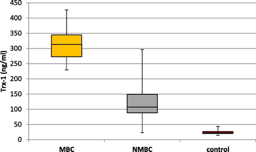
Fig. 2 The median and range of matrix metalloproteinase-9 level (MMP-9) (ng/ml) in metastatic breast cancer (MBC), non-metastatic breast cancer (NMBC) and control groups.
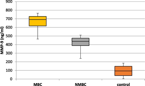
Fig. 3 The median and range of malondialdehyde level (MDA) (nmol/ml) in metastatic breast cancer (MBC), non-metastatic breast cancer (NMBC) and control groups.
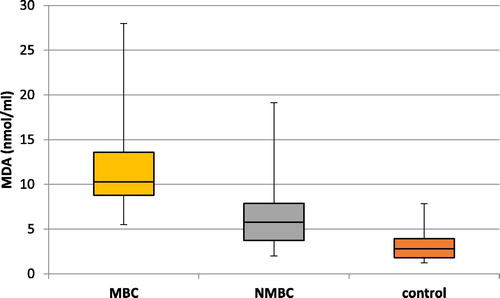
Table 2 Comparison of studied parameters in MBC vs. NMBC patients and control group [Median (Min-Max)].
3.3 Relations between measured parameters
The relation between serum Trx-1 level and MMP-9 level with clinicopathological parameters in all patients was analyzed and represented in . Serum Trx-1 level and MMP-9 level in patients of stage IV breast cancer were significantly increased than those of stage I, II or III (P < .001). Trx-1 level and MMP-9 level were significantly increased with tumor mass more than 5 cm (P < .001). Serum levels of both Trx-1 and MMP-9 were significantly higher in patients with lymph node metastasis than those without lymph node metastasis (P < .05). Serum Trx-1 level and MMP-9 level were significantly higher in grades 2 and 3 than grade 1 (P < .05). However, the serum levels of both Trx-1 and MMP-9 show no statistically significant difference between the different age groups in the breast cancer patients.
Table 3 Relation between serum thioredoxin-1 (Trx-1), matrix metalloproteinase 9 (MMP-9) and malondialdehyde (MDA) serum levels with clinicopathological parameters in breast cancer patients.
MDA level is significantly increased in patients with stage IV breast cancer than stages I, II and III (P < .001). MDA is significantly increased only with tumor mass more than 5 cm (P < .001). Furthermore MDA level in patients with lymph node metastasis was highly significant than those with no lymph node metastasis (P < .05). However, serum level of MDA shows no statistically significant difference between the different age groups in the breast cancer patients.
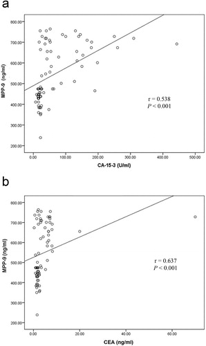
Spearman's ρ analysis revealed a highly significant positive correlation between Trx-1 and MMP-9 serum levels (r = 0.718, P < .001; ). Furthermore, there was a highly significant positive correlation between serum level of Trx-1 and serum level of CEA and CA15-3 (r = 0.581, P < .001 and r = 0.570, P < .001, respectively; ). Also, the analysis shows a significant positive correlation between serum level of MMP-9 and serum levels of CEA and CA15-3 (r = 0.538, P < .001 and r = 0.637, P < .001, respectively; ).
Fig. 4 Positive correlation between serum thioredoxin-1 (Trx-1) level and matrix metalloproteinase-9 (MMP-9) level in all breast cancer patients.
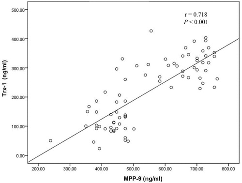
Fig. 5 Positive correlation between serum thioredoxin-1 (Trx-1) and cancer antigen 15-3 (CA15-3) levels (a) and serum Trx-1 and carcinoembryonic antigen (CEA) levels (b) in all breast cancer patients.
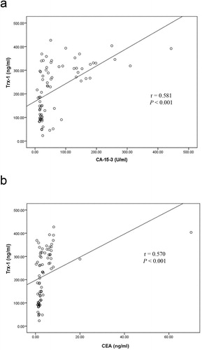
Fig. 6 Positive correlation between serum matrix metalloproteinase-9 (MMP-9) level and cancer antigen 15-3 (CA15-3) level (a) and serum MMP-9 level and carcinoembryonic antigen (CEA) level (b) in breast cancer patients.
3.4 Determination of diagnostic values of Trx-1 and MMP-9
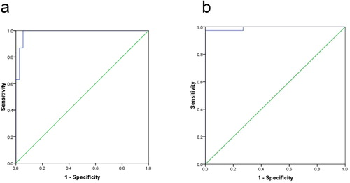
ROC analysis was adopted to detect the diagnostic values of serum Trx-1 and MMP-9 as biomarkers in metastatic breast cancer disease. Analysis of results suggested that; serum Trx-1 and MMP-9 could predict breast cancer metastasis at cutoff points; 231.1 ng/ml and 492.4 ng/ml, respectively. Trx-1 diagnostic specificity and sensitivity were found to be 94.6% and 97.4%, respectively, and MMP-9 diagnostic specificity and sensitivity were found to be 97.3% and 97.4% respectively. Moreover, the area under curve (AUC) was 0.986 for Trx-1 and 0.993 for MMP-9 ().
Fig. 7 Receiver operating characteristic curve (ROC) for serum thioredoxin-1 (Trx-1) level (a) and for serum matrix metalloproteinase-9 (MMP-9) level (b) for detection of breast cancer metastases.
4 Discussion
Breast cancer metastasis is the leader cause for breast cancer-related deaths [Citation4]. In spite of advances in treatment, 20–30% of patients with early breast cancer ultimately develop relapse with distant metastasis [Citation18]. CA 15-3 and CEA are broadly used markers for the early diagnosis of MBC in the clinical situations. The united measurement of both serum markers permits early diagnosis of metastases in up to 60–80% of breast cancer patients [Citation19]. Recent study shows that elevated CA 15-3 and CEA levels were identified in 163 (57.4%) and 97 (34.2%), respectively, of the 284 patients with distant metastasis [Citation20]. Thus, we studied the role of both Trx-1 and MMP-9 in diagnosis of breast cancer and their correlation with clinicopathological parameters of MBC and NMBC Egyptian patients, as well as, their sensitivity and specificity in the disease prognosis.
Breast cancer metastasis is the process by which tumor cells migrate and establish from their primary site to secondary tumors [Citation21]. This process requires degradation of the ECM in both primary and secondary tumor tissues via MMP-9 [Citation22]. Trx-1 stimulates MMP-9 expression and promotes MMP-9 dependent invasion in breast cancer cells [Citation12]. Thioredoxin system is highly expressed in breast tumors [Citation23,Citation24] and Trx-1 has been previously suggested as a suitable and diagnostic biomarker for breast cancer [Citation8,Citation13] . Our results extend these previous studies to show that Trx-1 level is significantly increased in breast cancer patients as compared to control. Moreover, Trx-1 correlates with distant metastasis and poor prognosis [Citation11]. Tumor metastasis and aggressiveness could be explained by the ability of Trx-1 to push the balance towards MMP-9 function. MMP-9 involvement in malignant behavior depends upon change in equilibrium with its tissue specific inhibitor, TIMP-1 [Citation12]. This suggests that Trx-1 could be a therapeutic target to reduce the tumor cell invasion and hence metastasis of breast cancer [Citation11]. To our knowledge, this is the first time to report the relationship between serum Trx-1 and breast cancer metastasis and progression. Serum Trx-1 level is significantly increased in metastatic breast cancer patients. It is correlated with tumor stage, tumor grading and lymph node metastasis. However, serum Trx-1 level is not related to tumor size. Since Trx-1values in MBC patients are significantly higher than those in NMBC patients, so serum Trx-1 may be used as a predictive biomarker for metastasis.
MMP-9 has an important role in breast cancer invasion and metastasis. Our study shows that serum MMP-9 level was significantly elevated in breast cancer patients as compared to control. This result agreed with that of Patel et al. [Citation25] and La Rocca et al. [Citation26]. In addition, MMP-9 appears to have a greater biological activity in carcinogenesis. Recent studies show that a down- regulation of MMP-9 prevents cancer cell growth, invasion and angiogenesis [Citation27,Citation28] . Moreover, we constitute that serum MMP-9 was significantly increased in MBC patients; subsequently MMP-9 was correlated with breast cancer stage as defined by AJCC-TNM system. Therefore, MMP-9 may play a vital role in breast cancer metastasis which agreed with the results of Patel et al. [Citation25] and Rashad et al. [Citation29].
We found that serum MMP-9 level predicts lymph node metastasis, which agreed with Zhang et al. [Citation30] and Heo et al. [Citation31]. In addition, serum MMP-9 level is significantly higher in grade 2 and 3 than grade 1. Zhang et al. [Citation30] stated that MMP-9 parallel with tumor size, on the other hand Daniele et al. [Citation32] and Rashad et al. [Citation29] studies show no correlation among serum MMP-9 and tumor size which agreed with our results. MMP-9 is associated with poor general condition (progressive disease stage and metastasis), so, we recommend that MMP-9 may be a possible therapeutic target in breast cancer. This was in agreement with Li et al. [Citation28], Park et al. [Citation33] and Sinha et al. [Citation34] who conclude that suppression of MMP-9, served as a possible target in treatment of breast cancer metastasis.
Malondialdehyde (MDA) is the end-product of lipid peroxidation (LPO) produced from the free radical degradation of polyunsaturated fatty acids. MDA promote breast cancer via interaction with amino groups of nucleic acid bases in DNA forming malondialdehyde-DNA adducts [Citation35].
Previous studies show that a serum MDA level was significantly elevated in breast cancer patients compared to healthy subjects [Citation13,Citation36Citation[37]Citation[38]–Citation39] . These results are in consistence with our study and confirm increase lipid peroxidation in breast cancer patients.
Moreover, Pande et al. shows that VEGF and MDA levels are increased in breast cancer patients and were positively correlated. VEGF is a significant stimulator of tumor angiogenesis and metastasis and up-regulated by oxidative stress conditions [Citation40]. In our study the serum MDA level was significantly elevated in metastatic breast cancer patients and correlated with clinical stage. This result agreed with Sadati Zarrini et al. [Citation36] Baskic et al. [Citation41] and Gonenc et al. [Citation42]. In addition, MDA level was correlated with tumor grading and lymph node metastasis but not related to tumor size.
Conclusion
| • | Serum Trx-1 level and MMP-9 level are significantly increased in metastatic breast cancer patients. Trx-1and MMP-9 elevation correlate with progression of the disease state. | ||||
| • | Trx-1 and MMP-9 are promising candidates as guide for relapse of breast cancer with distant metastases, as they linked with advanced disease stage. | ||||
| • | Trx-1 and MMP-9 are possible therapeutic targets for prevention of breast cancer metastasis. | ||||
| • | MDA as biomarker of oxidative stress is implicated in the progress of metastasis in breast cancer patients. | ||||
| • | These results should be confirmed in further prospective studies employing a large number of subjects for the use of Trx-1 and MMP-9 in early diagnosis and follow-up of metastatic breast cancer. | ||||
References
- J.FerlayI.SoerjomataramR.DikshitS.EserC.MathersM.Rebeloet al.Cancer incidence and mortality worldwide: sources, methods and major patterns in GLOBOCAN 2012Int J Cancer13652015E359E386
- K.ServickBreast cancer. Breast cancer: a world of differencesScience (New York, NY)3436178201414521453
- A.S.IbrahimH.M.KhaledN.N.MikhailH.BarakaH.KamelCancer incidence in Egypt: results of the national population-based cancer registry programJ Cancer Epidemiol20142014437971
- S.KimbungN.LomanI.HedenfalkClinical and molecular complexity of breast cancer metastasesSemin Cancer Biol3520158595
- R.L.SiegelK.D.MillerA.JemalCancer statistics, 2015CA Cancer J Clin6512015529
- A.R.NourazarianP.KangariA.SalmaninejadRoles of oxidative stress in the development and progression of breast cancerAsian Pac J Cancer Preven: APJCP1512201447454751
- I.S.HarrisA.E.TreloarS.InoueM.SasakiC.GorriniK.C.Leeet al.Glutathione and thioredoxin antioxidant pathways synergize to drive cancer initiation and progressionCancer Cell2722015211222
- B.J.ParkM.K.ChaI.H.KimThioredoxin 1 as a serum marker for breast cancer and its use in combination with CEA or CA15-3 for improving the sensitivity of breast cancer diagnosesBMC Res Notes720147
- A.HolmgrenThioredoxinAnnu Rev Biochem541985237271
- S.G.RheeH.Z.ChaeK.KimPeroxiredoxins: a historical overview and speculative preview of novel mechanisms and emerging concepts in cell signalingFree Radical Biol Med3812200515431552
- M.BhatiaK.L.McGrathG.Di TrapaniP.CharoentongF.ShahM.M.Kinget al.The thioredoxin system in breast cancer cell invasion and migrationRedox Biol820166878
- A.R.FarinaL.CappabiancaG.DeSantisN.Di IanniP.RuggeriM.Ragoneet al.Thioredoxin stimulates MMP-9 expression, de-regulates the MMP-9/TIMP-1 equilibrium and promotes MMP-9 dependent invasion in human MDA-MB-231 breast cancer cellsFEBS Lett58520201133283336
- N.KilicM.Yavuz TaslipinarY.GuneyE.TekinE.OnukAn investigation into the serum thioredoxin, superoxide dismutase, malondialdehyde, and advanced oxidation protein products in patients with breast cancerAnn Surg Oncol2113201441394143
- C.MehnerA.HocklaE.MillerS.RanD.C.RadiskyE.S.RadiskyTumor cell-produced matrix metalloproteinase 9 (MMP-9) drives malignant progression and metastasis of basal-like triple negative breast cancerOncotarget59201427362749
- J.M.SandL.LarsenC.HogaboamF.MartinezM.HanM.Rossel Larsenet al.MMP mediated degradation of type IV collagen alpha 1 and alpha 3 chains reflects basement membrane remodeling in experimental and clinical fibrosis–validation of two novel biomarker assaysPLoS One8122013e84934
- S.B.EdgeC.C.ComptonThe American joint committee on cancer: the 7th edition of the AJCC cancer staging manual and the future of TNMAnn Surg Oncol176201014711474
- T.YoshiokaK.KawadaT.ShimadaM.MoriLipid peroxidation in maternal and cord blood and protective mechanism against activated-oxygen toxicity in the bloodAm J Obstet Gynecol13531979372376
- Effects of chemotherapy and hormonal therapy for early breast cancer on recurrence and 15-year survival: an overview of the randomised trials. Lancet (London, England). 2005;365 (9472):1687–717.
- R.MolinaV.BarakA.van DalenM.J.DuffyR.EinarssonM.Gionet al.Tumor markers in breast cancer- European group on tumor markers recommendationsTumour Biology: J Int Soc Oncodev Biol Med2662005281293
- B.GengM.M.LiangX.B.YeW.Y.ZhaoAssociation of CA 15–3 and CEA with clinicopathological parameters in patients with metastatic breast cancerMol Clin Oncol312015232236
- I.J.FidlerCritical factors in the biology of human cancer metastasis: twenty-eighth G.H.A. Clowes memorial award lectureCancer Res5019199061306138
- H.YamaguchiJ.WyckoffJ.CondeelisCell migration in tumorsCurr Opin Cell Biol1752005559564
- D.T.LincolnE.M.Ali EmadiK.F.TonissenF.M.ClarkeThe thioredoxin-thioredoxin reductase system: over-expression in human cancerAnticancer Res233b200324252433
- M.K.ChaK.H.SuhI.H.KimOverexpression of peroxiredoxin I and thioredoxin1 in human breast carcinomaJ Exp Clin Cancer Res: CR28200993
- S.PatelG.SumitraB.C.KonerA.SaxenaRole of serum matrix metalloproteinase-2 and -9 to predict breast cancer progressionClin Biochem4410–112011869872
- G.La RoccaI.Pucci-MinafraA.MarrazzoP.TaorminaS.MinafraZymographic detection and clinical correlations of MMP-2 and MMP-9 in breast cancer seraBr J Cancer907200414141421
- J.GaoX.LiuF.YangT.LiuQ.YanX.YangBy inhibiting Ras/Raf/ERK and MMP-9, knockdown of EpCAM inhibits breast cancer cell growth and metastasisOncotarget62920152718727198
- J.LiJ.ZhangY.WangX.LiangZ.WusimanY.Yinet al.Synergistic inhibition of migration and invasion of breast cancer cells by dual docetaxel/quercetin-loaded nanoparticles via Akt/MMP-9 pathwayInt J Pharm52312017300309
- Y.A.RashadT.R.ElkhodaryA.M.El-GayarL.A.EissaEvaluation of serum levels of HER2, MMP-9, nitric oxide, and total antioxidant capacity in Egyptian breast cancer patients: correlation with clinico-pathological parametersSci Pharm8212014129145
- J.ZhangL.YinJ.WuY.ZhangT.XuR.Maet al.Detection of serum VEGF and MMP-9 levels by Luminex multiplexed assays in patients with breast infiltrative ductal carcinomaExp Ther Med812014175180
- D.S.HeoH.ChoiM.Y.YeomB.J.SongS.J.OhSerum levels of matrix metalloproteinase-9 predict lymph node metastasis in breast cancer patientsOncol Rep314201415671572
- A.DanieleA.F.ZitoG.GiannelliR.DivellaM.AsseltiA.Mazzoccaet al.Expression of metalloproteinases MMP-2 and MMP-9 in sentinel lymph node and serum of patients with metastatic and non-metastatic breast cancerAnticancer Res309201035213527
- J.H.ParkY.Y.ChoS.W.YoonB.ParkSuppression of MMP-9 and FAK expression by pomolic acid via blocking of NF-kappaB/ERK/mTOR signaling pathways in growth factor-stimulated human breast cancer cellsInt J Oncol493201612301240
- S.SinhaS.KhanS.ShuklaA.D.LakraS.KumarG.Daset al.Cucurbitacin B inhibits breast cancer metastasis and angiogenesis through VEGF-mediated suppression of FAK/MMP-9 signaling axisInt J Biochem Cell Biol7720164156 Pt A
- Wang M, Dhingra K, Hittelman WN, Liehr JG, de Andrade M, Li D. Lipid peroxidation-induced putative malondialdehyde-DNA adducts in human breast tissues. Cancer epidemiology, biomarkers & prevention: a publication of the American Association for Cancer Research, cosponsored by the American Society of Preventive Oncology. 1996;5(9):705–10
- A.Sadati ZarriniD.MoslemiH.ParsianM.VessalA.MosapourKelagari Z.ShirkhaniThe status of antioxidants, malondialdehyde and some trace elements in serum of patients with breast cancerCaspian J Internal Med7120163136
- C.P.RajneeshA.ManimaranK.R.SasikalaP.AdaikappanLipid peroxidation and antioxidant status in patients with breast cancerSingapore Med J4982008640643
- C.C.YehM.F.HouS.M.TsaiS.K.LinJ.K.HsiaoJ.C.Huanget al.Superoxide anion radical, lipid peroxides and antioxidant status in the blood of patients with breast cancerClin Chim Acta; Int J Clin Chem3611–22005104111
- A.GonencY.OzkanM.TorunB.SimsekPlasma malondialdehyde (MDA) levels in breast and lung cancer patientsJ Clin Pharm Ther2622001141144
- D.PandeR.NegiS.KhannaR.KhannaH.D.KhannaVascular endothelial growth factor levels in relation to oxidative damage and antioxidant status in patients with breast cancerJ Breast Cancer1432011181184
- D.BaskicS.PopovicD.BankovicA.ArsovicV.VukovicI.Zelenet al.Evaluation of inflammatory biomarkers as helping diagnostic tool in patients with breast cancerCancer Biomarkers: Sec A Dis Markers1462014401408
- A.GonencD.TokgozS.AslanM.TorunOxidative stress in relation to lipid profiles in different stages of breast cancerIndian J Biochem Biophys4232005190194