
 ?Mathematical formulae have been encoded as MathML and are displayed in this HTML version using MathJax in order to improve their display. Uncheck the box to turn MathJax off. This feature requires Javascript. Click on a formula to zoom.
Abstract
The Dielectric and Fourier Transform Infrared (FTIR) spectroscopy have been used to investigate the effect of low dose (10 cGy) and high dose (2 Gy) X-rays on the lung of whole-body irradiated rats. The dielectric permittivity ε′, dielectric loss ε″, electric modulus M′ and the imaginary part of the electric modulus M″ measured show changes in the molecular dynamics of the cellular membrane following 10 cGy and 2 Gy-irradiation, reflecting change in the fluidity of the cellular membrane following irradiation, with the most significant change recorded following 2 Gy-irradiation. The FTIR data further illustrate an increase in the fluidity cellular membrane following 10 cGy and 2 Gy-irradiations as marked by the shift and broadening of the CH2 asymmetric stretching vibration band. However, our findings show that 2 Gy-irradiation effects on the cellular membrane were much higher than 10 cGy but they also highlight the considerable effect of radiation dose as low as 10 cGy and its associated risk.
1 Introduction
Exposure to ionizing radiation has been proven to cause variety of cellular effects [Citation1]. Of the most deleterious cellular effects of ionizing radiation comes the cell membrane damage. The fluidity of the cell membrane bilayer plays an important role in its functionality. Thus, damage in the cell membrane affects its functionality and the whole cell integrity [Citation2]. Investigation of the cellular membrane damage is crucial when studying the cellular effects of ionizing radiation. The delectability of the radiation-induced damages relies essentially on the degree of the damage and the sensitivity of the detection techniques. Various physical techniques have been proven to be powerful, sensitive, and non-destructive techniques to investigate the radiation-induced damages at the cellular and molecular levels. Two of these techniques were used in this study, Dielectric spectroscopy and FTIR Spectroscopy.
Dielectric spectroscopy has widely been used to investigate molecular changes in biological tissue [Citation3Citation[4]Citation[5]–Citation6] . The dielectric properties of biological tissue arise from the interaction of electromagnetic radiation with the constituents of the biological tissue. The molecular dynamic at different applied radio-frequencies RF can be tracked and well determined using dielectric characterizations [Citation7Citation[8]Citation[9]–Citation10] . The application of RF around 1 MHz due to their interaction with biological tissues dates back to 1950s [Citation11,Citation12] .
Dielectric complex permittivity and its reciprocal as complex Electric Modulus
are useful tools in the study of biological tissues for basic and applied science applications [Citation10,Citation13] . The complex permittivity
parameters as dielectric constant
and losses
are always measured by parallel plate separated by the sample as capacitor upto 100 MHz. On the other hand, the complex electric modulus
is a calculus method used for eliminate the interfacial electrode polarization that might cover any of the main relaxation processes in dielectric loss plots. Through the permittivity parameters measurements, the influence of different doses of radiation will be investigated for lung tissues of irradiated rats.
In this paper, the radio frequency RF (100 Hz–10 MHz) was used to investigate the effect of low- and high-dose whole-body irradiation on the molecular dynamics of the cellular membrane of the lung tissue of rats. The range 100 Hz–10 MHz is known to be the site of β dispersion. The β dispersion site arises from a polarization mechanism known as Maxwell-Wagner effect. In this region, the interface represents a barrier for ionic charge flow and hence leads to polarization and subsequent relaxation. The β dispersion in tissues often occurs at the cell membrane. Thus, any alterations in the structure of the cell membrane would markedly affect this region [Citation14]. Additionally, the radiation-induced damage in the phospholipid membrane bilayer was further investigated using FTIR spectroscopy. FTIR is a non-destructive technique that provides in depth information about the molecular changes in the cell membrane [Citation15Citation[16]–Citation17] through providing a fingerprint of all molecules in the cells [Citation18].
2 Materials and methods
2.1 Experimental animals
Experimental animals, twenty male Sprague-Dawley rats approximately 4 months old weighing 160 ± 20 g, were maintained under the standard conditions of 22–24 °C and 12-h light/dark cycle with free access to food and water. The experimental procedures were approved by the animal care and ethics committee at the authors' institute (approval no:17-018). The animals were divided into 3 groups each containing 5 rats; The Sham-irradiated group (0 Gy), the animals were kept unirradiated control. The 2 Gy-irradiated group; Animals were 2 Gy whole-body irradiated (dose rate 4 Gy/min). The 10 cGy-irradiated group; Animals were 10 cGy whole-body irradiated (dose rate 4 Gy/min).
2.2 Irradiation
Rats were irradiated with x-rays using a 6 MV linear accelerator (Precise Linear Accelerator; Elekta, Stockholm, Sweden) at the oncology and nuclear medicine center at Mansoura University hospital, Mansoura, Egypt.
Before irradiation, the animals were anesthetized by intraperitoneal injection of Ketamine and Xylazine cocktail (75 mg/kg and 5 mg/Kg, respectively). In average, the animals were under anesthesia for 60 min during which the animals were kept at room temperature (20–25 °C). During irradiation, the animals were restrained in the sternal recumbent position on the irradiation table and the animals were centered in the radiation field.
2.3 Samples collection
At the end of the experimental period, all animals were anesthetized with halothane and sacrificed and sections from the lung were collected. All samples were then freeze-dried and stored until use.
2.4 The dielectric measurements
The dielectric permittivity parameters of lung tissues were measured in low frequencies from 100 Hz to 100 KHz by Ando AG-4311 LCR meter (United State). The capacitance C and dielectric loss tangent D were measured directly. Then, frequencies from 100 KHz to 120 MHz were applied by Hioki LCR 3535-Hi Tester meter (Hioki Co., Japan). The former LCR hi tester is upgraded using 9700 head amplifier unit by the manufacturer to minimize the effect of the measuring leads. The experiments were carried out using 9699 SMD test fixture which has two electrodes to be fitted with sample object sizes as width: 1.0 mm length: 4.0 mm and height: less than 1.5 mm. This test fixture has the operating frequency range from DC to 120 MHZ.
Both and
are calculated based on equations
and dielectric loss
where C is capacitance of the sample placed in between two electrodes as dielectrics, and d is thickness which is less than 1.5 mm. A is area of electrode, and
is permittivity of vacuum (8.8543 × 10⁻¹² F/m). Calculation of the permittivity in case of using parallel plates as a capacitor is performed by dividing the capacitance of the cell with dielectric material
to capacitance of empty cell
where its permittivity value for air
and the capacitance is equal
The complex electric modulus is used to shift up the frequencies of the relaxation peak and suppress the electrode polarization effect. The reciprocal values of complex permittivity as dielectric constant
and dielectric loss
determine the complex values of real and imaginary parts of electric modulus [Citation19]
The dc Conductivity
which is depending on the dielectric loss
and angular frequency
represent the electric charge mobility carries inside the sample matrix or network of the sample. Thus, the dissipated energy could be used in driving charges carriers under applied electric field as shown in the following equation
Moreover, the dielectric properties refer to the electric charge and their carries inside the tissues regarding to an external electric field. The dc conductivity of the tissues represents their free charge movement forced by the external field. Therefore, dielectric response of biological materials is always frequency dependent. Thus, a linear response means that the dielectric properties are independent of the external field strength, which is true when the external electric field is not very strong.
2.5 FTIR spectroscopy
All FTIR measurements of freeze-dried samples were performed at room temperature in the energy range 4000–400 cm−1 using a Bruker Vertex 70 Fourier transform spectrometer in combination with diamond ATR unit at 4 cm−1 spectral resolution.
3 Results and discussion
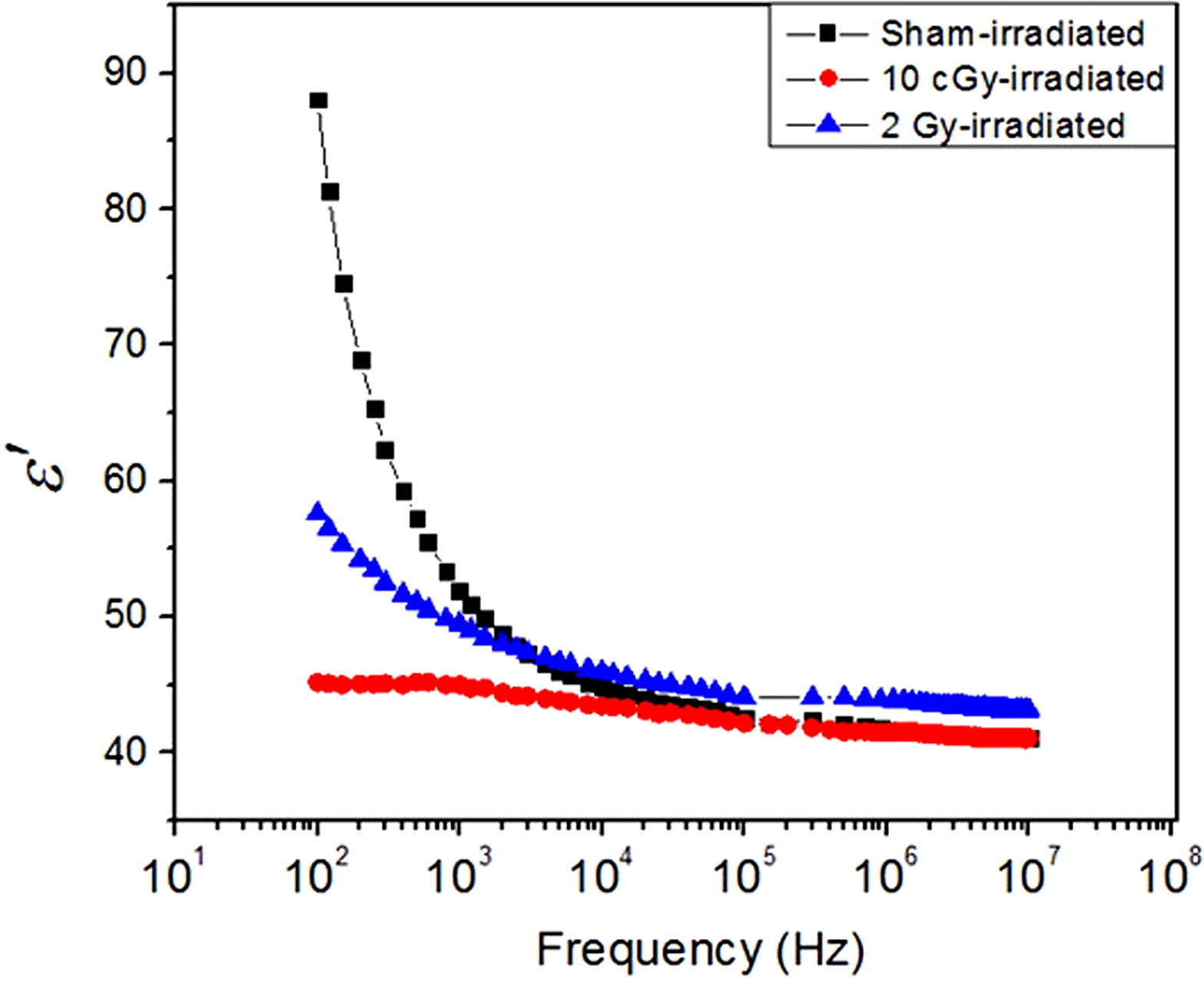
Dielectric spectroscopy has been an important physical tool for the modeling and evaluating the energy deposition in tissue exposed to radio-waves [Citation20]. The dielectric properties of the lung tissue of the sham, 10 cGy and 2 Gy-irradiated rats are represented in this section. shows the permittivity as dielectric constants versus the applied frequency at room temperature. Both complex permittivity parameters as dielectric constants
and dielectric loss
were measured from 100 Hz to 10 MHz. The value of permittivity
of sham-irradiated group starts at 88 and decrease rapidly over the frequency range 102 Hz–104 Hz. On the other hand, the permittivity value at the high frequency ranges 105 Hz–107 Hz, known as infinity dielectric constant
, was 41. The reason for lower values of
could be the due to freeze drying the samples which removed the water content from the tissues leaving behind the non-liquid components of the tissue including cell membrane and lipids. The difference between these values (88 and 41) refers to the dielectric strength of the intra-molecular dynamic process
[Citation9]. This is in agreement with data reported for bone tissue, whose dielectric properties were measured by Gabriel et al. in 1996 [Citation3]. However, other organs that have large content of water recorded very high permittivity values (order of 105–107) at low RF frequencies [Citation3]. Although the 10 cGy irradiated samples show lower permittivity values of 45 at low frequencies, at high frequencies there is a slight decrease to 41. The 2 Gy-irradiated samples show different trend with higher permittivity value of 57.6 at the low frequencies, followed by significant decrease down to 43 at the higher frequencies.
Fig. 1 The dielectric permittivity . Changes in the permittivity
of the sham, 10 cGy and 2 Gy-irradiated groups as a function of frequency.
The internal dynamic processes of the molecular structure resulting from the applied electric field can be investigated by measuring dielectric loss . shows the dielectric loss
for the sham, low- and high-dose irradiated groups. As shown in the figure, part of the relaxation processes appears at low RF frequencies for the samples with contribution of the interfacial or electrode polarizations especially in the sham and high-dose irradiated groups. Whereas, the low-dose irradiated group does not show any relaxation process. The overall trend shows the relaxation processes to be shifted towards the lower frequencies. Due to the interference of electrode polarization, it is hard to determine the relaxation processes at low RF region [Citation21]. Therefore, the electric modulus
was used to eliminate the effect of electrode polarization as it is considered a useful correlation to get the molecular dynamics and their related relaxation processes clearly and easily as shown in Figs. 3 and 4.
Fig. 2 The dielectric loss . Changes in the dielectric loss
of the sham, 10 cGy and 2 Gy-irradiated groups as a function of frequency.
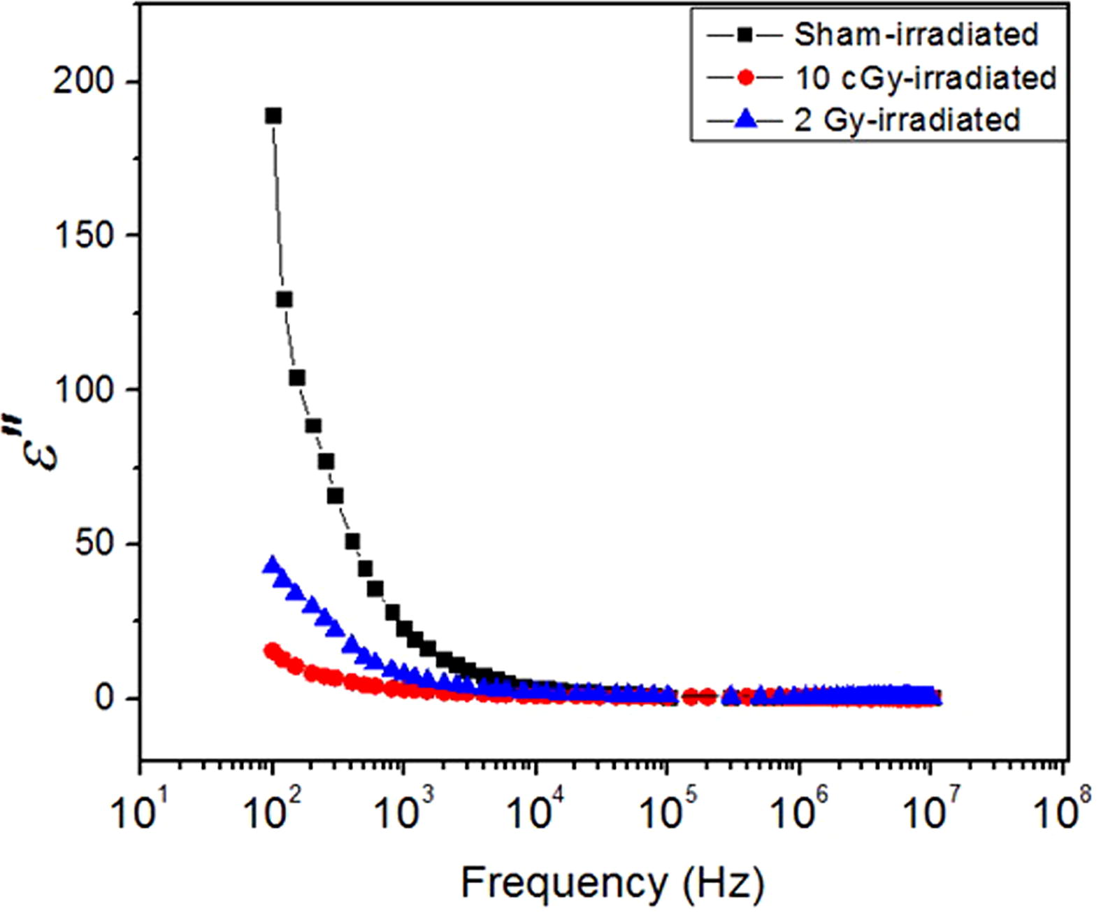
Fig. 3 The real part of the electric modulus . Changes in the real part of the electric modulus M′ of the sham, 10 cGy and 2 Gy-irradiated groups as a function of frequency.
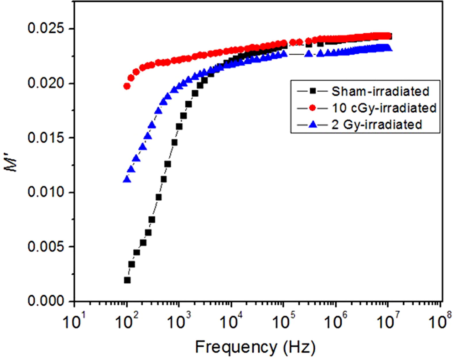
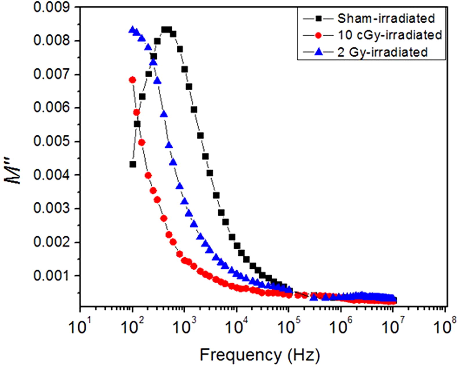
Fig. 4 The imaginary part of electric modulus . The imaginary part of electric modulus (M″) of the sham, 10 cGy and 2 Gy-irradiated groups measured versus frequency.
As shown in , in the low-dose irradiated group, the real part of the electric modulus slightly increases from 0.020 to 0.024 as a result of its high shift towards the low frequency region. However, the electric modulus
of the sham-irradiated group shows a dramatic increase from 0.002 to 0.022 before reaching the plateau level. This typical behavior of complete relaxation process reflects the changes in the molecular dynamics of the macromolecules and is attributed to the flexibility of the structure and enlarge the free volume in the texture [Citation14]. The electric modulus
of the high-dose irradiated group shows a shift starting from 0.0111 and reaching the plateau from 0.0214 to 0.0232 over the frequency range 103–107 Hz.
Thus, the imaginary part of electric modulus is useful to show the relaxation processes without any contribution of the AC conductivity due electrode polarization. shows the imaginary electric modulus
full relaxation process for the sham-irradiated group while the 2 Gy-irradiated group shows an intermediate relaxation process peak. This is an evidence of little shift in the main relaxation of the lung tissue cellular membrane as a result of 2 Gy-irradiation. However, for the 10 cGy-irradiation, shift towards very low frequencies was observed. These data clearly show the influence of ionizing radiation on the fluidity of the cellular membrane.
The free volume distribution depends on the temperature and frequency, and affects the relaxation peak frequency [Citation22]. Our results show 10 cGy-irradiation lead to greater shift in the relaxation peak towards low frequencies than the 2 Gy-irradiation. This implies that 10 cGy-irradiation leads to increased rigidity in the cell membrane as compared with 2 Gy-irradiation.
The dielectric relaxation process in the linear systems refers to the time delay needed by the molecule to regain its original state before applying the electric [Citation23]. The relaxation time of the 10 cGy-irradiated group is high as a result of the shift of the relaxation peak towards low frequencies. The more rigid the molecule is, the less free volume the molecule has and hence the more the time the relaxation process takes. Thus more shifts toward lower frequencies is observed [Citation22]. Changes in the temperature remarkably affect the dielectric relaxation process [Citation23]. In this study, all samples were measured at room temperature. Thus, the temperature had negligible effect on our results which indicates that the shift in the dielectric relaxation process to lower frequencies was solely due to the effect ionizing radiation on the macromolecules in the samples.
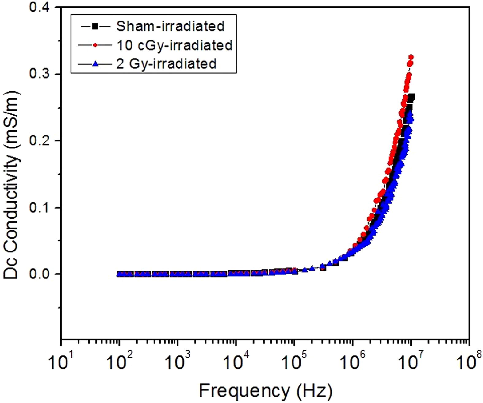
shows the dc conductivity as illustrated in the equation before. All irradiated and sham-irradiated groups show the same behavior of lipid and polymeric attributes. At low frequencies, no charge carriers movement through the samples can be detected, while the threshold of moving charges and ions through the samples start at 106 Hz, where high energy field is applied. The following dramatic and significant increase is due to the start of charge carries movements between the electrodes. There is no saturation level (plateau) reached at higher frequency, which means continuous increase in charges moving with increasing the applied field.
Fig. 5 The DC conductivity. Changes in the DC conductivity of the sham, 10 cGy and 2 Gy-irradiated groups as a function of frequency.
To further investigate the effect of low and high doses of ionizing radiation on the conformational disorder of the cell membrane, FTIR spectroscopy was used. shows the FTIR spectra of the sham, 10 cGy and 2 Gy-irradiated groups in the spectral range 4000–400 cm−1. As illustrated in and , the CH2 asymmetric stretching vibration band located at 2920 cm−1 in the sham-irradiated group. However, this band was slightly shifted towards higher wavenumber in both 10 cGy and 2 Gy-irradiated groups with the higher shift seen in the 2 Gy-irradiated group. The shift in this band indicates a decrease in the order of the system which can be correlated to the increase in the membrane fluidity [Citation17,Citation24] with the 2 Gy-irradiation having a higher effect on membrane fluidity. These results are in agreement with our dielectric data showing that the 2 Gy-irradiated group shows more flexibility (due to increased fluidity) than the 10 cGy-irradiated group as indicated by the difference in the dielectric relaxation processes between the two groups.
Fig. 6 FTIR spectra. The FTIR spectra of the rat lung of the sham, 10 cGy and 2 Gy-irradiated groups.
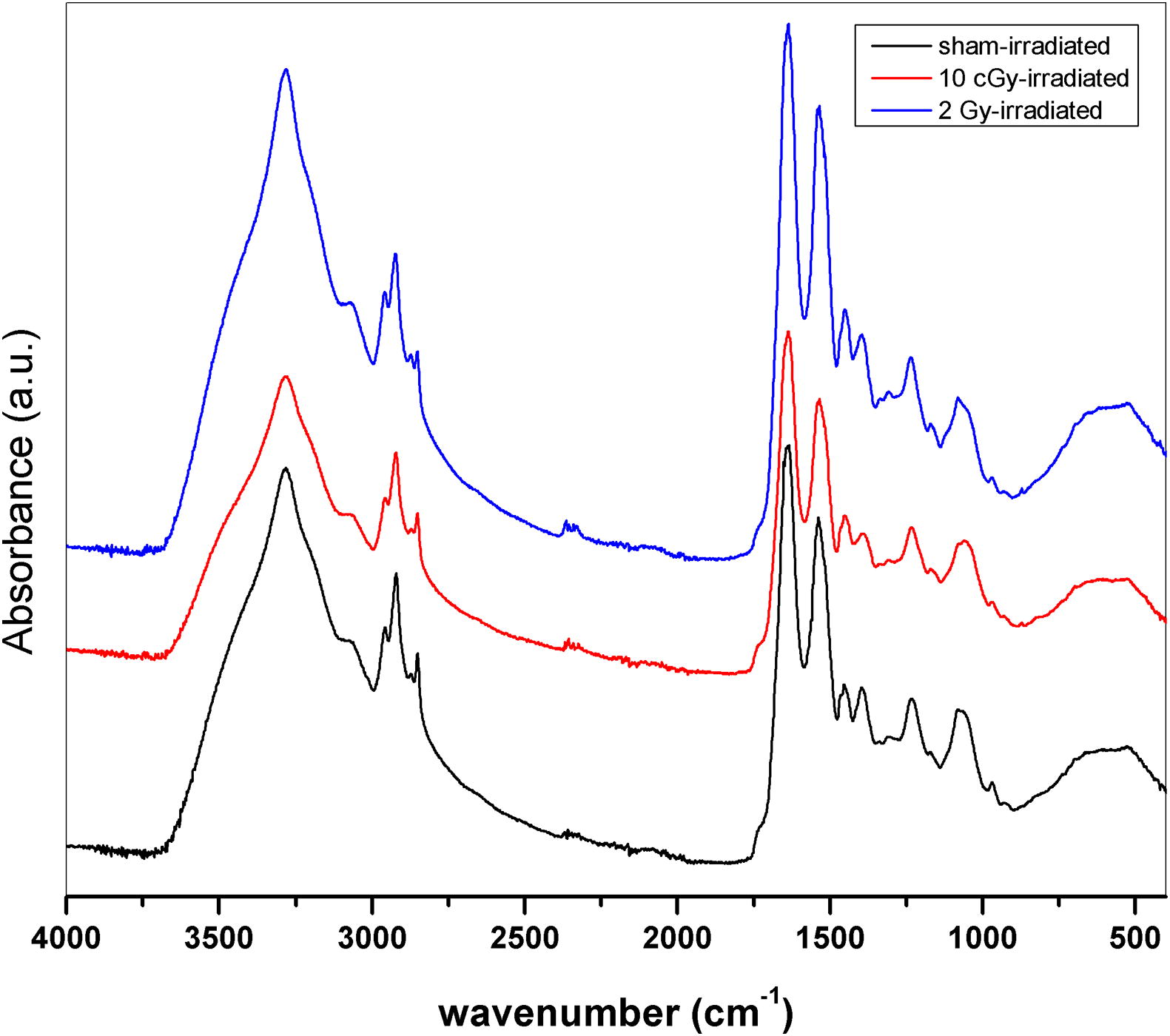
Table 1 Changes in the band position and full width at half maximum (FWHM) values of CH2 asymmetric stretching FTIR band of sham, 10 cGy and 2 Gy-irradiated groups.
Furthermore, an increase of the bandwidth of the CH2 asymmetric stretching vibration band was also recorded (). This indicates an increase in the membrane fluidity on the irradiated groups [Citation18]. The increase in the membrane fluidity is always accompanied by a decrease in order as a result of free rotation of lipid acyl chains and lateral lipid motion [Citation15,Citation25] . These data all together suggest varying degrees of disorders in the molecular structure of the cell membrane as a result of radiation with the highest degree of disorder is seen following 2 Gy-irradiation. Our findings however seem to be logical, that a higher radiation dose induces the higher effect, they point out the risk associated with low-dose radiation and its implications on human health.
4 Conclusions
Our findings reveal significant conformational changes in the cell membrane of the rat lung cells following 10 cGy and 2 Gy-irradiations. Measuring the various dielectric parameters of permittivity , dielectric loss
, electric modulus
, the imaginary part of the electric modulus
and DC conductivity divulge that both irradiations increased the membrane fluidity. The remarkable shifts of the relaxation peaks toward low frequencies following irradiation indicate changes in the fluidity of the cellular membranes. The dielectric constant values are considerably high even in absence of water contents after freeze drying. 2 Gy-irradiation-induced membrane fluidity was way higher than that recorded following 10 cGy-irradiation. Furthermore, FTIR data confirms the remarkable conformational changes in the cell membrane following irradiation with the 2 Gy-irradiation having the major effect. The data also emphasize the risk associated with low-dose radiation exposure through its damaging effect at the cellular level.
5 Role of funding
This research did not receive any specific grant from funding agencies in the public, commercial, or not-for-profit sectors.
References
- E.J.HallA.J.GiacciaRadiobiology for the Radiologist2006Lippincott Williams & Wilkins Philadelphia
- M.M.GaschlerB.R.StockwellLipid peroxidation in cell deathBiochem Biophys Res Commun48232017419425
- S.GabrielR.LauC.GabrielThe dielectric properties of biological tissues: II. Measurements in the frequency range 10 Hz to 20 GHzPhys Med Biol411119962251
- A.PeymanS.HoldenC.GabrielDielectric properties of tissues at microwave frequenciesMobile Telecommun Health Res Program Final Report2005
- K.SasakiK.WakeS.WatanabeMeasurement of the dielectric properties of the epidermis and dermis at frequencies from 0.5 GHz to 110 GHzPhys Med Biol591620144739
- G.SchmidG.NeubauerU.M.IllievichF.AleschDielectric properties of porcine brain tissue in the transition from life to death at frequencies from 800 to 1900 MHzBioelectromagnetics2462003413422
- E.WassermanB.WoodJ.BrodholtMolecular dynamics study of the dielectric constant of water under high pressure and temperature conditionsBer Bunsenges Phys Chem9871994906911
- M.GolioThe RF and microwave handbook2000CRC Press
- M.WübbenhorstBroadband dielectric spectroscopy. Edited by Friedrich Kremer and Andreas Schonhals2004Wiley Online Library
- O.GerebenL.PusztaiOn the accurate calculation of the dielectric constant from molecular dynamics simulations: the case of SPC/E and SWM4-DP waterChem Phys Lett5071–320118083
- B.CosmanE.CosmanGuide to radio frequency lesion generation in neurosurgery1974RadionicsBurlington, MA
- Cosman B, Cosman E. Guide to radiofrequency lesion generation in neurosurgery. Radionics Procedure Technique Series Monographs. Burlington (MA): Radionics, Inc; 1974.
- H.WagnerR.RichertDielectric relaxation of the electric field in poly (vinyl acetate): a time domain study in the range 10−3–106 sPolymer3821997255261
- C.GabrielDielectric properties of biological tissue: variation with ageBioelectromagnetics26S72005
- O.BozkurtM.SevercanF.SevercanDiabetes induces compositional, structural and functional alterations on rat skeletal soleus muscle revealed by FTIR spectroscopy: a comparative study with EDL muscleAnalyst13512201031103119
- G.CakmakI.ToganF.Severcan17β-Estradiol induced compositional, structural and functional changes in rainbow trout liver, revealed by FT-IR spectroscopy: a comparative study with nonylphenolAquat Toxicol77120065363
- F.SevercanN.KaptanB.TuranFourier transform infrared spectroscopic studies of diabetic rat heart crude membranesJ Spectroscopy172–32003569577
- R.GasperJ.DewelleR.KissT.MijatovicE.GoormaghtighIR spectroscopy as a new tool for evidencing antitumor drug signatures, Biochimica et BiophysicaActa (BBA)-Biomembranes17886200912631270
- McCrum NG, Read BE, Williams G. Anelastic and dielectric effects in polymeric solids, 1967.
- A.ShahzadS.KhanM.JonesR.M.DwyerM.O’HalloranInvestigation of the effect of dehydration on tissue dielectric properties in ex vivo measurementsBiomed Phys Eng Express342017 045001
- P.B.IshaiM.S.TalaryA.CaduffE.LevyY.FeldmanElectrode polarization in dielectric measurements: a reviewMeas Sci Technol24102013 102001
- M.KakizakiY.AbeT.HideshimaDistribution of free volume and dielectric relaxation due to segmental motion of amorphous chains in chlorinated poly (ethylene)Jpn J Appl Phys253R1986485
- E.RiandeR.Díaz-CallejaElectrical properties of polymersWiley Online Library2004
- F.SevercanI.SahinN.KazancıMelatonin strongly interacts with zwitterionic model membranes—evidence from Fourier transform infrared spectroscopy and differential scanning calorimetryBiochimica et Biophysica Acta (BBA)-Biomembranes166822005215222
- W.K.SubczynskiJ.WidomskaJ.B.FeixPhysical properties of lipid bilayers from EPR spin labeling and their influence on chemical reactions in a membrane environmentFree Radical Biol Med4662009707718