
Abstract
The objective of this study was to evaluate the gross and histo-pathological lesions caused by plastic bags in the rumen of sheep. Sixteen (16) castrated, one-year old Dorper sheep were used for the study. The animals were divided into 4 groups each consisting of 4 sheep. Three of the groups had 129 g, 258 g and 387 g of plastic bags, respectively, introduced into the rumen through rumenotomy, while the fourth group, without implants served as control. All the animals were observed daily for 6 weeks following implantation. All animals were euthanized on day 42 and subjected to gross and histopathological examination. Gross changes observed at post mortem included; atrophy of the muscle and body fat, atrophy and fibrosis of the spleen, liver, kidneys and hydropericardium. Gross lesions in the rumen included stunting, atrophy, thinning and loss of ruminal papillae, erosion, ulcerations and nodular formation on the ruminal mucosa. Histopathological examination revealed atrophy, ulceration, erosion and disruptions of the stratified epithelial layer of the papillae. Other changes included parakeratosis, hyperkeratosis, prominent rete pegs, oedema and severe hydropic degeneration of different parts of the mucosal layer. There was increased mononuclear cell infiltration, increase in the number of lymphatic vessels and lymphangiectasis in the submucosa and oedema in the muscularis and serosal layers. The pathological changes observed contributed to clinical signs and may interfere with the absorption of nutrients resulting in poor condition of the animal, and loss of production and productivity.
1 Introduction
Sheep allowed to roam and scavenge for food ingest various indigestible foreign bodies [Citation1]. These materials, particularly, used plastic bags pollute the grazing fields due to indiscriminate disposal of waste [Citation2,Citation3]. The risk factors that predispose sheep to ingest these non-food materials include: deficiency of minerals such as calcium, phosphorus and other micronutrients [Citation4], lack of feed [Citation5], pica [Citation6], lack of owner care, poverty among animal owners hence inability to provide feed, and increased number of animals per given land space [Citation7].
Presence of indigestible foreign bodies in the digestive tract may cause complications in the rumen as well as other organs of the body [Citation8]. The complications caused by the ingested foreign materials vary with the type of material, duration in the body, location and the degree of interference with flow of ingesta [Citation9]. Some of the gross and histopathological lesions in tissues of sheep with indigestible rumen foreign materials observed at slaughter or post mortem include, sloughed and hyperplastic epithelia; atrophy and loss of ruminal papillae; rumenitis; erosion, ulceration and scarred ruminal pillars and epithelia [Citation10–Citation12]. However, the types of foreign bodies, quantities of those materials, the duration of their presence in the rumen, as well as the degree of obstruction that produced the reported effects in the sheep are unknown, though those of goats have been evaluated [Citation8].
Plastic bags are the most predominant indigestible materials found in the rumen of sheep at slaughter and at necropsy [Citation1,Citation13–Citation15]. However, gross and histopathological changes in sheep due specifically to plastic bags in the rumen have not been previously evaluated as has been done for goats. The current study was aimed at evaluating gross and histopathological changes in sheep experimentally implanted with specific quantities of plastic bags in the rumen over a period of 6 weeks.
2 Material and methods
2.1 Ethical approval
Animal use was approved by the Biosafety, Animal use and Ethics Committee (BAEC), of the Faculty of Veterinary Medicine, University of Nairobi, Kenya, according to international standards of animal use in research; clearance certificate number: 11250313.
2.2 Experimental animals
Sixteen Dorper sheep with a mean body weight of 26.7 ± 1.6 kg and a body condition score of 3.5 ± 0.5 (on a scale of 1–5) were used. The animals were housed in 4 groups in the stalls at the Large Animal Unit of the Department of Clinical Studies, University of Nairobi, for the whole period of the experiment. The animals were allowed 6 weeks to acclimatize to the environment and the feed. They were fed on chopped Rhodes grass hay and supplemented with commercially produced small stock concentrate meal (UNGA AFYA Meal, UNGA Farm Care Ltd, Nairobi, Kenya). Feed and drinking water were provided ad libitum. The sheep were treated against endo-parasites with 2.5% Albendazole (Alfabas® Norbrook, Kenya) administered at a dose rate of 4 mL/kg of body weight, P/O. They were also treated against ecto-parasites with Ivermectin (Supermec ®, Assia Pharmaceuticals, Kenya) at a dose rate of 1 mL/50 kg of body weight, S/C. All the animals were administered 20% injectable Oxytetracycline HCl (Alamycin LA 20®, Norbrook, Ireland) at a dose rate of 20 mg/10 kg of body weight, I/M as a prophylactic measure against stress induced by transportation and any bacterial infections. The animals were subjected to routine physical examination over the acclimatization period.
The sheep were assigned into 4 groups of 4 animals each and designated SE1, SE2, SE3 and SC4. Three groups of sheep (SE1, SE2 and SE3) were implanted with 4831 mg/kg live weight (129 g), 9663 mg/kg live weight (258 g) and 14,494 mg/kg live weight (387 g) respectively, of soft plastic bags into the rumen through rumenotomy as previously described by Hendrickson [Citation16]. The plastic bags implanted were the non-perforated small soft polythene bags (KEBS Industries Ltd, Nairobi, Kenya). Each poly bag measured 167 mm × 290 mm in size, 30 µm thick and a packet of 100 pieces weighed 129 g. These were the most common type of plastic bags found in the rumen of sheep and goats slaughtered at two abattoirs in Nairobi [Citation1]. Sheep in the fourth group (SC4) served as control on which rumenotomy was done but with no plastic bags implanted. All animals were routinely monitored for a period of 6 weeks.
2.3 Euthanasia of experimental animals
Six weeks post-implantation of plastic bags in the rumen, all the surviving animals were euthanized for postmortem examination. Euthanasia was carried out humanely by sedation using Xylazine hydrochloride at a dosage of 0.2 mg/kg body weight, followed by stunning with a captive bolt pistol after which the animals were exsanguinated.
2.4 Postmortem examination
Postmortem examination was done through inspection of individual carcasses and organs of both the control and experimental animals. The carcasses were weighed, then flayed to examine the state of the musculature and to note any abnormalities. The carcasses were then opened and the gastrointestinal tract was examined for gross pathological lesions. The rumen was examined while intact with the contents, and then incised to inspect the contents and the nature of the implanted plastic bags as previously reported [Citation8]. The contents were removed and the rumen thoroughly washed for better inspection of the wall, mucosa, the papillae and the pillars for any abnormalities. Findings in all tissues were recorded on data collection sheets. Photographs of carcasses and tissues of both experimental and control animals were taken using an iPAD 4 with retina display application (Apple Computers Inc, USA).
2.5 Histopathological examination
Rumen tissues of 1–2 cm thickness were collected. Twenty (20) samples were collected from each group of sheep from areas with gross pathological lesions as well as areas with no visible pathological lesions. The samples were immediately preserved in 10% buffered formalin and allowed to fix before processing. The formalin fixed tissue specimens were processed for histological examination as previously described by Smith and Bruton [Citation17]. They were then stained with haematoxylin and eosin (H&E) and examined under the light microscope using ×4, ×10, ×40 and ×100 objective lenses. The results were recorded and where necessary, photomicrographs were taken using the photomicroscope (Olympus CXSF1, Olympus Corporation, Tokyo. Japan).
3 Results
3.1 Gross pathological findings
Gross pathological findings observed in carcasses and gastrointestinal tract in sheep implanted with plastic bags for 6 weeks are presented in . Two out of four sheep implanted with 258 g and 387 g of plastic bags had ascites and generalized oedema and emphysema of the subcutaneous tissues throughout the carcass. All sheep implanted with plastic bags had atrophy of the muscles and body fat which increased in severity with the weight of implanted plastic bags Sheep implanted with 387 g of plastic bags had atrophy and degeneration of the omental and mesenteric fat.
Table 1 Gross pathological changes observed on carcasses and gastrointestinal tract of sheep implanted with plastic bags into the rumen.
The rumen had variable lesions including congestion, haemorrhages as well as stunting, bending, atrophy and thinning of the papillae and the pillars (ruminal folds). Areas of erosion and ulcerations were also observed in the rumen of all the sheep implanted with 258 g and 387 g of plastic bags. Scars and nodular lesions were present particularly on the ruminal pillars in all sheep implanted with 258 g and 387 g of plastic bags. The rumen wall was thinner than normal in all sheep implanted with plastic bags, with the thinnest in those with 387 g. The reticular and omasal mucosa was congested and haemorrhagic and the papillae were stunted and thin. In some cases were absent. Abomasal mucosa was congested and haemorrhagic. Overall, pathological changes were more severe in sheep implanted with 387 g of plastic bags than those with 258 g. Two sheep died before the end of 6 weeks, one in each of the groups implanted with 258 g and 387 g of plastic bags respectively. These animals had similar pathological changes as described for the rest. Apart from a slight mottled appearance of the pillars and compressed papillae of the rumen observed in 2 of the sheep implanted with 129 g of plastic bags, the remaining 2 and the control sheep did not show gross pathology at the end of the 6 weeks. The gastrointestinal tract distal to the reticulum was empty of any ingesta in all the sheep implanted with 258 g and 387 g of plastic bags.
3.2 Histopathological findings
Histopathological findings of rumen tissues from sheep in both control and experimental groups are presented in –. In the control sheep all the papillae were present, well developed and elongated. The long, intermediate and short papillae were all present. The long papillae from their base to the tip were 2–3 microscope (magnification 40×) fields long. The stratified epithelia of the papillae were thin and continuous without disruptions. The mucosa was intact and about 3–5 cells thick with no projections into the underlying submucosa. The submucosa had good connective tissues with few lymphatic ducts. The muscular layer and the serosa had no lesion ().
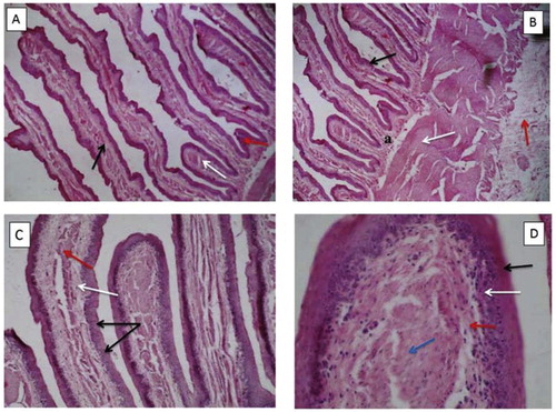
Tissues from the rumen of sheep implanted with 129 g of plastic bags showed patchy or segmental rupture, destruction, necrosis, degeneration and focal hyperplasia of keratinized epithelia. Papillae were shortened, broadened, atrophied, bent and compressed. The length of the papillae was about three quarter (3/4) of the microscope field (Magnification 40×) ( and ). The rumen mucosa was thick, and some areas were disrupted, degenerated and hyperplastic, forming prominent rete pegs of variable length that projected into the lamina propria and the underlying submucosa. The mucosa was 9–14 cells in thickness. These mucosal cells had hydrophic degeneration, vacuolation and were oedematous. The submucosa was widened with dilated lymphatics and prominent oedema mainly between the rete pegs. Its connective tissue had areas of degeneration and necrosis and some fibrosis with increased mononuclear cell infiltration. The muscular layer was hypertrophic and oedematous with separation of muscle fibers ( and ).
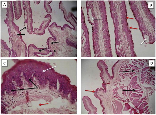
The rumen of sheep implanted with 258 g of plastic bags had more severe lesions than those implanted with 129 g. The lesions included segmental rupture, degeneration, erosion, necrosis ( and ), karyolysis and focal hyperplasia of the keratinized epithelia. These lesions were observed mainly at the pressure points where the plastic bags were lodged. There were also areas of shredding and sloughing of the keratinized epithelia (–). In some areas, the rumen papillae were shortened, atrophied, compressed and flattened ( and ). In other areas they were broadened but shortened to less than a quarter (1/4) of the microscope field (Magnification 40×). There were clefts at the tips of the atrophied papillae in all the sheep in this group (). There was segmental hyperplasia of the mucosal layer with very prominent rete pegs projecting into the submucosa (). The mucosa epithelium was 20–22 cells in thickness with both extracellular and intracellular epithelial cell oedema characteristic of hydrophic degeneration, cellular vacuolation and spongiosis of these cells of the mucosa. Areas of regeneration of the mucosa were prominent in some parts. Submucosa oedema with prominent dilated lymphatics and increased mononuclear cell infiltration mainly macrophages, mast cell and lymphocytes, and increased vascularization, were observed. The muscular layer was atrophied with separated muscle fibers due to oedema. The serosa was stretched, widened and oedematous ().
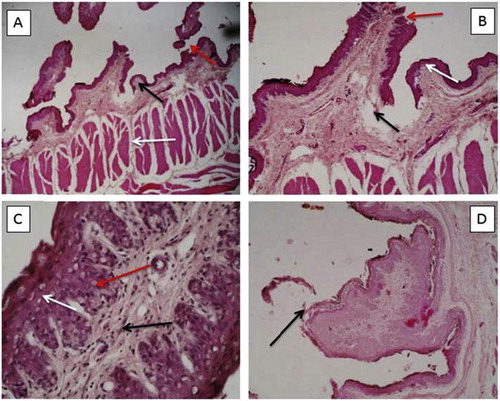
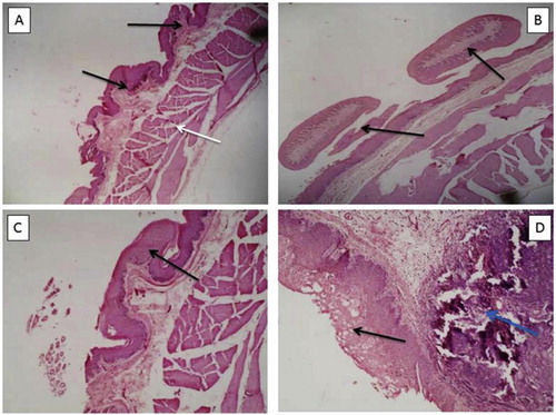
The rumen of sheep implanted with 387 g of plastic bags had shortened, stunted or completely atrophied papillae leaving only stumps. There was patchy or focal thickening of the ruminal wall, degeneration, erosion and necrosis of the stratified epithelia in many areas of the rumen. Areas of degeneration, ulcerations, erosion and necrosis, were also observed on the keratinized epithelia of the stunted papillae. Partial or complete shredding of the epithelial layer in hypertrophied and parakeratotic parts was common ().
The mucosal epithelium was thickened and papilloma-like structure due to hyperplasia with prominent interlocking rete pegs projecting into the underlying submucosa. The epithelium was 30–33 cells thick, when present. It had hydrophic degeneration, cellular vacuolation and spongiosis of the cells due to oedema. The mucosal epithelium had areas that were ruptured, degenerated and necrotic with a new layer forming below the damaged one (). The submucosa was widened and oedematous with increased infiltration of both mononuclear and polymorphonuclear inflammatory cells, with exostosis of few polymorphs within the epithelium. There were prominent interlocking and inter-connecting rete pegs, increased vascularization with congestion of the blood capillaries, prominent and dilated lymphatics, and increased oedema in the submucosa. The muscular layer was also edematous ().
4 Discussion
Generally, severity of the gross and histopathological changes observed in this study varied with the quantity of plastic bags present in the rumen of the sheep as was the case in the goats reported previously [Citation8]. The larger the quantity of plastic bags in the rumen, the more severe the pathological changes and lesions and vice versa. It has also been reported that severity of pathological changes depends on the type of foreign body, duration of presence in the rumen and the degree of obstruction caused by the foreign body [Citation9]. However, quantitative damage by the same type of foreign body and differences between two species has not being previously reported. The finding of more severe gross and histopathological changes in goats than sheep may imply that goats are more affected by plastic bags and more susceptible to stress than sheep [Citation8].
Generalized muscle and organ atrophy; degeneration of body, omental and mesenteric fat observed in the studied sheep may be due to the anorexia that caused reduced feed intake, physiological nutrient imbalances, emaciation and dehydration. Factors that determine feed intake by ruminants are related to the capacity of the gastro-intestinal tract, digestibility of the feed and the rate of passage through the gastro-intestinal tract [Citation4]. The impaction of the rumen and obstruction of the passage of the gastrointestinal tract due to plastic bags observed in the sheep may have hindered effective fermentation and functioning of the rumen microflora [Citation4]. Chronic wasting diseases, malnutrition and cachexia result in mobilization of fat deposits [Citation18], hence the degeneration and atrophy. Loss of body mass has also been related to reduction in total body water [Citation19,Citation20].
Consistent findings in previous reports on clinical and abattoir studies involving different types of foreign materials included, shortening, stunting and atrophy of the papillae; erosion, excoriation of the papillae as well as oedema of the ruminal pillars, ulcerations, scars, nodules, hyperemia and haemorrhages of the rumen mucosa and thinning of the ruminal wall [Citation3,Citation8,Citation21]. However, these changes varied in the sheep compared to that earlier reported in goats and also with the quantity of plastic bags implanted in the rumen [Citation8]. The degree of papillae and pillar atrophy was dependent on the quantity of plastic bags and species of animal. These changes may be attributed to the pressure exerted against the ruminal walls by the plastic bags and perhaps toxicants from the plastic bags and the ruminal contents as previously suggested by [Citation11]. Furthermore, constant irritation of the wall of the rumen by continuous movement of the plastic bags may lead to erosion and excoriations of the rumen papillae, ruminal pillars and mucosa, resulting in inflammation and hyperplasia of the epithelial mucosa [Citation10]. It is also possible that some of the lesions may result from poisonous substances released from the plastic bags and toxicants absorbed from the rumen as speculated before [Citation8]. Hyperemia, haemorrhages and oedema observed in the reticulum, omasum, abomasum and intestines could be due to irritation, circulatory disturbances and inflammation as a result of toxic substances released from the plastic bags and absorbed from the gastrointestinal tract [Citation4].
Histopathological changes observed in the rumen involving the papillae, pillars and the epithelia could be attributed to excessive pressure exerted on the ruminal wall by the plastic bags impacting the rumen. These changes included shortening, broadening, atrophy, patchy or segmental rupture, erosion, necrosis and degeneration of the stratified epithelia. Similar findings have been reported by other authors [Citation10,Citation11] who found shortening, broadening, atrophy, rupture, erosion, necrosis and degeneration of the stratified epithelia in clinical and abattoir studies, although the time span and quantities of foreign bodies were not known or evaluated as in these studies. The degree of atrophy and estimation of cell numbers in hyperplastic tissue have also not been previously reported in rumen with foreign bodies. The observed histopathological changes could be attributed to mechanical irritation induced by the plastic bags or chemical substances released by these bags as suggested previously suggested by Bakhiet [Citation11]. Similar lesions as found in the current study have also been reported in cattle [Citation21] and in goats [Citation8].
The hyperplasia, hydrophic degeneration, cellular vacuolation and karyolysis observed in the mucosa of the rumen may be attributed to mechanical irritation and pressure caused by the plastic bags or some chemical substances released from the plastic bags as suggested by Bakhiet [Citation11] or absorbed toxicants from the gastrointestinal tract. According to Robbins et al. [Citation22], pathologic hyperplasia constitutes precursors from which cancerous proliferation may eventually arise. Hence the papilloma-like hyperplasia of the mucosa of the rumen with prominent interlocking rete pegs projecting into the underlying submucosa and nodules observed grossly in the current study could be an indication of an early process. Further investigations are however needed to elucidate this assertion.
The prominent mucosal and submucosal oedema observed in this study may be due to fluid seeping through the ruminal wall into the tissues of the rumen as a result of the physical damage caused by the presence of plastic bags as suggested previously by Bakhiet [Citation11]. It could also be due to generalized oedema resulting from the severe starvation and cachexia due to the plastic bags [Citation18]. The presence of oedema is the reason for spongiosis in the cells of the mucosa and submucosa observed in the sheep implanted with 258 g and 387 g of plastic bags. Cellular oedema could be as a result of epithelial damage resulting in of hydrophic cellular degeneration.
The degeneration, necrosis and fibrosis of the connective tissue with increased mononuclear and polymorphonuclear cell infiltrations into the submucosa could be due to progressive cellular change attracting phagocytic cell to remove destroyed ruminal tissue and provide stimuli for tissue repair after irritation and cellular damage by the plastic bags [Citation8]. The exostosis of the polymorphonuclear cells and increased vascularization with congestion of the blood capillaries in the submucosa can occur due to secondary bacteria invasion or in the early stages of regeneration of the epithelia in response to chronic irritation by the plastic bags. Hyperplasia, oedema and separation of the muscular layer and serosa observed could be due to the pressure exerted on the ruminal wall by the plastic bags.
5 Conclusions
Several pathological changes in the ruminal tissue of sheep are directly proportional to the quantities of plastic bags in the rumen. These changes are resulted from drastic reduction of water and feed intake over time due to impaction and obstruction of the gastrointestinal tract leading to dehydration and emaciation. This may contribute to economic losses in production of sheep. Plastic bags pollution is a serious threat to sheep production, especially in urban and peri-urban farming and needs to be controlled.
Competing interests
There is no conflict of interest regarding this manuscript.
Acknowledgment
The authors are grateful to Transdisciplinary Training for Resource Efficiency and Climate Change Adaptation in Africa (TRECCAfrica) and the University of Ghana for providing the funds for this work, research assistants who assisted in data collection and the University of Nairobi for the support.
Notes
Peer review under responsibility of Faculty of Veterinary Medicine, Cairo University.
References
- H.R.OtsyinaJ.Nguhiu-MwangiE.G.M.MogoaP.G.MbuthiaW.O.OgaraPrevalence of indigestible rumen foreign bodies in sheep and goats at Dagoretti and Kiserian Abattoirs, KenyaInter J Vet Med320157580
- B.D.Remi-AdewunmiE.O.GyangA.O.OsinowoAbattoir survey of foreign body rumen impaction in small ruminantsNig Vet J2520043238
- M.A.H.GhurashiH.I.SeriA.H.BakheitE.A.M.AshwagEffect of surgical removal of foreign body from goat’s rumen with special reference to the prevalence of foreign body in goats in Southern DafurAust J Basic Appl Sci32009664668
- Radostitis OM, Gray CC, Blood DC, Hinchelift KW. Veterinary medicine: a textbook of the diseases of cattle, sheep, pig, goats and horses, 10th ed. New York, USA: Saunders Elsevier, 2009; p. 189–382, 296 and 313.
- I.O.IgbokweM.Y.KoloG.O.EgwuRumen impaction in sheep with indigestible foreign body in the semi-arid region of NigeriaSmall Rum Res492003141147
- V.VanithaA.P.NambiP.GowriS.KavithRumen impaction of cattle with indigestible foreign bodiesTamil J Vet Anim Sci62010138140
- FAO. Spotlight issues in urban agriculture studies suggest that up to two-thirds of city and peri-urban households are involved in farming. 1999: http://www.fao.org/ag/magazine/9901sp2.htm (05/06/2012).
- Otsyina HR, Nguhiu-Mwangi J, Mogoa EGM, Mbuthia PG, Ogara WO. Gross and histo-pathologic findings in goats with plastic bags in the rumen. J Basic Appl Sci 2017; 1 (In press).
- D.TesgayeM.ChanieStudy on rumen and reticulum foreign bodies in cattle slaughtered at Jimma Municipal abattoir, South west EthiopiaAm-Eurasian J Sci Res72012160167
- N.HailatA.Al-DarrajiS.LafiS.A.F.BarakatF.Al-AniH.El-MagrhabyK.Al-QudahS.GharaibehM.RousanM.Al-SmadiPathology of the rumen in goats caused by plastic foreign bodies with reference to its prevalence in JordanSmall Rum Res3019987783
- O.M.BakhietStudies on the rumen pathology of Sudanese desert sheep in slaughter houseSci Res Ess32008294298
- AHPitrodaDKTiwariMehraj-u-din DarPVParikhUltrasonographic diagnosis and treatment of rumen impaction in a goatIntas Polivet22010251252
- F.AbebeM.NuruPrevalence of indigestible foreign bodies ingested by small ruminants slaughtered at Luna export abattoir, East Shoa, EthiopiaJ Anim Vet Adv10201115981602
- M.A.SaulawaS.UkashatuM.G.GarbaA.A.MagajiM.B.BelloA.S.MagajiPrevalence of indigestible substances in rumen and reticulum of small ruminants slaughtered at Katsina central abattoir, Katsina State, NigeriaSci J Pure Appl Sci120121721
- HROtsyinaJNguhiu-MwangiEGMMogoaPGMbuthiaWOOgaraA retrospective study on the prevalence of plastic materials in the rumen of sheep and goats in Nairobi, KenyaBull Anim Hlth Prod Afr622014197205
- D.A.HendricksonTechniques in large animal surgeryThird edition2007Blackwell Publishing LtdOxford, England223226
- ASmithJBrutonHistopathological staining techniques1977Wolfe Medical Publications Ltd.London, England
- Jubb KVF, Kennedy PC, Palmer N. Pathology of domestic animals. Vol 2 and Vol 3, 7rd edition. Acadamy Press Inc, London 2014. p. 5, 241–289, 306–64, 448.
- A.A.DegenM.KamBody mass loss and body fluid shifts during dehydration in Dorper sheepJ Agric Sci1191992419422
- M.A.AbdallaA.E.SalwaM.H.YahiaEffect of state of hydration on body weight, blood constituents and urine excretion in Nubian goats (Capra hircus)Wld J Agric Sci62010178188
- I.TanimotoY.OtisukiY.NoinuraRumenoabomasal lesions in steers induced by naturally ingested hairVet Path311994280282
- S.L.RobbinsS.CotranV.KumaPathologic basis of disease3rd Ed.1984W.S. Saimders. PhiladelphiaUSA32