

 ?Mathematical formulae have been encoded as MathML and are displayed in this HTML version using MathJax in order to improve their display. Uncheck the box to turn MathJax off. This feature requires Javascript. Click on a formula to zoom.
Abstract
Background
Inhibition of Rho/ROCK signalling pathway related proteins can alleviate left ventricular hypertrophy. Berberine hydrochloride (BBR) can effectively inhibit left ventricular hypertrophy. The purpose of this study is to explore the relationship between BBR and Rho/ROCK signalling pathway.
Methods
Isoproterenol (ISO) was used to induce left ventricular hypertrophy in rats. Two-dimensional speckle tracking technique (2D-STE) was used to evaluate rats in each group (group A: normal control group; Group B: isoproterenol model group; Group C: ISO + 5mg/kg BBR group; Group D: ISO + 10mg/kg BBR group) Heart structure and systolic function. HE staining and Masson staining were used to observe the pathological changes in four groups of rats. The expression levels of RhoA, ROCK-1, TGF-β1 and PTEN protein in myocardial tissues were detected by immunohistochemistry.
Result
The interventricular septum (IVS) of rats in groups C and D was thinner than that in group B (4.05 ± 0.16 mm vs. 3.50 ± 0.29 mm vs. 4.41 ± 0.23 mm, respectively, p < 0.05), and the global radial strain (GRS) of rats in groups C and D was higher than that in group B, especially in group D (26.05 ± 1.41 vs. 30.64 ± 1.63 vs. 19.40 ± 1.05, respectively, p < 0.05). Compared with group B, the expression levels of RhoA, ROCK-1 and TGF-β1 in groups C and D decreased, while the expression level of PTEN increased, especially in group D (all p < 0.05).
Conclusion
BBR can improve the cardiac structure and systolic function of rats with left ventricular hypertrophy, and the improvement mechanism may be related to Rho/ROCK signalling pathway.
Introduction
Cardiac remodelling is an essential pathological mechanism in the process of heart failure. Cardiac hypertrophy plays a vital role in cardiac remodelling. It is a critical stage in the development of cardiac structure and function from compensation to decompensation, as well as an independent risk factor for the increased prevalence and mortality of cardiovascular diseases [Citation1]. Early detection and treatment of cardiac hypertrophy are critical. Rho/ROCK signalling pathway plays an essential role in the development of cardiovascular diseases. Overexpression of Rho and ROCK can aggravate cardiac hypertrophy; inhibition of Rho/ROCK signalling pathway-related proteins can effectively alleviate left ventricular hypertrophy and prevent cardiac remodelling from progressing to heart failure [Citation2–4]. BBR is a natural plant alkaloid that can inhibit pathological cardiac hypertrophy by up to 50% [Citation5–7]. However, whether berberine hydrochloride can improve cardiac hypertrophy by regulating the Rho/ROCK signalling pathway is unclear. 2D-STE is a relatively new detection technology, which is more accurate than traditional echocardiography in the early detection of abnormal changes in cardiac function [Citation8–11]. There are few studies on applying 2 D-STE in evaluating left ventricular function in rat models of myocardial hypertrophy.
Therefore, this study investigated whether 2D-STE could monitor early myocardial function impairment in rats with myocardial hypertrophy, evaluate the effect of BBR on the left ventricular structure and systolic function, and explore the role of the Rho/ROCK signalling pathway in rats with myocardial hypertrophy.
Methods
Laboratory animal
Sixty male Sprague Dawley rats (240–260 g, about 10 weeks) were obtained from Chengdu Dashuo Laboratory Animal Company. The rats were fed at the Experimental Animal Center of Sichuan Provincial People's Hospital with free access to diet and tap water. This work has been approved by the Animal Protection and Use Committee of Sichuan Provincial People's Hospital and abides by the recognised the NIH guidelines for the use of animals in experiments.
Animal grouping
Sixty male Sprague Dawley rats were completely randomly divided into four groups: group A: normal control group; group B: ISO model group; group C: ISO + 5mg/kg BBR group; group D: ISO + 10mg/kg BBR group, 15 in each group. Rats in group B, group C and group D were given intraperitoneal injection of ISO solution at a dose of 2 mg/kg for 14 days, while rats in group A were given intraperitoneal injection of the same amount of normal saline. The rats in groups B, C, and D were injected with ISO to establish a rat model of myocardial hypertrophy. From the modelling day, the rats in groups C and D were given 5 mg/kg and 10 mg/kg of BBR solution by gavage. The rats in group A and group B were given the same amount of normal saline by gavage once a day. BBR (Lot number HY18258) was purchased from Seville Biotechnology Co., Ltd.
Echocardiography
Rats were anaesthetised by 10% chloral hydrate, and the ultrasonic images of left ventricular short axis papillary muscle level were collected at baseline, 7th day and 14th day after administration by GE Vivid E9 echocardiography (GE Healthcare). 12-MHz linear transducer (GE Healthcare) and 12 s paediatric probe were used for detection, and the imaging display condition was set to rodent cardiology. papillary level short axis B-mode images were used to measure interventricular septum thickness (IVS), left ventricular posterior wall thickness (LVPW) in end-diastole. In the two-dimensional image, the time when the left ventricle diameter was the smallest was the end time of systole, and the time when the left ventricle diameter was the largest was the end time of diastole. At the level of the papillary muscle, M-mode sampling lines perpendicular to the interventricular septum and the posterior wall of the left ventricle were set to obtain the corresponding M-mode echocardiogram. EF values were measured on the M-mode echocardiogram. Dynamic images were analysed using Echo PAC (GE Medical Systems), the peak systolic radial strain (RS) of the six myocardial segments was recorded, and the global radial strain (GRS) was the average of the 6 RS values. All data were measured three times and averaged by echocardiographic experts who were blinded to the animal groups.
Pathological observation
On the 14th day after echocardiography, five rats in each group were sacrificed for HE and Masson staining. The volume fraction of collagen fibres (CVF) in heart tissue (CVF % = collagen area/total visual field area) was calculated, and the correlation between CVF and GRS value was investigated. The immunohistochemical technique was used to detect the expression of RhoA (Ras homolog gene family member A), ROCK-1 (Rho-related helix forming protein kinase 1), TGF-β1 (Transforming factor -β1), and PTEN (Phosphatase and Tensin Homologue Deleted OH Chromosome Ten) in four groups of rats’ myocardial tissues. Image-Pro Plus 6.0 image processing software was used to calculate the average optical density (AOD) of each image, which was used to reflect the intensity of immunostaining. RhoA (Lot number, AF6352), ROCK-1(Lot number, AF7016), TGF-β1 (Lot-number, BS-20411R), and PTEN (Lot number, AB31392) are both purchased from Beijing boaosen biotechnology company.
Statistics
SPSS 22.0 software was used for statistical analysis, and the data are expressed as mean ± standard deviation (±S). Paired-samples t-test was used within groups, a one-way analysis of variance was used between groups, and pairwise comparisons were performed using the LSD (least significant difference) test. Spearman analysis or Spearman’s method was used to determining the data’s correlation. p < 0.05 was regarded as a statistically significant difference. Ten ultrasound images were randomly selected to assess intra- and inter-observer reproducibility of the anterior septal myocardial RS values.
Results
In the experiment, the rats in group A showed no apparent abnormalities. Sudden death occurred in two rats in group B, one rat in group C, and one rat in group D was abandoned due to unclear image display. Finally, the number of rats included in the four groups was 15, 13, 14, and 14.
Baseline data
There was no statistical difference in the baseline data of the four groups of rats. The intra- and inter-observer correlation coefficients of variation were 0.921 (95% confidence interval, 0.918–0.945) and 0.934 (95% confidence interval, 0.923–0.962), respectively, indicating that the RS values were reproducible ().
Table 1. Baseline data of the four groups of rats (±S).
Data of rats in four groups on the 7th day of administration
On the 7th day, the IVS values of rats in groups B, C, and D were higher than those in group A, and the IVS values of rats in groups C and D were lower than those in group B, with group D as significant. The LVPW values of group B and C were increased compared with those of group A (p < 0.05), while the LVPW values of group D were increased compared with those of group A, but there was no statistical difference. There was no significant difference in EF values among the four groups (p > 0.05). Compared with the rats in group A, the RS and GRS values of six cardiac segments in group B were decreased, most of the cardiac segments in group C were decreased, and only A few cardiac segments in group D were decreased. The RS values of some myocardium segments of rats in groups C and D were higher than those in group B, and were more significant in group D (p < 0.05) (, ).
Figure 1. Radial strain values at the level of left ventricular short-axis papillary muscle on the 7th day of administration in four groups of rats. The radial strain of rats in group B decreased significantly.
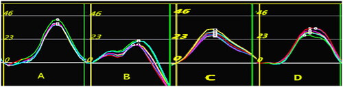
Table 2. Data on the 7th day of administration of four groups of rats(±S).
Data of rats in four groups on the 14th day of administration
With the prolongation of the course of cardiac hypertrophy, the structural changes of the rat heart are more prominent, and the left ventricular systolic function is significantly reduced. The wall thickness of rats in groups B, C, and D was further increased than that in group A, but the wall thickness of rats in groups C and D was still lower than that in group B (p < 0.05). At this time, EF values of rats in groups B, C, and D were significantly lower than those in group A. EF values of rats in groups C and D were higher than those in group B, especially in group D (p < 0.05). In addition, the GRS and the RS values of six myocardial segments in groups B, C, and D were significantly lower than those in group A. RS values of most myocardial segments in groups C and D were higher than those in group B, especially in group D (p < 0.05) ().
Table 3. Data on the 14th day of administration of the four groups of rats (±S).
Self-contrast observation of the data of the four groups of rats
During the whole experimental period, there was no significant change in the ultrasound data of rats in group A. Compared with the baseline data, the IVS values and LVPW values of rats in groups B, C, and D increased on the 7th day of administration. There was no significant difference in EF values among the four groups. On the 14th day after administration, the wall thickness of the three groups of rats increased further, and at this time, the values of EF decreased significantly. By observing rats in groups B, C, and D at three-time points in the experiment, we found that with the progress of myocardial hypertrophy, the left ventricular systolic function of rats gradually decreased, which was lower than the previous experimental time point. RS value is more sensitive than EF value in reflecting early myocardial systolic dysfunction. BBR can reduce the myocardium’s thickness and improve the left ventricle’s systolic function, but it can’t completely reverse the occurrence and development of myocardial injury.
Pathological results
The cardiomyocytes of rats in A were homogeneous in morphology and regular in fibre arrangement, and the myocardial CVF value was 1.87 ± 0.95%. Cardiomyocytes in group B were significantly enlarged and irregular, and myocardial fibrosis was aggravated. The CVF value was 30.74 ± 9.65%. The above myocardial pathological changes were alleviated in group C and group D compared with group B, and the myocardial CVF values were 22.53 ± 8.48% and 11.98 ± 5.58%, respectively. We also observed that with the increase of CVF value, GRS showed a decreasing trend, and CVF was negatively correlated with GRS value (r= −0.79, p < 0.05) (, and ).
Figure 2. The effect of BBR on the myocardial structure of ISO-induced myocardial hypertrophy in rats. (a) Normal control group of rats. (b) Myocardial cells in group B were significantly hypertrophied, and the degree of fibrosis was the highest. (c) Cardiomyocyte hypertrophy and fibrosis in group C were less severe than in group B. (d) The pathological results of rats in group D were less severe than those in groups B and C.
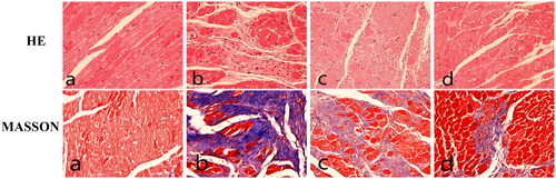
Figure 3. Correlation analysis of myocardial collagen volume fraction (CVF) and GRS value in rats.GRS was negatively correlated with CVF.
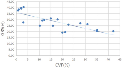
Table 4. AOD and CVF values of myocardial tissue-related proteins in four groups of rats(±S).
Protein expression levels of RhoA, ROCK-1, PTEN, and TGF-β1 in myocardial tissue of rats in four groups
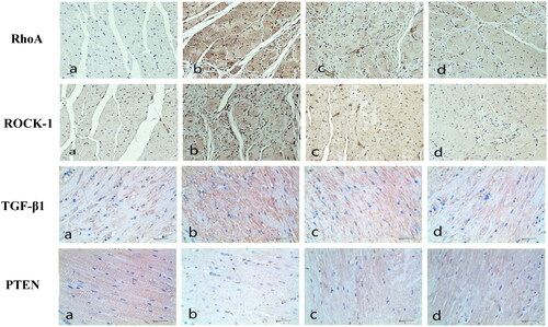
In the experiment, the expression levels of ROCK-1, RhoA, and TGF-β1 in the myocardial tissue of rats in group B were significantly higher than those in group A, and the expression level of PTEN was considerably lower than that in group A. The expression levels of ROCK-1, RhoA, and TGF-β1 in myocardial tissue of rats in groups C and D were lower than those in group B, and PTEN was higher than those in group B, especially in group D (p < 0.05) (, ).
Figure 4. Immunohistochemistry results. (a) Normal control group of rats. (b) RhoA, ROCK-1and TGF-β1 of rats in group B were significantly higher than those in the other three groups, while PTEN protein was significantly lower than those in the other three groups. (c) Compared with group B, the expression of RhoA, ROCK-1and TGF-β1 in group C decreased, and PTEN protein increased. (d) Compared with group C, the expression of RhoA, ROCK-1and TGF-β1in group D further decreased, and PTEN protein increased.
Discussion
To the best of our knowledge, we are the first study to evaluate the improvement of left ventricular myocardial hypertrophy by berberine hydrochloride using ultrasonic speckle tracking technology in rats. We also found that the mechanism of berberine hydrochloride improving cardiac hypertrophy and protecting heart systolic function may be related to the Rho/ROCK signalling pathway regulation.
Our study successfully induced a typical rat model of cardiac hypertrophy using ISO (2 mg/kg/day for 14 days). Cardiac hypertrophy is an independent risk factor for increased cardiovascular morbidity and mortality [Citation12–16]. ISO is a catecholamine that can stimulate β-adrenergic receptors. Previous studies have confirmed that injection of ISO can induce myocardial hypertrophy, and ISO-induced left ventricular remodelling is characterised by ventricular hypertrophy and myocardial fibrosis [Citation17–18]. In our study, the ventricular wall thickness of rats was significantly increased (IVS and LVPW values were increased considerably) on conventional ultrasound, cardiomyocytes were increased significantly by pathological HE staining, and the degree of myocardial fibrosis was increased dramatically by MASSON staining. We used ultrasound technology combined with pathological observation to establish the cardiac hypertrophy model successfully.
In this study, the changes in left ventricular structure and systolic function in rats with cardiac hypertrophy were dynamically monitored by conventional ultrasound and two-dimensional speckle tracking techniques. The two-dimensional speckle tracking technique is a relatively new method for quantifying left ventricular function. It is validated by comparison with different gold standard methods. The results show that the two-dimensional speckle tracking technique is reliable and has been widely used in various animal models [Citation19]. In this experiment, the radial motion was selected as the research object. Radial motion, the heart’s short axis movement, reflects the extent of the thickening of the systolic ventricular wall. The radial strain values of six cardiac muscle segments at the level of papillary muscle in rats with myocardial hypertrophy were generally decreased, indicating that the deformation ability of the myocardium in the radial direction was reduced, and contractile dysfunction occurred. In addition, we found that the radial parameters of six cardiac segments were heterogeneous in the rats with cardiac hypertrophy. In our experiment, the thickness of the rat ventricular wall was aggravating, and the extent of the injury was gradually expanding. It seems that using berberine hydrochloride could not completely reverse the myocardial ventricular structure and left ventricular systolic function of rats to normal. However, the myocardial wall thickness of rats treated with berberine hydrochloride was relatively reduced, and the left ventricular systolic function increased somewhat. Our results indicate that berberine hydrochloride can improve cardiac hypertrophy and protect cardiac function to a certain extent, and the intensity of improvement may be dose-dependent.
Our study found that RS can detect left ventricular systolic dysfunction earlier and sensitively. Injection of ISO can cause hypertrophy of cardiomyocytes and aggravation of myocardial interstitial fibrosis in rats, manifested as an increase of myocardial wall thickness in ultrasound parameters. Myocardial wall thickening compensates to some extent for increased left ventricular afterload. Therefore, although the local systolic function of the left ventricle was reduced, the regular operation of the rat heart’s overall cardiac systolic function and ejection function can still be maintained, and the macroscopically manifested EF values do not change significantly. However, the hypertrophic myocardium has been ischaemic and hypoxic, and the systolic function has been reduced. In our experiment, we found that the thickness of the left ventricular wall increased on day 7, but there was no statistical difference in the conventional EF values. We speculated that the cardiac hypertrophy rats were in a compensatory state at this time, and the overall contractile function of the heart was normal. However, the contractile function of some myocardium was reduced, and the change of radial strain parameters could reflect this situation. These results seem to suggest that RS is more sensitive than traditional EF values in reflecting left ventricular systolic function, which is consistent with the findings of Shih et al. [Citation20], Julien Ternacle et al. [Citation21] and Yu Kang et al. [Citation22]
Our results indicate that berberine hydrochloride has a protective effect on myocardial fibrosis. In this study, injection of ISO can significantly increase the content of myocardial collagen, and berberine hydrochloride can inhibit myocardial fibrosis induced by ISO, which is shown by the decrease of CVF value of rat myocardium under MASSON staining. In addition, the GRS value was verified to be negatively correlated with CVF, which was also consistent with previous findings: myocardial mechanical abnormalities were associated with myocardial fibrosis, and the RS value decreased with the increase of fibrosis degree [Citation23,Citation24]. Therefore, we hypothesised that the improvement of myocardial systolic function may be related to the reduction of myocardial interstitial fibrosis.
Our study suggests that BBR may improve cardiac hypertrophy and protect cardiac systolic function by regulating Rho/ROCK signalling pathway. The Rho/Rock signalling pathway is an important signal transduction pathway in the body. There are two subtypes of ROCK: ROCK1 and ROCK2, of which ROCK-1 is involved in the process of cardiac hypertrophy and myocardial fibrosis [Citation4]. TGF-β1 is an upstream activator of the Rho/ROCK signalling pathway, and it is also one of the most apparent cytokines to induce fibroblast proliferation and collagen synthesis [Citation25]. PTEN can regulate cardiomyocyte hypertrophy and cell survival. Specific inactivation of PTEN will lead to cardiac hypertrophy and reduced myocardial contractile function [Citation26]. In this study, the protein expressions of RhoA, ROCK-1, and TGF-β1 in the myocardium of ISO-induced cardiac hypertrophy rats were increased, while the protein expression of PTEN was significantly decreased. These results indicated that ISO increased the expression of RhoA, ROCK-1, and TGF-β1 protein and inhibited the expression of PTEN protein. However, in the myocardial tissue of rats treated with berberine hydrochloride, we can see that the protein expression of RhoA, ROCK-1 and TGF-β1 decreased, and the protein expression of PTEN was relatively increased. The higher the dose of berberine hydrochloride, the greater the effect on protein expression. We speculated that BBR might improve cardiac hypertrophy by regulating Rho/ROCK signalling pathway, and reduce the expression of TGF-β1 to reduce the secretion of extracellular matrix and alleviating myocardial fibrosis [Citation25], and inhibiting the hypertrophy process of cardiomyocytes by up-regulating the expression of PTEN protein. There may be a dose-dependent effect of BBR on improving myocardial hypertrophy. Our experimental results were confirmed at three levels: ultrasonic parameters, pathological detection, and protein expression.
Limitations
First, due to the small size of the rats and the poor quality of the ultrasound images in the longitudinal section, the data related to the long axis of the left ventricle were not obtained. Secondly, due to the limited experimental conditions, we did not monitor the liver and kidney function and blood pressure of rats; In addition, we used immunohistochemistry to detect the expression of proteins in rat myocardial tissue. Real-time PCR and Western Blot detection methods can more accurately detect the expression levels of related proteins and mRNA in cardiomyocytes.
Conclusion
This is the first study to explore the effect of berberine hydrochloride on left ventricular structure and systolic function in rats with myocardial hypertrophy by two-dimensional speckle tracking ultrasound. Two-dimensional speckle tracking technology can detect the changes of left ventricular systolic function earlier and dynamically. The mechanism of berberine hydrochloride in improving myocardial hypertrophy may be related to the regulation of Rho/ROCK signalling pathway. The study of Rho/ROCK signalling pathway may become a new direction for the treatment of myocardial hypertrophy, which has broad research and application prospects. In the future research, it is necessary to use more advanced experimental methods to measure protein expression level.
Ethical approval
All animal studies have been approved by the Ethics Committee of Sichuan Provincial People’s Hospital and are therefore conducted in accordance with the ethical standards stipulated in the 1964 Declaration of Helsinki and subsequent amendments. decision date: 3 February 2021. No.: Ethical Review (Research) No.26, 2021.
Author contributions
Conception and design of the research: HL and TK and Yang Shen; Acquisition of data and Analysis and interpretation of the data and Statistical analysis: HL and TK and Ye Su; Writing of the manuscript: HL and TK and Yang Shen; Critical revision of the manuscript for intellectual content: HL and TK and Yang Shen; LY, the corresponding author of this paper, interpreted the data and substantially contributed to critical revision of the intellectual content.
Acknowledgements
The authors express their gratitude to Sichuan provincial people’s hospital for their cooperation and assistance.
Disclosure statement
No potential conflict of interest was reported by the author(s).
Additional information
Funding
References
- Qi J, Tan Y, Fan D, et al. Songling Xuemaikang capsule inhibits isoproterenol-induced cardiac hypertrophy via CaMKIIδ and ERK1/2 pathways. J Ethnopharmacol. 2020;253:112660.
- Yan X, Jiao K, Song X. Shen’ge powder decreases the cardiomyocyte hypertrophy in chronic heart failure by activating the rho protein/rho-associated coiledcoil forming protein kinase signalling pathway. J Cell Biochem. 2019;120(3):3038–3045.
- Olgar Y, Celen MC, Yamasan BE, et al. Rho-kinase inhibition reverses impaired Ca2+ handling and associated left ventricular dysfunction in pressure overload-induced cardiac hypertrophy. Cell Calcium. 2017;67:81–90.
- Bailey KE, MacGowan GA, Tual-Chalot S, et al. Disruption of embryonic ROCK signalling reproduces the sarcomeric phenotype of hypertrophic cardiomyopathy. JCI Insight. 2020;5(24):e146654.
- Lai S, Wei Y, Wu Q, et al. Liposomes for effective drug delivery to the ocular posterior chamber. J Nanobiotechnology. 2019;17(1):64.
- Rezaeian L, Kalalian-Moghaddam H, Mohseni F, et al. Effects of berberine hydrochloride on methamphetamine-induced anxiety behaviors and relapse in rats. Iran J Basic Med Sci. 2020;23(11):1480–1488.
- Evans L, Shen Y, Bender A, et al. Divergent and overlapping roles for selected phytochemicals in the regulation of pathological cardiac hypertrophy. Molecules. 2021;26(5):1210.
- Stevens AL, Ferferieva V, Bito V, et al. Exercise improves cardiac function and attenuates insulin resistance in dahl salt-sensitive rats. Int J Cardiol. 2015;186:154–160.
- Niu P, Li L, Yin Z, et al. Speckle tracking echocardiography could detect the difference of pressure overload-induced myocardial remodelling between young and adult rats. J R Soc Interface. 2020;17(163):20190808.
- Zhang H, Sun Y, Liu X, et al. Speckle tracking echocardiography in early detection of myocardial injury in a rat model with stress cardiomyopathy. Med Ultrason. 2019;21(4):441–448.
- Mátyás C, Kovács A, Németh BT, et al. Comparison of speckle-tracking echocardiography with invasive hemodynamics for the detection of characteristic cardiac dysfunction in type-1 and type-2 diabetic rat models. Cardiovasc Diabetol. 2018;17(1):13.
- Xu Y, Liang C, Luo Y, et al. MBNL1 regulates isoproterenol-induced myocardial remodelling in vitro and in vivo. J Cell Mol Med. 2021;25(2):1100–1115.
- Yang L, He J, Xia G, et al. Crim1 suppresses left ventricular hypertrophy. Biomed Rep. 2019;1(1):1–5.
- Shen L, Gan M, Tan Z, et al. A Novel class of tRNA-Derived small Non-Coding RNAs respond to myocardial hypertrophy and contribute to intergenerational inheritance. Biomolecules. 2018;8(3):54.
- Ma D, Zhang J, Zhang Y, et al. Inhibition of myocardial hypertrophy by magnesium isoglycyrrhizinate through the TLR4/NF-κB signaling pathway in mice. Int Immunopharmacol. 2018;55:237–244.
- Niu J, Zeng M, Wang Y, et al. Sensitive marker for evaluation of hypertensive heart disease: extracellular volume and myocardial strain. BMC Cardiovasc Disord. 2020;20(1):292.
- Aluja D, Inserte J, Penela P, et al. Calpains mediate isoproterenol-induced hypertrophy through modulation of GRK2. Basic Res Cardiol. 2019;114(3):21.
- Simko F, Baka T, Repova K, et al. Ivabradine improves survival and attenuates cardiac remodelling in isoproterenol-induced myocardial injury. Fundam Clin Pharmacol. 2021;35(4):744–748.
- Bachner N, Tsadok Y, Adam D. Increase in endocardial rotation during doxorubicin treatment. Ann NY Acad Sci. 2010;1188:128–132.
- Shi J, Pan C, Shu X, et al. The role of speckle tracking imaging in the noninvasive detection of acute rejection after heterotopic cardiac transplantation in rats. Acta Cardiol. 2011;66(6):779–785.
- Ternacle J, Wan F, Sawaki D, et al. Short-term high-fat diet compromises myocardial function: a radial strain rate imaging study. Eur Heart J Cardiovasc Imaging. 2017;18(11):1283–1291.
- Kang Y, Wang W, Zhao H, et al. Assessment of subclinical doxorubicin-induced cardiotoxicity in a rat model by speckle-tracking imaging. Arq Bras Cardiol. 2017;109(2):0.
- Wang X, Qiao W, Xiao Y, et al. Experimental Research on the evaluation of left ventricular function by layered speckle tracking in a constrictive pericarditis rat model. J Ultrasound Med. 2020;39(11):2219–2229.
- Morishita T, Takeishi N, Ii S, et al. Effects of left ventricular hypertrophy and myocardial stiffness on myocardial strain under preserved ejection fraction. Ann Biomed Eng. 2021;49(7):1670–1687.
- Fu S, Li YL, Wu YT, et al. Icariside II attenuates myocardial fibrosis by inhibiting nuclear factor-κB and the TGF-β1/Smad2 signalling pathway in spontaneously hypertensive rats. Biomed Pharmacother. 2018;100:64–71.
- Yang X, Chen G, Chen Z. MicroRNA-200a-3p is a positive regulator in cardiac hypertrophy through directly targeting WDR1 as well as modulating PTEN/PI3K/AKT/CREB/WDR1 signalling. J Cardiovasc Pharmacol. 2019;74(5):453–461.

