
ABSTRACT
Ceratocystis wilt and canker disease has severely compromised the profitability of Acacia mangium plantations in Southeast Asia. The focus of this review is on Ceratocystis wilt and canker disease in Acacia trees. Its aim is to synthesise information about this fungal pathogen that can be used to inform development of suitable disease-control strategies in forest plantations. The last 20 years have seen many taxonomic changes in Ceratocystis, with some disagreement as to species boundaries. Therefore, an understanding of the origins and development of this disease requires reference to other species, particularly in the context of the biology and fungal taxonomy, disease symptoms and mechanisms of fungal dispersal. The risks and impacts of the disease on the sustainability of Acacia wood production are examined. Observing or surveying disease symptoms in plantations, selecting and planting tolerant or resistant Acacia trees, and the potential of endophytic bacteria as biological control agents are also included in this review.
Introduction
Acacia species have been exploited for wood, including fuel wood, animal forage, human food, tannin and land rehabilitation (Midgley & Turnbull Citation2003). In southern and eastern Africa, they have been grown for more than 100 years for the control of sand drift, and for timber, plywood and pulp and paper industries (Roux et al. Citation2005; Griffin et al. Citation2011). In the Asia-Pacific region, tropical species have been grown for over 80 years. Acacia auriculiformis A. Cunn. ex Benth. was introduced from Australia to Malaysia in 1932, to Thailand in 1935 and subsequently to India and China (Midgley & Turnbull Citation2003). Acacia mangium Willd. and Acacia crassicarpa A. Cunn. ex Benth. have formed the basis of large plantation estates for pulpwood production in Southeast Asian countries for at least three decades (Griffin et al. Citation2011). Acacia mangium was first introduced from the humid tropical forests of north-eastern Australia to Malaysia in 1966 and subsequently to Papua New Guinea, Indonesia, Bangladesh, China, India, the Philippines, Sri Lanka, Thailand and Vietnam (Midgley & Turnbull Citation2003; Krisnawati et al. Citation2011). Acacia crassicarpa was first planted in the early 1980s in Thailand and soon after in Indonesia where it is very well-adapted to peatlands in Sumatra (Midgley & Turnbull Citation2003). Acacia hybrid (A. mangium × A. auriculiformis) was developed over 20 years ago (Kha Citation2000) and is now the most commonly planted Acacia ‘species’ in Vietnam (Griffin et al. Citation2011). All species are fast growing, have good wood quality, are able to grow up to 30 m in height and can adapt to many types of soil and environmental conditions (Griffin et al. Citation2011; Krisnawati et al. Citation2011).
The utilisation potential of these tropical Acacia species for pulp and paper led to the rapid expansion of their planting and use by large industrial companies in Southeast Asia; the total area planted in this region had reached over 2000 000 ha by 2014 (Harwood & Nambiar Citation2014), though by then, some estates reliant on A. mangium had already become non-commercial (Mohammed et al. Citation2014). This was because climate change and operational systems had combined to reinforce the threats of pathogens to forest production (Witzell et al. Citation2014). In particular, intensively managed short-rotation monoculture plantations based on seedlings or clonal stock of a single species had affected forest biodiversity and led to the emergence of ‘new’ diseases, often in the form of more aggressive strains of pathogens (Anderson et al. Citation2004; Martín-García et al. Citation2011).
In particular, wood yields and the sustainability of production of A. mangium in Indonesia and Malaysia have been compromised by two diseases. The first was red root-rot disease caused by Ganoderma philippii (Bres. & Henn. ex Sacc.) Bres. (Lee Citation2004; Coetzee et al. Citation2011; Francis et al. Citation2014; Mohammed et al. Citation2014). Acacias became infected when planted in areas where the pathogen was already present, and losses in wood production increased with each rotation (Lee Citation2004; Francis et al. Citation2014). The second was wilt and canker disease caused by a species of Ceratocystis (Tarigan et al. Citation2011a; Fourie et al. Citation2015). The same pathogen is also a new disease threat in Vietnam (Thu et al. Citation2012). In Indonesia and Malaysia, this resulted in even greater mortality and loss of productivity in A. mangium than with root rot, and quickly led to the demise of this species as a source of pulpwood in Sumatra (Tarigan et al. Citation2011a).
The focus of this review is on Ceratocystis wilt and canker disease in Acacia trees. Its aim is to synthesise information about this fungal pathogen that can be used to inform development of suitable disease-control strategies in forest plantations. However, an understanding of the origins and development of this disease requires reference to other species, particularly in the context of the biology and fungal taxonomy, disease symptoms and mechanisms of fungal dispersal. Some of the examples given below are no longer considered species of Ceratocystis due to recent taxonomic revisions but are still relevant in the discussion of disease transmission and control strategies. The risks and impacts of the disease on the sustainability of Acacia wood production are then examined. Observing or surveying disease symptoms in plantations, selecting and planting tolerant or resistant Acacia trees, and the potential of endophytic bacteria as biological control agents (BCA) also form part of this review.
Biology and disease dynamics
Ceratocystis is a fungal genus with several species that cause rot diseases of agricultural crops and vascular wilt and canker stain of woody plants (Van Wyk et al. Citation2009a; Harrington Citation2013), though application of the term vascular wilt to diseases caused by Ceratocystis and related species is controversial (Kile Citation1993). A typical vascular wilt pathogen such as Fusarium or Verticillium moves through the xylem but does not invade xylem parenchyma or ray cells until the host metabolism is disrupted and the tissues surrounding xylem vessels die (Talboys Citation1972). Bretziella fagacearum (Bretz) Z.W. de Beer, Marinc., T.A. Duong & M.J. Wingf. (syn. Ceratocystis fagacearum (Bretz) J. Hunt) is the only member of Ceratocystidaceae that fits this classical definition (Juzwik Citation2008). By contrast, species such as Ceratocystis platani (J.M. Walter) Engelbrecht & Harrington and Ceratocystis albifundus M.J. Wingf., de Beer & M.J. Morris kill parenchyma tissue and also kill cambium and bark tissue, resulting in cankers (Morris et al. Citation1993; Lehtijärvi et al. Citation2018). Ceratocystis species invade their hosts through wounds, which may be caused by human activity, other mammals such as monkeys, elephants and squirrels, wind or boring insects (Harrington Citation2007; Tarigan Citation2011b).
The emergence of Ceratocystis wilt and canker disease in commercial plantings of Acacia and also Eucalyptus spp. has been associated with a period of rapid growth of plantation estates based on these species following their introduction as non-native trees (Wingfield et al. Citation2001b; Roux & Wingfield Citation2009). Disease development is associated with discolouration of woody tissues, leaf yellowing, wilting and canker, and levels of mortality that affect the commercial viability of plantations (Barnes et al. Citation2003a; Roux & Wingfield Citation2009; Brawner et al. Citation2015). In Acacias, these symptoms have been reported for Acacia mearnsii De Wild. in South Africa and Uganda (Wingfield et al. Citation2001a), and for A. mangium in Indonesia (Tarigan et al. Citation2011a), Malaysia (Brawner et al. Citation2015) and Vietnam (Thu et al. Citation2012). These symptoms have also been reported for Eucalypts in Africa (Roux et al. Citation2000a; Roux et al. Citation2000b; Roux et al. Citation2001a; Roux et al., Citation2004b; Van Wyk et al. Citation2010a) and South America (Laia et al. Citation2000; Barnes et al. Citation2003b). The incidence of Ceratocystis disease in Eucalypts in Indonesia is confined to a small number of susceptible clones (Heru Indrayadi pers. comm.) and it has not yet been determined whether the pathogen on Eucalypts is identical to that on Acacias though this work is in progress (Istiana Prihatini pers. comm.).
Taxonomy and plant hosts
The genus Ceratocystis and the genus Ophiostoma were previously placed in the same order, Ophiostomatales, based on the similarity of their conidial morphology. Ascocarps of both genera have a similar pattern of development and ecological niche; their necks are elongated and able to bear masses of sticky spores that easily stick to the legs and bodies of insects that feed on these fungi, facilitating spore dispersal to other trees (Malloch & Blackwell Citation1993). However, species in these genera can be distinguished through an examination of the anamorph (asexual) stage and their sensitivity to the antibiotic cycloheximide. Ophiostoma species are tolerant to cycloheximide, while Ceratocystis species are sensitive to cycloheximide (Samuels Citation1993). This difference and DNA sequence analyses have resulted in Ceratocystis being placed in the order Microascales (Spatafora & Blackwell Citation1994) and family Ceratocystidaceae (Réblová et al. Citation2011), distinct from Ophiostomatales.
A taxonomic revision of the family, supported by phylogenetic analyses (De Beer et al. Citation2014), resulted in the erection of two new genera, Davidsoniella and Huntiella, as well as emended descriptions for Ambrosiella, Ceratocystis, Chalaropsis, Endoconidiophora and Thielaviopsis. Species of Ambrosiella and Endoconidiophora are associates of ambrosia and bark beetles (Coleoptera: Scolytinae), with minimal direct impact on plant health. Most of the species pathogenic to dicotyledonous plants were placed into two of the emended genera, Ceratocystis and Davidsoniella, while pathogens of monocotyledonous plants fell into a single clade that corresponded to the emended description of Thielaviopsis. No sexual state is known for species retained or transferred to Chalaropsis, and the asexual state is indistinguishable from those of Ceratocystis spp. These species are found on woody substrates but are not known to have any ecological or economic significance (De Beer et al. Citation2014). Huntiella includes wound-colonising saprobes or mild pathogens that may be responsible for sapstain in timber. Some species, including the oak wilt pathogen C. fagacearum, did not fit into a well-defined clade. Subsequently, another new genus, Bretziella, was erected to accommodate B. fagacearum, syn. C. fagacearum (De Beer et al. Citation2017).
The species remaining in the redefined genus Ceratocystis consist of those conforming to the previous species concept of Ceratocystis fimbriata Ellis & Halst. (De Beer et al. Citation2014; Liu et al. Citation2018). Species delineations in this group are controversial and some authors maintain that many of the newly described species are conspecific with C. fimbriata (Oliveira et al. Citation2015).
The pathogenic association of a species of Ceratocystis with a cultivated crop was first reported in 1890 in New Jersey, USA, where it was associated with tuber black rot on Ipomoea batatas (L.) Lam. (sweet potato) (Halsted & Fairchild Citation1891). The causal fungus was described as the new species C. fimbriata. Since then, a large number of agricultural crops and woody trees, both gymnosperms and angiosperms, have been reported as hosts of C. fimbriata and related species (Roux & Wingfield Citation2009; Al Adawi et al. Citation2013). Baker et al. (Citation2003) list 31 species of plants from 14 families as hosts of Ceratocystis spp. As well as many tree species, this includes root crops, edible aroids (Araceae), Coffea arabica L. and Theobroma cacao L. In Colocasia esculenta (L.) Schott (taro), Ceratcystis fimbriata causes a post-harvest rot, similar to that on sweet potato (Harrington et al. Citation2015).
Tree hosts include conifers such as Picea abies (L.) H. Karst. (Norway spruce), Pinus spp. and hardwoods: Quercus spp., Ulmus spp., Platanus spp., Eucalyptus spp. and Acacia spp.; Hevea brasiliensis Müll. Arg. and other cultivated trees are also affected (Kile Citation1993; Harrington Citation2007). Economically important levels of damage can be caused, for instance, tree death of Mangifera indica L. (mango) caused by Ceratocystis manginecans M. van Wyk, Al Adawi & M.J. Wingf., and wilt disease of Punica granatum L. (pomegranate) by C. fimbriata (Huang et al. Citation2003). Environmental impacts of Ceratocystis species may also be dramatic; for example, rapid ohia death (ROD) in Metrosideros polymorpha Gauch. (Keith et al. Citation2015).
Initially identified as C. fimbriata (Morris et al. Citation1993; Roux & Wingfield Citation2009) several new Ceratocystis species have been described as causing serious damage on Acacia spp., for example, C. albifundus wilt and canker disease in A. mearnsii (Barnes et al. Citation2005). The Ceratocystis species infecting Acacia spp. in Indonesia was initially identified as belonging to two species, C. manginecans, originally described from M. indica in Oman (van Wyk et al. Citation2007a), and a new species, Ceratocystis acaciivora Tarigan & M. van Wyk (Tarigan et al. Citation2010), the two being differentiated by slight differences in their rDNA internal transcribed spacer (ITS) sequence. Subsequent phylogenetic analyses showed that the ITS marker was unreliable (Al Adawi et al. Citation2013; Naidoo et al. Citation2013) and phylogenetic analyses based on four informative gene regions and single nucleotide polymorphism (SNP) markers, could not distinguish these two species (Fourie et al. Citation2015). As a result, C. acaciivora was considered synonymous with C. manginecans (Fourie et al. Citation2015). Ceratocystis manginecans infects Lansium parasiticum (Osbeck) K.C. Sahni & Bennet (duku) as well as Acacia spp. in Indonesia (Irsan et al. Citation2016), though has not been reported from mango in Indonesia. In Oman and Pakistan, it infects native legume trees (Al Adawi et al. Citation2013) as well as mango (Van Wyk et al. Citation2007a; ). Two new species, Ceratocystis mangicola M. van Wyk & M.J. Wingf. and Ceratocystis mangivora, M. van Wyk & M.J. Wingf. were described from mango in Brazil (Van Wyk et al. Citation2011a), however, pathologists in Brazil continue to regard the pathogens on mango, Eucalyptus, Hevea, Tectona and many other host species as members of a single species, C. fimbriata, (Firmino et al. Citation2012; Harrington et al. Citation2014; Valdetaro et al. Citation2015; Oliveira et al. Citation2016; Zhang et al. Citation2017). Recent population genetic studies provide some evidence to support this view; isolates from kiwifruit (Actinidia spp.) have been separated into three distinct groups, with one group closely related to isolates from Eucalypts and another closely related to isolates from mango and taro (Ferreira et al. Citation2017). The majority of isolates belonged to a third group, labelled PM, that was linked to the nursery that supplied kiwifruit plants to the other sampled farms. The high level of clonal replication indicated a strong likelihood of vegetative reproduction, either in infected scion material or by contaminated tools. The relative pathogenic aggressiveness of isolates from the three groups was not tested, though all groups were isolated from multiple diseased kiwifruit vines. Phylogenetic analysis of the mating type genes provided additional support for groupings based on microsatellite data (Ferreira et al. Citation2017), with the PM population the most divergent.
Table 1. Ceratocystis pathogens of woody plants, their hosts and geographic locations
Morphology and reproduction
Ceratocystis spp. have teleomorph (sexual) and anamorph (asexual) stages of reproduction. The teleomorph stage can be recognised through the presence of small, dark- through to light-coloured, sub-globose, globose or spherical fruiting bodies known as ascocarps (or ascomata) which are typically ostiolate or perithecial. The base of the ascocarps is enlarged and they have long necks with ostiolar hyphae on the tip (see fig. 2 in De Beer et al. Citation2014). Deliquescent asci emerge from the centrum of the ascocarp and produce sticky, hat-shaped ascospores (sexual spores; Upadhyay Citation1993). The ascospores are hyaline and lack germ-pores. The ascospores exude from the ascocarp as a sticky droplet through the ostiolar hyphae at the top of the ascomatal necks. These spores adhere easily to insects (Upadhyay Citation1993; De Beer et al. Citation2014).
The anamorph stage of Ceratocystis is morphologically like that of Chalara species. All Ceratocystis species have a Chalara-like anamorph (Paulin-Mahady et al. Citation2002; Harrington Citation2013). Simple tubular conidiogenous cells called phialides typically taper towards their apices, and produce either chains of rectangular conidia or dark barrel-shaped secondary conidia as asexual spores (De Beer et al. Citation2014). Production of chlamydospores or aleuroconidia by some species of Ceratocystis facilitates survival in and transmission through soil. Aleurioconidia are pigmented, thick-walled, chlamydospore-like spores that are produced in specialised conidiophores (see fig. 3 in Harrington Citation2013; De Beer et al. Citation2014).
Geographic distribution of Acacia and Eucalyptus pathogens
The genus Ceratocystis has a wide geographic distribution and can be found causing serious disease to a range of species in tropical, subtropical and temperate climates and in all continents (). In the previous century, many reports of damage to woody hosts were ascribed to C. fimbriata, but these pathogens have subsequently been described as new species, e.g. wilt disease in A. mearnsii in South Africa was initially ascribed to C. fimbriata (Morris et al. Citation1993) and later described as C. albifundus (Wingfield et al. Citation1996). The same pathogen was found in A. mearnsii in Uganda (Roux et al. Citation2001b), Malawi, Zambia (Roux et al. Citation2004a), Kenya and Tanzania (Roux et al. Citation2005). It also affects Acacia caffra (Thunb.) Willd., Acacia decurrens Willd., and Acacia nigra Clos, syn. Calliandra chilensis Benth. (Roux et al. Citation2007; Nkuekam et al. Citation2008; Roux & Wingfield Citation2009) as well as Protea spp. and Terminalia sericea Burch. ex DC. in South Africa (Crous et al. Citation2013; Pornsuriya & Sunpapao Citation2015). Ceratocystis obpyriformis R.N. Heath & Jol. Roux, Ceratocystis pirilliformis I. Barnes & M.J. Wingf. and Ceratocystis polyconidia R.N. Heath & Jol. Roux, are also found on A. mearnsii in South Africa (Heath et al. Citation2009b; Lee et al. Citation2016). Unfortunately, fewer publications focus on the relative damage caused by these different species or methods of disease control than on the fungal taxonomy.
Of these Acacia-infecting species, only C. pirilliformis has been recorded from Australia, where it causes sap-stain in Eucalyptus nitens (H. Deane & Maiden) Maiden (Barnes et al. Citation2003a), but has not been reported in association with Acacia species. A molecular and phylogenetic study using polymorphic simple sequence repeat (SSR) markers on C. pirilliformis indicated that this pathogen is more diverse in Australia than in South Africa and therefore presumed to be native to Australia (Nkuekam et al. Citation2009). In South Africa, it causes vascular stain in Eucalyptus grandis W. Hill (Barnes et al. Citation2003a; Nkuekam et al. Citation2009) in addition to A. mearnsii. Two other species, Ceratocystis atrox M. van Wyk & M.J. Wingf. and Ceratocystis corymbiicola Kamgan-Nkuek., occur on Eucalypts in Australia, while another six have been described from Eucalyptus spp. in South Africa and South America ().
The most severe outbreaks of Ceratocystis wilt and canker of Acacia species have been in Southeast Asia where C. manginecans infects Acacia species, including A. auriculiformis, A. crassicarpa and A. mangium (Tarigan et al. Citation2010; Thu et al. Citation2012; Brawner et al. Citation2015). This species was first described from mango trees in Oman and Pakistan (Van Wyk et al. Citation2007a). Other hosts include Dalbergia sissoo DC. in Pakistan (Al Adawi et al. Citation2013), L. parasiticum in Indonesia (Irsan et al. Citation2016), Mimusops elengi L. in Thailand (Pornsuriya & Sunpapao Citation2015) and Prosopis cineraria (L.) Druce in Oman (Al Adawi et al. Citation2013). Infections of Eucalyptus spp. also occur in Indonesia but the species identification has not yet been confirmed (Indrayadi pers. comm.). Ceratocystis fimbriata has recently been identified on Eucalyptus in Pakistan (Alam et al. Citation2017), but this identification did not rely on DNA sequencing.
Many of the recently described Ceratocystis species lived in equilibrium with indigenous hosts in their native environment and attracted little scientific attention until they moved onto a susceptible, exotic host species (Baker et al. Citation2003). Subsequent isolation and DNA sequencing enabled identification as novel species (Barnes et al. Citation2003a), for example, Ceratocystis fimbriotomima M. van Wyk & M.J. Wingf. in Venezuela that infected introduced Eucalyptus spp. as well as native species (van Wyk et al. Citation2009a). Similarly, Ceratocystis colombiana M. van Wyk & M.J. Wingf. that infects native trees in Colombia was identified as a new pathogen infecting introduced coffee, cacao and citrus (van Wyk et al. Citation2010b), and Ceratocystis ficicola Kajitani & Masuya that infects native trees in Japan, as a new pathogen that infects Ficus drupacea Thunb. (syn. Ficus indica L.) (Kajitani & Masuya Citation2011). Morphologically these three species are very close to C. fimbriata; support for their separation into distinct taxa depends on comparison of carefully selected DNA sequence data (van Wyk et al. Citation2010b; Kajitani & Masuya Citation2011).
Discrimination among many Ceratocystis species is highly dependent on an enthusiastic application of phylogenetic species recognition concepts where every grouping that forms a well-supported clade is deemed to be a separate species. There is a risk that this approach may identify geographically or environmentally isolated sub-populations as distinct species (Harrington et al. Citation2014; Fourie et al. Citation2015). While these new ‘species’ are shown to be distinct, host-adapted lineages, it is not clear whether they fit the classical biological species concept (Oliveira et al. Citation2015); some may be in the process of speciation (Baker et al. Citation2003). Fungi with both sexual and asexual methods of reproduction can have complicated population genetics that may lead to confusion over species boundaries, e.g. C. acaciivora. The ITS region is commonly used for phylogenetic analyses, but this region can produce misleading results in Ceratocystis species; a single haploid isolate has been shown to contain two widely divergent ITS sequences (Harrington et al. Citation2014).
Mating studies have confirmed species boundaries among C. fimbriata s.s., C. cacaofunesta and C. platani (Engelbrecht & Harrington Citation2005), with interspecific pairings mainly infertile, though some produced perithecia either lacking ascospore masses or with transparent or milky ascospore masses and the few, if any, ascospores observed were misshapen. Few ‘interspecific’ mating tests have been conducted in Ceratocystis, but success in mating C. manginecans with C. fimbriata s.s. has been demonstrated (Fourie et al. Citation2018). Of the 70 hybrid offspring produced, 11 were pathogenic to sweet potato, seven were pathogenic to A. mangium and three were pathogenic to both. The authors concluded that host specificity was governed by a small number of genes.
Symptoms
Rot in roots or stems, vascular wilt, sapwood discolouration and cankers are symptomatic of plants infected by Ceratocystis species (Kile Citation1993). Vascular wilt, wood stain and stem cankers are the most characteristic symptoms of infection by Ceratocystis species in woody trees (Harrington Citation2013). Tuber crops such as I. batatas and Colocasia esculenta commonly exhibit black rot symptoms (Muramoto et al. Citation2012; Harrington et al. Citation2015).
The sudden wilting of leaves is the earliest visible symptom associated with ‘true’ vascular wilt. In this case, the pathogen travels through the non-living water-conducting vessels and tracheids, colonising the host away from the wound. An example is B. fagacearum that attacks Quercus species (Kile Citation1993). This wilting develops as the hyphae grow through and then plug the vessels, blocking the conducting system above the site of infection, thereby desiccating the plant (Mace et al. Citation1981).
In Ceratocystis infections, sapwood discolouration is induced after the pathogen attacks living parenchyma cells (Harrington Citation2007). Staining or sap streaks can start from where spores have invaded freshly damaged tissue; they rapidly germinate and colonise the xylem and phloem (Johnson et al. Citation2005), absorbing nutrients from the xylem parenchyma (Mace et al. Citation1981). The discolouration is caused by a combination of host response chemicals and the pigmentation of the spores and hyphae of the Ceratocystis (Harrington Citation2013). Once present in the vascular cylinder, the hyphae of Ceratocystis species can then move systematically into the cambium and inner bark; killing these tissues causes a canker (Kile Citation1993; Harrington Citation2013).
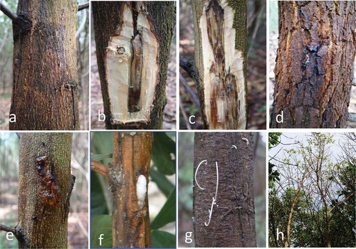
In Acacias, Ceratocystis species cause vascular stain, canker on the stem and eventually wilting. Once infected, the symptoms are first expressed as black or red lesions on the bark, blackened streaks within vascular tissue or sapwood discolouration (). Cankers on the stem and cracked or sunken bark above cankers emerge as further symptoms. This is followed with yellowing of the leaves, wilting, and death of the tree due to lack of nutrient supply (Roux et al. Citation2001b; Tarigan et al. Citation2011a; Brawner et al. Citation2015). The foam or fermentation exudate of yeasts or bacteria also often emerge from the lesions or from entrance holes made by stem borer or fungal feeding insects near to stem cankers (). In particular, this exudate attracts fungivorous nitidulid beetles (Coleoptera: Nitidulidae) which are associated with fungal dispersal (Brawner et al. Citation2015).
Figure 1. Ceratocystis wilt and canker disease symptoms on Acacia mangium. External discolouration (a); internal discolouration (b, c); canker on stem (d); gummosis (e); fermented exudate with fruity odours (f); frass indicating activity of boring insects (g); and yellowing leaves and dieback of tree (h) (Photos: Aswardi Nasution 2017)
Epidemiology
Disease spread depends upon inoculum production and dispersal, the presence of susceptible hosts and suitable environmental conditions. Ceratocystis species produce several different kinds of spore inoculum and exploit a range of dispersal mechanisms which may vary according to the type of spores produced. Like many filamentous fungi, the spores of Ceratocystis species infect their hosts through an infection court, often a wound, where the spores germinate after deposition (Kile Citation1993; Harrington Citation2013); wounding is therefore critical for infection to occur. Root grafting may also contribute to disease development (Harrington Citation2013). For woody trees, the most common mechanism of disease spread is through spore dispersal, vector activity and mechanical transmission. Spore types may include endoconidia, aleurioconidia, chlamydospores (formed by thickening of the walls of endoconidia) and ascospores. The first three are asexual spores while ascospores are the products of sexual recombination. Sporulating mats on exposed wood or in the bark of infected trees produce all spore types (Friday et al. Citation2016) while aleurioconidia are particularly abundant in the infected wood (Araujo et al. Citation2014a). Conidia and ascospores have a short period of viability whereas aleurioconidia have thicker, more durable walls and can likely survive for years, especially inside wood, and increase the likelihood of the disease being soil-borne (Paulin-Mahady et al. Citation2002).
Aleurioconidia are typically soil-borne, though may also be present in insect frass which is ejected from trees and wind-dispersed (Souza et al. Citation2013). Species of Ceratocystis and Thielaviopsis produce aleurioconidia and are known soil-borne pathogens (Harrington Citation2007). The thick-walled aleurioconidia can survive for years in the soil (Upadhyay Citation1981). Subsequent germination then has the potential to infect new plant material brought into the area (Moutia & Saumtally Citation2001; Marín Montoya & Wingfield Citation2006). The status of C. manginecans as a soil-borne pathogen is unclear: it has been isolated from soil in Vietnamese Acacia plantations (Thu et al. Citation2016) and attempts to isolate from soil in Indonesian plantations have recently been successful (Marthin Tarigan pers. comm.), though DNA sequencing is yet to confirm accurate identification of the soil isolates. Ascospores produced by fruiting bodies on woody stems or branches may be transmitted through an insect vector, mechanical tools, wind and water (Kile Citation1993).
Disease spread can also occur via natural root grafts between infected and healthy roots, a phenomenon that occurs in Quercus rubra L. and that facilitates the transmission of B. fagacearum without first passing through the root collar (Juzwik Citation2008). This same pathogen was isolated from around one quarter of natural root grafts in Quercus ellipsoidalis E.J. Hill, the rate of transmission being influenced by host density, soil depth, soil texture and the occurrence of other species (Juzwik et al. Citation2011).
Dispersal by water was reported by Vigouroux and Stojadinovic (Citation1990) who established that wounded roots of Platanus orientalis L. were infected by C. platani spores carried in irrigation water. Dispersal by wind or water occurs more often in the asexual stage because conidia have rounded surfaces, allowing easy removal from the conidiophores. Conversely in the sexual stage, ascospores are produced on perithecia and held together by a sticky, hydrophobic matrix; their concave surfaces also promote adherence to each other (Malloch & Blackwell Citation1993). The sticky matrix facilitates dispersal by insects or other vectors though reduces transmission by wind and rain, at least when the intensity of wind and rain are low (Harrington Citation2007).
Wounds created by insects, animals, or human activities through the use of pruning implements or tapping knives, are the most common infection courts for Ceratocystis spp. (Kile Citation1993; Brawner et al. Citation2015). For example, monkeys strip bark from A. mangium to feed on the sweet-tasting cambium and outer wood of young trees (Hardie et al. Citation2017). The wounds created act as the initial entry point for the fungal spores into the plant tissues. Direct transmission occurs via known associations with fungal feeding insects (Kile Citation1993; Harrington Citation2013), particularly nitidulid beetles and flies (Diptera) (Heath et al. Citation2009b), which then play an important role as disease vectors (Harrington Citation2007). These insects feed on mycelial mats in the infected plants which contain abundant perithecia where ascospores are produced (Harrington Citation2007).
The relationship between fungal feeding insects and Ceratocystis is facilitated by the production of fruity odours by Ceratocystis and a fermentation exudate (Kile Citation1993; Harrington Citation2007; Brawner et al. Citation2015). The fruity aromas contain fatty acids and esters that are toxic to insects which do not rely on fungi for their nutrition. Only insects with a high tolerance of mycotoxins, like nitidulid beetles, are attracted to and feed on the mycelial mats of Ceratocystis spp. (Kile Citation1993; Dowd Citation1995). When the insects feed on the fungi, the sticky ascospores adhere to their bodies; these spores are then transmitted to healthy trees with fresh wounds (Heath et al. Citation2009a). However, even though the relationship between these insects and mycelial mats is well-recognised, most species of Ceratocystis do not have specific insect vectors for their dispersal (Kile Citation1993).
Although disease transmission in Acacia plantations can be caused by all or a combination of the above mechanisms, the initial development of the disease in Indonesia was linked to singling and removal of lower branches to facilitate silvicultural operations (Anthony Francis pers. comm.). Acacia mangium trees were singled in plantations at age 4–8 months to reduce the incidence of multiple stems and to increase tree diameter. An investigation showed that these practices increased Ceratocystis transmission and significantly increased rates of tree death (Tarigan et al. Citation2010).
Biosecurity
Many Ceratocystis species represent biosecurity threats to regions where they are not already present, including to Australian forestry and horticulture. Ceratocystis manginecans poses a threat to the forestry and horticulture sectors, though the threat to Acacias and Eucalypts in natural ecosystems is unclear as the susceptibility of plantation Acacias and mangoes is undoubtedly enhanced by planting at close spacing in large monocultures. The close proximity of Indonesia to Australia has encouraged a high level of tourist traffic and carved wooden products are popular souvenirs. Ceratocystis, though not identified to species level, has been isolated from Balinese wooden statues carved from Acacia wood (Proborini Citation2016).
Similarly, Ceratocystis species infecting Eucalypts pose a threat to Australian forestry and potentially to natural ecosystems. The threat to horticulture is less clear as the mechanism for host adaptation has not been elucidated, though recent studies indicate that a small number of genes are involved (Fourie at al. 2018). Species infecting Eucalypts include C. curvata, C. diversiconidia and C. ecuadoriana, all present in Colombia and Ecuador, C. neglecta in Colombia, C. eucalypticola, C. pirilliformis and C. tsitsikammensis in South Africa, C. fimbriatomima in Venezuela, and C. fimbriata s.l. in Brazil (). The host ranges of these species have not been delineated though C. fimbriata has an extremely broad host range in Brazil. Biosecurity is underpinned by the knowledge of which plant pathogenic species occur in which areas, thus taxonomic certainty, or at least agreement, is critical to biosecurity processes. The name of a pathogen is expected to provide information as to which host plants may be threatened should the pathogen invade a new region; thus, regulations are generally invoked against pathogens at the species level. This is a strong driver for taxonomic research and delineation of new fungal species. The picture can become a little blurred when a genetically diverse species is composed of strains adapted to different hosts, such as occur in Austropuccinia psidii (Graça et al. Citation2013), C. fimbriata s.l., (Harrington et al. Citation2014) and even in C. manginecans, which causes a severe disease of mango trees in Oman but not in Indonesia, where it is prevalent on Acacias. It is unclear whether this disparity in host range is due to environmental, genetic or epigenetic factors. A similar puzzle is posed by A. psidii in Brazil; in this case, the different host-adapted lineages occur in the same geographic location and can be distinguished only by microsatellite analyses (Graça et al. Citation2013; Stewart et al. Citation2018).
Disease management and control
Strategies for managing forest diseases and their vectors parallel those used in agriculture: avoidance, exclusion, eradication, protection, host resistance, curative treatments and integrated management (Edmonds Citation2013; Harrington Citation2013). However, as plantation forests are usually grown in monoculture over larger areas for several years before harvest and attain a much greater biomass than agricultural crops, the range of viable strategies is often more limited. For environmental and economic reasons, control using pesticides is rarely used (Wingfield et al. Citation2001a; Harrington Citation2013). As is the case for Acacia in Southeast Asia, the species planted is often an exotic, and Ceratocystis has emerged as a new disease. As a result, disease management strategies must first be based on a clear understanding of the biology of the host and the behaviour of the pathogen (Wingfield et al. Citation2001a).
Detection, surveying and monitoring
Diseases can be detected from an on-ground examination of individual trees (Bechtold & Patterson Citation2005; Carnegie et al. Citation2018), or at the landscape level using aerial photography and aerial sketch mapping, satellite imagery and LiDAR (Stone & Mohammed Citation2017). Landscape-level detection can be followed up by ground-based surveying and monitoring. For Ceratocystis wilt disease, ground monitoring is an important first step in developing strategies for its control. Importantly, it is also necessary for detecting the presence of insect vectors (Juzwik Citation2008; Heath et al. Citation2009a) as there can be a significant correlation between vector activity and disease severity (Hayslett et al. Citation2008).
Remote sensing and geographic information systems (GIS)
Remote sensing of forest diseases has been applied since aerial photography first became available in the 1930s (Coppin & Bauer Citation1996). Combined with GIS and ground-based surveys, both spatial coverage and objective assessments of forest health can be realised (Wulder et al. Citation2006). Remote sensing using Colour Infra-Red (CIR) digital imagery detected oak wilt disease caused by B. fagacearum (Everitt et al. Citation1999). The imagery was first interpreted from the radiometric reflectance measurements and then verified by ground observations. To distinguish between healthy, infected and dead oak trees, the data was linked to leaf chlorophyll concentration using three spectral bands: visible green (0.52–0.60 µm), visible red (0.63–0.69 µm) and near-infrared (0.63–0.69); infected trees had veinal necrosis and tip burn (Everitt et al. Citation1999). In a second study (Souza et al. Citation2015), a camera mounted in an unmanned aerial vehicle (UAV) detected wilt disease caused by C. fimbriata on Eucalyptus spp. The images were analysed and compared using four distinct machine learning techniques: K-Nearest Neighbours (K-NN), Random Forest (RF), Artificial Neural Network (ANN) and Gaussian Processes (GP). The last, which is a Bayesian non-parametric tool that learns the input-output transformation function based on training data, was best able to reliably and accurately distinguish between healthy and infected trees, and was suitable for detecting wilt disease in other species and other diseases affecting large-scale plantations. A spectral signature of Ceratocystis-infected Metrosideros polymorpha has been detected at leaf and canopy level and provides a basis for mapping and monitoring of disease spread in Hawaii (Asner et al. Citation2018). A clear timeline of symptom development is the first requirement for aerial assessment of Ceratocystis disease in Acacias. A preliminary study indicates that a reduction in leaf area index is associated with early external visible symptoms of Ceratocystis disease in A. mangium (Nasution unpubl.) and this could be exploited for aerial detection.
Silvicultural and chemical control of Ceratocystis
The most frequently suggested strategy for preventing infection by Ceratocystis is wound avoidance (Kile Citation1993; Harrington Citation2013), and efforts to manage Ceratocystis wilt and canker disease in Acacia plantations have focused on better managing the causes of wounding, minimising wound size and limiting pruning and singling activities to periods when insect vectors are less active (Heath et al. Citation2009a). The role of wounds in disease epidemics is well-established, particularly when linked to pruning (Hayslett et al. Citation2008; Roux & Wingfield Citation2009). In Indonesia, wilt and canker symptoms and mortality in A. mangium plantations caused by C. manginecans occurred after trees were singled at 6–8 months old using poor pruning practice (Tarigan et al. Citation2010; Tarigan et al. Citation2011b). Timing of singling or pruning may also be important, with lower disease incidence if conducted during dry than wet weather (Pilotti et al. Citation2016). Climate is also correlated with insect-vector activity; in South Africa beetle activity is much higher during spring and early summer than winter (Heath et al. Citation2009a). Silvicultural interventions that lead to wounds should therefore be avoided during periods of high insect activity (Hayslett et al. Citation2008; Heath et al. Citation2009b). In tropical Southeast Asia, nitidulid and ambrosia beetles are present in Acacia plantations and considered important vectors of C. manginecans, though their activity has not been monitored (Tarigan et al. Citation2011a; Thu et al. Citation2012; Brawner et al. Citation2015). There is an expectation that insect activity will occur year round (Wolda Citation1988) though, and because of the dynamics of parasitism and plant investment in anti-herbivore defences, populations may be higher during the dry than wet season (Dyer et al. Citation2012).
Wound dressings provide a physical barrier to Ceratocystis infection as well as inhibiting fungal growth (Harrington Citation2013) and low toxicity treatments have been developed. Application of latex paint to pruning wounds in Quercus spp. has been shown to be non-toxic and to reduce wilt disease (French & Juzwik Citation1999; Camilli et al. Citation2007).
Removal of symptomatic trees and stumps as well as neighbouring trees that may have been infected by Ceratocystis may help to limit disease spread (Kile Citation1993). This approach aims to minimise the inoculum load by reducing the potential for fungal dispersal (Kile Citation1993; Harrington Citation2013).
Stem injecting 20 ml of 14.3% propiconazole into the root collar of young Q. rubra two weeks before inoculation by B. fagacearum delayed disease development and extended the life of infected trees for at least two years (Blaedow Citation2009). Two years after injection, the fungicide could still be detected in the primary root but its concentration decreased with distance from the injection point which would have reduced its ability to combat the infection (Blaedow Citation2009). Chemical control can be a high-cost strategy, however and as in this case, may only delay the expression of disease symptoms; the fungicides may also be toxic to other organisms. The use of fungicides in this way, as well as wound dressings and removal of symptomatic trees and stumps are also not suitable for large-scale plantation estates being managed for pulpwood. Because of these limitations, the selection of disease resistant and tolerant host materials is currently considered the most effective and economic strategy for managing Ceratocystis disease (Kile Citation1993).
Genetic control of Ceratocystis
The first line of defence to pest and disease invasion is avoidance which includes physical barriers such as hairs, thorns and resin ducts; these generally work as a defence mechanism against animal attack (Vale et al. Citation2001) and may decrease the incidence of wounding caused by animals. Resistance and tolerance mechanisms come into play after contact has been established and are more common for protecting plants from infection by fungal pathogens (Vale et al. Citation2001). Schafer (Citation1971) has defined tolerance as the ability of a cultivar to minimise loss of yield or quality, in comparison with other cultivars, after pathogen infection, whereas resistance is the ability of a plant to overcome the infection process and prevent development of the pathogen, resulting in a disease-free plant. Resistance is considered more important for plant defence than avoidance and tolerance (Vale et al. Citation2001) and the best way to deal with plant disease problems in forest trees, including Ceratocystis (Wingfield et al. Citation2001a; Harrington Citation2013). Such an approach requires genetic variation within the host species that has the potential to be exploited to increase the resistance of trees to disease through breeding. A successful example is Platanus acerifolia (Aiton) Willd. (London plane); trees resistant to C. platani that remain free of canker and sap stain symptoms were obtained in this way (Harrington Citation2013). The adoption of mango cultivars resistant to C. fimbriata has been the most effective strategy for disease control in mangoes in Brazil (Araujo et al. Citation2014b). In Eucalypts, four out of 18 commercial clones of E. grandis × Eucalyptus urophylla S.T.Blake hybrid expressed no discolouration symptoms on the stem and showed an ability to overcome fungal infection caused by C. fimbriata (Zauza et al. Citation2004).
Genetic resistance to disease in plants can be monogenic and controlled by a single major gene which is race-specific and relatively easy to identify and manipulate in a breeding program; it can be expected to provide complete resistance (Poland et al. Citation2009), but this only provides protection against a specific strain or race of the pathogen; within a short time, it may easily be broken down (Maramorosch & Loebenstein Citation2009). Genetic resistance can also be polygenic and controlled by a number of genes (Brun et al. Citation2010). This provides a similar level of protection against all the races of a pathogen, although it may be difficult to identify and manipulate the genes in a breeding program to confer this type of resistance; if successful it is not easily broken down by the pathogen and should give the host long-lasting protection (Lindhout Citation2002).
Polygenic resistance is better suited to Acacia forestry as breeders work with populations rather than genotypes of both host and pathogen in order to protect a range of genotypes of a given host species from all known races of a pathogen (Carson & Carson Citation1989). As plantation growing cycles are usually several years, polygenic resistance against disease is expected to have a long-term utility (Sniezko Citation2006). The development of resistant clones may be expedited by mapping of quantitative trait loci, as has been done for Ceratocystis in Eucalypts in Brazil (Rosado et al. Citation2016).
To source the breeding material requires selecting a range of phenotypes in natural forests or forest plantations and conducting pathogenicity tests either in situ or, more commonly, by using artificial inoculation under controlled conditions (Sniezko Citation2006; Tarigan et al. Citation2010). This enables selection of putative genes that may confer disease resistance before the breeding program commences. This is currently considered the most effective method for controlling Ceratocystis vascular wilt and canker diseases in forest plantations (Kile Citation1993; Wingfield et al. Citation2001a). The trees with the highest rates of survival and lowest levels of disease symptoms are selected for inclusion in breeding programs (Sniezko Citation2006).
Inoculation tests in situ (natural) can provide the best information for a given location because the influence of the natural environment on host-pathogen interactions is captured (Brawner et al. Citation2015). However, there is inevitably a risk of disease spread from the introduced pathogen. This approach also first requires trees rather than seedlings, which slows the screening process (Tarigan et al. Citation2010; Sniezko & Koch Citation2017).
Artificial inoculations using fungal isolates are conducted by wounding tree stems or branches before placing a plug of mycelium or spore suspension of the pathogen under the bark. This is normally carried out on juvenile plant materials under controlled conditions (Green et al. Citation1985; Tarigan et al. Citation2010). Levels of resistance to Ceratocystis wilt and canker disease are evaluated by measuring the length of xylem discolouration (Zauza et al. Citation2004). This screening method is time-consuming as disease resistance is only expected to occur in a low percentage of the population. As large numbers of cultivars or clones must be used, rapid screening technologies are required (Brawner et al. Citation2015). One possible option for Ceratocystis is inoculation of detached leaves; this has been shown to give a similar result to tree wounding in screening assays using leaves of Castanea dentata (Marshall) Borkh., Castanea molissima Blume and Castanea pumila (L.) Mill. against Cryphonectria parasitica (Murrill) M.E. Barr (the chestnut blight pathogen; Newhouse et al. Citation2014). This approach is currently being tested for Acacia (Nasution et al. Citation2016b).
To date, no strains of A. mangium resistant to Ceratocystis have been identified, though there is evidence of some variation in disease tolerance (Brawner 2015). Acacia auriculiformis shows higher levels of tolerance to C. manginecans, and this tolerance may also be expressed in A. mangium × A. auriculiformis hybrids (Trang et al. Citation2018).
Biological control
Biological control is the use of fungi, bacteria, actinomycetes and viruses to suppress or decrease the population density of a plant pathogen (Bale et al. Citation2008). These microorganisms or BCAs may also have beneficial interactions internally or externally with their host plants (McInroy & Kloepper Citation1994). The attraction of biological control is that it can be applied to broad or narrow targets depending on the biocontrol organism, is less site-specific and less prone to build-up of resistance, and is cost-effective for specialised applications; it can also be integrated with other control strategies, and may enhance plant growth (Whipps & Lumsden Citation2001).
In practice, there are few examples of successful biological control in forest trees (Wingfield et al. Citation2001a; Garnas et al. Citation2012). This may be related to their large biomass, complex anatomy and longevity. The inoculum load of the pathogen also has an opportunity to build up during each rotation. Thus, repeated infection through a large root system by a soil borne pathogen allows the disease to persist in soils as dormant, quiescent or resistant propagules such as chlamydospores, aleurioconidia or microsclerotia after tree death and after the trees are harvested (Cazorla & Mercado-Blanco Citation2016). These propagules can then infect new trees or any root tissues that are not reached by the BCA, thereby reducing its effectiveness.
A notable success story has been use of the fungus Phlebiopsis gigantea (Fr.) Jülich for controlling root-rot disease in pine trees caused by Heterobasidion annosum (Fr.) Bref. This fungal pathogen can colonise and survive in the stumps of harvested trees which then act as an inoculum source. Spores of P. gigantea applied to the surface of freshly cut stumps rapidly germinate and colonise the wood, preventing subsequent growth of H. annosum and thus reducing disease incidence in the next rotation (Rishbeth Citation1979). A commercially produced BCA based on P. gigantea is used routinely to control root-rot disease in pine plantations (Pratt et al. Citation2000). While a potential Phlebiopsis sp. has been isolated that shows a microparasitic reaction against G. philippii, a root-rot pathogen that kills A. mangium (Agustini et al. Citation2014), further refinement of application methods is required to commercialise this fungus as a BCA.
Do BCAs have any potential application in controlling vascular wilt diseases? Injection of 100–400 ml (108 cells ml−1) of Pseudomonas syringae Van Hall using a specially developed ‘gouge-pistol’ into the trunk base of Ulmus sp. suppressed the expression of Ophiostoma ulmi (Buisman) Melin & Nannf. (Dutch elm disease) and resulted in a high proportion of healthy trees (Scheffer Citation1983). However, due to a lack of correlation between in vitro and field tests, it was concluded that P. syringae had triggered plant defence against Ceratocystis infection through induced resistance rather than antagonism (Scheffer Citation1990; Scheffer et al. Citation2008). Another potential BCA, Verticillium dahliae Kleb. isolate WCS850 was injected into mature clonal trees of Ulmus minor Mill., (syn. Ulmus carpinifolia Rupp. Ex Suckow) that were either resistant or susceptible to C. ulmi resulting in disease suppression in susceptible as well as resistant trees (Scheffer Citation1990). Injecting V. dahliae isolate Vd-48 into 6–7-year-old U. minor reduced wilting symptoms associated with Ophiostoma infection (Solla & Gil Citation2003). In both studies V. dahliae could be re-isolated only from at or near the injecting point, an indication that translocation of this fungus had been minimal and any contact with C. ulmi unlikely. Thus, it was concluded that V. dahliae acts by triggering host defences against Ceratocystis infection.
Bacteria that live internally in plant tissue without causing any negative impact on their host are recognised as endophytic bacteria (Schulz & Boyle Citation2006). Some have mutualistic relationships with their host plants and an ability to live as obligate or facultative endophytes at different stages in their life cycle (Hardoim et al. Citation2008). The majority of research into endophytic bacteria has been with agricultural and horticultural crops (Kobayashi & Palumbo Citation2000), nevertheless, several species of endophytic bacteria have been isolated from woody species (Kobayashi & Palumbo Citation2000; Bacon & Hinton Citation2006; Izumi Citation2011). The large biomass and perennial nature of trees potentially provide a stable habitat for a diverse range of endophytes (Izumi Citation2011) and raise the possibility that they could play a role as BCAs against vascular diseases such as Ceratocystis (Yadeta & Thomma Citation2013).
Bacterial endophytes suppress plant diseases through the production of enzymes, antifungal and antibacterial compounds (allellochemicals), by competition with pathogens for nutrients or niches and stimulation of induced systemic resistance (ISR) (Compant et al. Citation2005; Bacon & Hinton Citation2006). The main focus for the beneficial effects of endophytic bacteria in woody trees has been their role in promoting growth; it just happens that plant growth promoting rhizobacteria also produce enzymes and antibiotics that can prevent or reduce pathogen infection. In this way rhizobia can play an indirect role as BCAs (Ryan et al. Citation2008). However, there are few reports of endophytes acting as BCAs in forest trees (Chanway Citation1998). This delay in their development and application is in part because their performance in the field has not matched their apparent potential when tested in assay. Thus, a selected endophyte may be replaced by or act differently in the presence of other endophytes already present in the host. To be effective, assay-based selection techniques are required that capture the environment where the introduced endophyte is to be inoculated (Newcombe Citation2011; Hilszczańska Citation2016).
Nevertheless, a potential for using endophytes in managing vascular diseases in forest trees has been demonstrated. As both endophytes and pathogen live in the vascular system, endophytic bacteria can potentially act as antagonists (Yadeta & Thomma Citation2013). Application of Pseudomonas fluorescens Migula and Psuedomonas putida (Trevisan) Migula into the roots of E. urophylla reduced by 45% the incidence of bacterial wilt caused by Ralstonia solanacearum (Smith) Yabuuchi et al. emend Safni et al. (Ran et al. Citation2005). Endophytic bacteria have been applied to control wilt disease caused by B. fagacearum. A total of 889 endophytic bacterial isolates were obtained from healthy Quercus fusiformis Small; 183 isolates of mostly Bacillus spp. and Pseudomonas spp. showed high inhibition through chitinase activity against B. fagacearum in an in vitro assay. Two species, Pseudomonas denitrificans (nomen rejiciendum) isolate 1–15 and P. putida isolate 5–48 were selected and injected into potted oak trees which were then inoculated with a spore suspension of B. fagacearum. The experiment showed that P. denitrificans 1–15 was able to reduce disease incidence by 50% and the proportion of crown loss by 17% (Brooks et al. Citation1994).
This indicates that there may be potential for exploiting endophytes in the control of Ceratocyctis wilt and canker diseases in Acacia plantations. Their slow movement in plant tissues is a potential problem as contact with the pathogen may be limited in an organism as large as a tree (Hallmann et al. Citation1997). However, the ability of endophytic bacteria to elicit ISR in plants or to produce inhibitory secondary compounds suggests this may be a complementary approach to breeding strategies (Percival Citation2001; Hossain et al. Citation2016). Endophytic bacteria that produce secondary metabolites which restrict the growth of C. manginecans in vitro have been isolated from A. mangium and methods for inoculating seedlings or cuttings are under investigation (Nasution et al. Citation2016a).
Preliminary investigations into application of endophytic bacteria and fungi as external treatments for the control of Ceratocystis wilt and canker diseases in Acacia plantations have also been carried out (Tran et al. Citation2018). In this study, treatments were applied externally following inoculation of one-year-old seedlings with C. manginecans. All three biological products reduced the disease index compared to controls after 40 days, though none were as effective as the fungicides tested.
Conclusion
An integrated approach incorporating several strategies is necessary to make A. mangium once again a viable plantation species in Southeast Asia. The development of hybrid Acacias that retain the growth and pulping properties of A. mangium but incorporate the increased levels of resistance to C. mangenicans demonstrated by A. auriculiformis is one approach. Appropriate silvicultural techniques, augmented by deployment of BCAs would provide an extra level of protection that may keep pathogen inoculum levels low and thus help extend the lifespan of any resistant clones that may be developed. Some progress towards each of these aims is being made in Indonesia and Vietnam with the support of ACIAR.
Acknowledgements
Aswardi Nasution is the recipient of a John Allwright Fellowship. This work was supported by ACIAR project FST2014–068, Management strategies for Acacia plantation diseases in Indonesia and Vietnam.
Disclosure statement
No potential conflict of interest was reported by the authors.
Additional information
Funding
References
- Agustini L, Wahyuno D, Indrayadi H, Glen M. 2014. In vitro interaction between Phlebiopsis sp. and Ganoderma philippii isolates. Forest Pathology. 44:472–476.
- Al Adawi A, Barnes I, Khan I, Al Subh A, Al Jahwari A, Deadman M, Wingfield B, Wingfield M. 2013. Ceratocystis manginecans associated with a serious wilt disease of two native legume trees in Oman and Pakistan. Australasian Plant Pathology. 42:179–193.
- Alam MW, Rehman A, Iqbal M, Saira M, Aslam S, Muhammad S, Hameed A, Gleason ML. 2017. First report of Ceratocystis fimbriata causing Eucalyptus wilt in Pakistan. Plant Disease. 101:1050.
- Anderson PK, Cunningham AA, Patel NG, Morales FJ, Epstein PR, Daszak P. 2004. Emerging infectious diseases of plants: pathogen pollution, climate change and agrotechnology drivers. Trends in Ecology & Evolution. 19:535–544.
- Araujo L, Bispo WMS, Cacique IS, Moreira WR, Rodrigues FÁ. 2014a. Resistance in mango against infection by Ceratocystis fimbriata. Phytopathology. 104:820–833.
- Araujo L, Bispo WMS, Cacique IS, Cruz MFA, Rodrigues FA. 2014b. Histopathological aspects of mango resistance to the infection process of Ceratocystis fimbriata. Plant Pathology. 63:1282–1295.
- Asner GP, Martin RE, Keith LM, Heller WP, Hughes MA, Vaughn NR, Hughes RF, Balzotti C. 2018. A spectral mapping signature for the rapid ohia death (ROD) pathogen in Hawaiian forests. Remote Sensing. 10:404–414. doi: 10.3390/rs10030404.
- Bacon CW, Hinton DM. 2006. Bacterial endophytes: the endophytic niche, its occupants, and its utility. In: Gnanamanickam SS, editor. Plant-associated bacteria. Dordrecht: Springer Publishing; p. 155–194.
- Baker CJ, Harrington TC, Krauss U, Alfenas AC. 2003. Genetic variability and host specialization in the Latin American clade of Ceratocystis fimbriata. Phytopathology. 93:1274–1284.
- Bale J, Van Lenteren J, Bigler F. 2008. Biological control and sustainable food production. Philosophical Transactions of the Royal Society B: Biological Sciences. 363:761–776.
- Barnes I, Fourie A, Wingfield MJ, Harrington TC, McNew DL, Sugiyama LS, Luiz BC, Heller WP, Keith LM. 2018. New Ceratocystis species associated with rapid death of Metrosideros polymorpha in Hawai`i. Persoonia. 40:154–181.
- Barnes I, Nakabonge G, Roux J, Wingfield B, Wingfield M. 2005. Comparison of populations of the wilt pathogen Ceratocystis albifundus in South Africa and Uganda. Plant Pathology. 54:189–195.
- Barnes I, Roux J, Wingfield B, O’Neill M, Wingfield M. 2003b. Ceratocystis fimbriata infecting Eucalyptus grandis in Uruguay. Australasian Plant Pathology. 32:361–366.
- Barnes I, Roux J, Wingfield BD, Dudzinski MJ, Old KM, Wingfield MJ. 2003a. Ceratocystis pirilliformis, a new species from Eucalyptus nitens in Australia. Mycologia. 95:865–871.
- Bechtold WA, Patterson PL. 2005. The enhanced forest inventory and analysis program: national sampling design and estimation procedures, vol. 80. Asheville (NC): Southern Research Station, US Department of Agriculture Forest Service.
- Blaedow RA. 2009. Use of the systemic fungicide propiconazole for oak wilt management: an assessment of uncharacterized host-pathogen-fungicide interactions [dissertation]. Minneapolis-St. Paul (MN): University of Minnesota.
- Brawner J, Japarudin Y, Lapammu M, Rauf R, Boden D, Wingfield MJ. 2015. Evaluating the inheritance of Ceratocystis acaciivora symptom expression in a diverse Acacia mangium breeding population. Southern Forests. 77:83–90.
- Brooks DS, Gonzalez CF, Appel DN, Filer T. 1994. Evaluation of endophytic bacteria as potential biological-control agents for Oak Wilt. Biological Control. 4:373–381.
- Brun H, Chèvre A-M, Fitt BD, Powers S, Besnard A-L, Ermel M, Huteau V, Marquer B, Eber F, Renard M, et al. 2010. Quantitative resistance increases the durability of qualitative resistance to Leptosphaeria maculans in Brassica napus. New Phytologist. 185:285–299.
- Camilli K, Appel DN, Watson WT. 2007. Studies on pruning cuts and wound dressings for oak wilt control. Arboriculture and Urban Forestry. 33:132–139.
- Carnegie AJ, Lawson S, Wardlaw T, Cameron N, Venn T. 2018. Benchmarking forest health surveillance and biosecurity activities for managing Australia’s exotic forest pest and pathogen risks. Australian Forestry. 81:14–23.
- Carson S, Carson M. 1989. Breeding for resistance in forest trees – a quantitative genetic approach. Annual Review of Phytopathology. 27:373–395.
- Cazorla FM, Mercado-Blanco J. 2016. Biological control of tree and woody plant diseases: an impossible task? BioControl. 61:233–242.
- Chanway C. 1998. Bacterial endophytes: ecological and practical implications. Sydowia-Horn. 50:149–170.
- Coetzee MPA, Wingfield BD, Golani GD, Tjahjono B, Gafur A, Wingfield MJ. 2011. A single dominant Ganoderma species is responsible for root rot of Acacia mangium and Eucalyptus in Sumatra. Southern Forests. 73:175–180.
- Compant S, Duffy B, Nowak J, Clément C, Barka EA. 2005. Use of plant growth-promoting bacteria for biocontrol of plant diseases: principles, mechanisms of action, and future prospects. Applied and Environmental Microbiology. 71:4951–4959.
- Coppin PR, Bauer ME. 1996. Digital change detection in forest ecosystems with remote sensing imagery. Remote Sensing Reviews. 13:207–234.
- Crous PW, Denman S, Taylor JE, Swart L, Bezuidenhout CM, Hoffman L, Palm ME, Groenewald JZ. 2013. Cultivation and diseases of Proteaceae: Leucadendron, Leucospermum, and Protea. 2nd ed. Utrecht: CBS Biodiversity Series. N/A: 360.
- De Beer Z, Duong T, Barnes I, Wingfield B, Wingfield M. 2014. Redefining Ceratocystis and allied genera. Studies in Mycology. 79:187–219.
- De Beer ZW, Marincowitz S, Duong TA, Wingfield MJ. 2017. Bretziella, a new genus to accommodate the oak wilt fungus, Ceratocystis fagacearum (Microascales, Ascomycota). MycoKeys. 27:1–19.
- Dowd PF. 1995. Sap beetles and mycotoxins in maize. Food Additives & Contaminants. 12:497–508.
- Dyer LA, Carson WP, Leigh EG Jr. 2012. Insect outbreaks in tropical forests: patterns, mechanisms, and consequences. In: Barbosa P, Letourneau DK, Agrawal AA, editors. Insect outbreaks revisited. Hoboken (NJ): Wiley-Blackwell; p. 219–245.
- Edmonds RL. 2013. General strategies of forest disease management. infectious forest diseases. Wallingford: CABI; p. 29–49.
- Engelbrecht CJ, Harrington TC, Alfenas AC, Suarez C. 2007b. Genetic variation in populations of the cacao wilt pathogen, Ceratocystis cacaofunesta. Plant Pathology. 56:923–933.
- Engelbrecht CJB, Harrington TC. 2005. Intersterility, morphology and taxonomy of Ceratocystis fimbriata on sweet potato, cacao and sycamore. Mycologia. 97:57–69.
- Engelbrecht CJB, Harrington TC, Alfenas A. 2007a. Ceratocystis wilt of cacao – a disease of increasing importance. Phytopathology. 97:1648–1649.
- Everitt J, Escobar D, Appel D, Riggs W, Davis M. 1999. Using airborne digital imagery for detecting oak wilt disease. Plant Disease. 83:502–505.
- Ferreira MA, Harrington TC, Piveta G, Alfenas AC. 2017. Genetic variability suggests that three populations of Ceratocystis fimbriata are responsible for the Ceratocystis wilt epidemic on kiwifruit in Brazil. Tropical Plant Pathology. 42:86–95.
- Firmino AC, Tozze HJ Jr, Furtado EL. 2012. First report of Ceratocystis fimbriata causing wilt in Tectona grandis in Brazil. New Disease Reports. 25:24.
- Fourie A, Wingfield MJ, Wingfield BD, Barnes I. 2015. Molecular markers delimit cryptic species in Ceratocystis sensu stricto. Mycological Progress. 14:1–18.
- Fourie A, Wingfield MJ, Wingfield BD, van der Nest MA, Loots MT, Barnes I. 2018. Inheritance of phenotypic traits in the progeny of a Ceratocystis interspecific cross. Fungal Biology. 122:717–729.
- Francis A, Beadle C, Puspitasari D, Irianto R, Agustini L, Rimbawanto A, Gafur A, Hardiyanto E, Hidyati N, Tjahjono B. 2014. Disease progression in plantations of Acacia mangium affected by red root rot (Ganoderma philippii). Forest Pathology. 44:447–459.
- French D, Juzwik J. 1999. Oak wilt in Minnesota. University of Minnesota extension service, MI-3174-Z.
- Friday JB, Keith L, Hughes F, Cannon P. 2016. Ceratocystis wilt of oh’ia (rapid Oh’ia death): a new disease in Hawaii. Workshop Proceedings: Ceratocystis in tropical hardwood plantations; Feb 15–16; Yogyakarta. Available from: http://www.forestryhealth.org/
- Garnas JR, Hurley BP, Slippers B, Wingfield MJ. 2012. Biological control of forest plantation pests in an interconnected world requires greater international focus. International Journal of Pest Management. 58:211–223.
- Graça RN, Ross-Davies AL, Klopfenstein N, Kim M-S, Peever TL, Cannon PG, Aun CP, Mizubuti ESG, Alfenas AC. 2013. Rust disease of eucalypts, caused by Puccinia psidii, did not originate via host jump from guava in Brazil. Molecular Ecology. 22:6033–6047.
- Green CE, Guries R, Smalley E. 1985. Early screening of elms for resistance to Ceratocystis ulmi. Plant Disease. 69:60–63.
- Griffin A, Midgley S, Bush D, Cunningham P, Rinaudo A. 2011. Global uses of Australian acacias – recent trends and future prospects. Diversity and Distributions. 17:837–847.
- Hallmann J, Quadt-Hallmann A, Mahaffee W, Kloepper J. 1997. Bacterial endophytes in agricultural crops. Canadian Journal of Microbiology. 43:895–914.
- Halsted BD, Fairchild D. 1891. Sweet-potato black rot (Ceratocystis fimbriata Ell& Hals.). The Journal of Mycology. 7:1–11.
- Hardie M, Akhmad N, Mohammed C, Mendham D, Corkrey R, Gafur A, Siregar S. 2017. Role of site in the mortality and production of Acacia mangium plantations in Indonesia. Southern Forests. 80:37–50.
- Hardoim PR, van Overbeek LS, van Elsas JD. 2008. Properties of bacterial endophytes and their proposed role in plant growth. Trends in Microbiology. 16:463–471.
- Harrington TC. 2007. The genus Ceratocystis. where does the oak wilt fungus fit? In: Billings RF, Appel DN, editors. Proceedings of the 2nd National Oak Wilt Symposium; 2007 June 4–7; Austin, Texas.
- Harrington TC. 2013. Ceratocystis diseases. In: Gonthier P, editor. Infectious forest diseases. Wallingford: CABI; p. 230–255.
- Harrington TC, Huang Q, Ferreira MA, Alfenas AC. 2015. Genetic analyses trace the Yunnan, China population of Ceratocystis fimbriata on pomegranate and taro to populations on Eucalyptus in Brazil. Plant Disease. 99:106–111.
- Harrington TC, Kazmi MR, Al-Sadi AM, Ismail SI. 2014. Intraspecific and intragenomic variability of ITS rDNA sequences reveals taxonomic problems in Ceratocystis fimbriata sensu stricto. Mycologia. 106:224–242.
- Harwood C, Nambiar K. 2014. Productivity of acacia and eucalypt plantations in Southeast Asia. 2. Trends and variations. International Forestry Review. 16:249–260.
- Hayslett M, Juzwik J, Moltzan B, Appel D, Camilli K. 2008. Insect vectors of the oak wilt fungus in Missouri and Texas. In: Proceedings of the 2nd National Oak Wilt Symposium; Austin (Texas). p. 109–120.
- Heath RN, Wingfield MJ, Van Wyk M, Roux J. 2009a. Insect associates of Ceratocystis albifundus and patterns of association in a native savanna ecosystem in South Africa. Environmental Entomology. 38:356–364.
- Heath RN, Wingfield MJ, Wingfield BD, Meke G, Mbaga A, Roux J. 2009b. Ceratocystis species on Acacia mearnsii and Eucalyptus spp in eastern and southern Africa including six new species. Fungal Diversity. 34:41–67.
- Hilszczańska D. 2016. Endophytes – characteristics and possibilities of application in forest management. Forest Research Papers. 77:276–282.
- Hossain MT, Chung EJ, Chung YR. 2016. Biological control of rice bakanae by an endophytic Bacillus oryzicola YC7007. The Plant Pathology Journal. 32:228–242.
- Huang Q, Zhu Y, Chen H, Wang Y, Liu Y, Lu W, Ruan X. 2003. First report of pomegranate wilt caused by Ceratocystis fimbriata in Yunnan, China. Plant Disease. 87:1150.
- Irsan C, Suwandi AKD, Umayah A, Hamidson H. 2016. Ceratocystis wilt of Lansium tree: new disease and threat to duku fruit production in Indonesia. Workshop Proceedings: Ceratocystis in tropical hardwood plantations; Feb 15–16; Yogyakarta. Available from: http://www.forestryhealth.org/
- Izumi H. 2011. Diversity of endophytic bacteria in forest trees. In: Pirttilä AM, Frank C, editors. Endophytes of forest trees. Dordrecht: Springer; p. 95–105.
- Johnson JA, Harrington TC, Engelbrecht C. 2005. Phylogeny and taxonomy of the North American clade of the Ceratocystis fimbriata complex. Mycologia. 97:1067–1092.
- Juzwik J. 2008. Epidemiology and occurrence of oak wilt in Midwestern, Middle, and South Atlantic states. In: Billings RF, Appel DN, editors. Proceedings of the 2nd National Oak Wilt Symposium; 2007 Jun 4–7; Austin, Texas. p. 55–67.
- Juzwik J, Appel DN, MacDonald WL, Burks S. 2011. Challenges and successes in managing oak wilt in the United States. Plant Disease. 95:888–900.
- Kajitani Y, Masuya H. 2011. Ceratocystis ficicola sp. nov., a causal fungus of fig canker in Japan. Mycoscience. 52:349–353.
- Keith LM, Hughes RF, Sugiyama LS, Heller WP, Bushe BC, Friday JB. 2015. First report of Ceratocystis wilt on Ohia (Metrosideros polymorpha). Plant Disease. 99:1277.
- Kha LD. 2000. Studies on natural hybrids of Acacia mangium and A. auriculiformis in Vietnam. Journal of Tropical Forest Science. 12:794–803.
- Kile G. 1993. Plant diseases caused by species of Ceratocystis sensu stricto and Chalara. In: Wingfield MJ, Seifert KA, Webber JF, editors. Ceratocystis and Ophiostoma: taxonomy, ecology, and pathogenicity. St Paul (MN): APS Press; p. 173–183.
- Kobayashi D, Palumbo J. 2000. Bacterial endophytes and their effects on plants and uses in agriculture. In: Bacon CW, White J, editors. Microbial endophytes. Boca Raton (FL): CRC Press; p. 199–233.
- Krisnawati H, Kallio M, Kanninen M. 2011. Acacia mangium Willd.: ecology, silviculture and productivity. Bogor (Indonesia): CIFOR.
- Laia M, Alfenas A, Harrington T. 2000. Isolation, detection in soil, and inoculation of Ceratocystis fimbriata, causal agent of wilting, die-back and canker in Eucalyptus. In: Proceedings of the 12th Biennial Conference of the Australasian Plant Pathology Society; 1991 September 27–30; Canberra, Australia.
- Lee DH, Roux J, Wingfield BD, Barnes I, Wingfeld MJ. 2016. New host range and distribution Ceratocystis pirilliformis in South Africa. European Journal of Plant Pathology. 146:483–496.
- Lee S. 2004. Diseases and potential threats to Acacia mangium plantations in Malaysia. Unasylva. 217:31–35.
- Lehtijärvi A, Oskay F, Doğmus-Lehtijärvi H, Aday Kaya AG, Pecori F, Santini A, Woodward S. 2018. Ceratocystis platani is killing plane trees in Istanbul (Turkey). Forest Pathology. 48:e12375.
- Li Q, Harrington TC, McNew D, Li J. 2017. Ceratocystis uchidae, a new species on Araceae in Hawaii and Fiji. Mycoscience. 58:398–412.
- Lindhout P. 2002. The perspectives of polygenic resistance in breeding for durable disease resistance. Euphytica. 124:217–226.
- Linnakoski R, de Beer ZW, Rousi M, Niemela P, Pappinen A, Wingfield MJ. 2008. Fungi, including Ophiostoma karelicum sp. nov., associated with Scolytus ratzeburgi infesting birch in Finland and Russia. Mycological Research. 112:1475–1488.
- Liu FF, Barnes I, Roux J, Wingfield MJ, Chen SF. 2018. Molecular phylogenetics and microsatellite analysis reveal a new pathogenic Ceratocystis species in the Asian-Australian clade. Plant Pathology. 67:1097–1113.
- Liu FF, Mbenoun M, Barnes I, Roux J, Wingfield MJ, Li GQ, Li JQ, Chen SF. 2015. New Ceratocystis species from Eucalyptus and Cunninghamia in South China. Mycoscience. 107:1451–1473.
- Mace ME, Bell AA, Beckman CH. 1981. Fungal wilt diseases of plants. New York (London): Academic Press.
- Malloch D, Blackwell M. 1993. Dispersal biology of the ophiostomatoid fungi. In: Wingfield MJ, Seifert KA, Webber JF, editors. Ceratocystis and Ophiostoma: taxonomy, ecology, and pathogenicity. St Paul (MN): APS Press; p. 195–206.
- Maramorosch K, Loebenstein G. 2009. Plant disease resistance: natural, non-host innate or inducible. In: Schaechter M, editor. Encyclopaedia of Microbiology. Amsterdam (Netherlands): Elsevier Science; p. 589–596.
- Marín Montoya M, Wingfield MJ. 2006. A review of Ceratocystis sensu stricto with special reference to the species complexes C. coerulescens and C. fimbriata. Revista Facultad Nacional de Agronomía Medellín. 59:3045–3375.
- Martín-García J, Espiga E, Pando V, Diez JJ. 2011. Factors influencing endophytic communities in poplar plantations. Silva Fennica. 45:169–180.
- Mbenoun M, de Beer ZW, Wingfield MJ, Wingfield BD, Roux J. 2014. Reconsidering species boundaries in the Ceratocystis paradoxa complex, including a new species from oil palm and cacao in Cameroon. Mycologia. 106:757–784.
- McInroy J, Kloepper J. 1994. Novel bacterial taxa inhabiting internal tissues of sweet corn and cotton. In: Ryder MH, Stephens PM, Bowen GD, editors. Improving plant productivity with rhizosphere bacteria. Melbourne (Australia): CSIRO; p. 190.
- Midgley S, Turnbull J. 2003. Domestication and use of Australian acacias: case studies of five important species. Australian Systematic Botany. 16:89–102.
- Misse AC, Barnes I, Roets F, Mbenoun M, Wingfield MJ, Roux J. 2017. Ecology and population structure of a tree wound-infecting fungus in a native South African forest environment. Fungal Biology. 121:69–81.
- Mohammed C, Rimbawanto A, Page D. 2014. Management of basidiomycete root- and stem-rot diseases in oil palm, rubber and tropical hardwood plantation crops. Forest Pathology. 44:428–446.
- Morris M, Wingfield M, de Beer C. 1993. Gummosis and wilt of Acacia mearnsii in South Africa caused by Ceratocystis fimbriata. Plant Pathology. 42:814–817.
- Moutia Y, Saumtally S. 2001. Detection from soil and distribution of Ceratocystis paradoxa Moreau, causal agent of the pineapple disease of sugarcane. Reduit (Mauritius): AMAS Sugar Industry Research Institute, Food And Agricultural Research Council; p. 75.
- Muramoto N, Tanaka T, Shimamura T, Mitsukawa N, Hori E, Koda K, Otani M, Hirai M, Nakamura K, Imaeda T. 2012. Transgenic sweet potato expressing thionin from barley gives resistance to black rot disease caused by Ceratocystis fimbriata in leaves and storage roots. Plant Cell Reports. 31:987–997.
- Naidoo K, Steenkamp ET, Coetzee MP, Wingfield MJ, Wingfield BD. 2013. Concerted evolution in the ribosomal RNA cistron. PloS One. 8:e59355.
- Nasution A, Glen M, Gafur A, Evans K, Mohammed CL 2016a. Endophytic bacteria from Acacia mangium and their potential antagonism against Ceratocystis sp. Workshop Proceedings: Ceratocystis in tropical hardwood plantations; Feb 15–16; Yogyakarta. Available from: http://www.forestryhealth.org/
- Nasution A, Glen M, Gafur A, Evans K, Mohammed CL 2016b. Developing a rapid screening protocol for resistance of Acacia mangium against Ceratocystis wilt and canker disease. Workshop Proceedings: Ceratocystis in tropical hardwood plantations; Feb 15–16;Yogyakarta. Available from: http://www.forestryhealth.org/
- Newcombe G. 2011. Endophytes in forest management: four challenges. In: Pirttalä AM, Frank C, editors. Endophytes of forest trees. Dordrecht: Springer Publishing; p. 251–262.
- Newhouse AE, Spitzer JE, Maynard CA, Powell WA. 2014. Chestnut leaf inoculation assay as a rapid predictor of blight susceptibility. Plant Disease. 98:4–9.
- Nkuekam GK, Barnes I, Wingfield MJ, Roux J. 2009. Distribution and population diversity of Ceratocystis pirilliformis in South Africa. Mycologia. 101:17–25.
- Nkuekam GK, Jacobs K, de Beer ZW, Wingfield MJ, Roux J. 2008. Ceratocystis and Ophiostoma species, including three new taxa, associated with wounds on native South African trees. Fungal Diversity. 29:37–59.
- Nkuekam GK, Wingfield MJ, Mohammed C, Carnegie AJ, Pegg GS, Roux J. 2012. Ceratocystis species, including two new species associated with nitidulid beetles, on eucalypts in Australia. Antonie van Leeuwenhoek. Suppl. 101:217–241.
- Nkuekam GK, Wingfield MJ, Roux J. 2013. Ceratocystis species, including two new taxa, from Eucalyptus trees in South Africa. Australasian Plant Pathology. 42:283–311.
- Oliveira LS, Damacena MB, Guimarães LM, Siqueira DL, Alfenas AC. 2016. Ceratocystis fimbriata isolates on Mangifera indica have different levels of aggressiveness. European Journal of Plant Pathology. 145:847–856.
- Oliveira LSS, Harrington TC, Ferreira MA, Damacena MB, Al-Sadi AM, Al-Mahmooli HIS, Alfenas AC. 2015. Species or genotypes? Reassessment of four recently described species of the Ceratocystis wilt pathogen, Ceratocystis fimbriata, on Mangifera indica. Phytopathology. 105:1229–1244.
- Paulin-Mahady AE, Harrington TC, McNew D. 2002. Phylogenetic and taxonomic evaluation of Chalara, Chalaropsis, and Thielaviopsis anamorphs associated with Ceratocystis. Mycologia. 94:62–72.
- Percival GC. 2001. Induction of systemic acquired disease resistance in plants: potential implications for disease management in urban forestry. Journal of Arboriculture. 27:181–192.
- Pilotti M, Di Lernia G, Modesti V, Lumia V, Brunetti A. 2016. Outcome of Ceratocystis platani inoculations in Platanus × acerifolia in relation to season and inoculum dose. iForest. 9:608–617.
- Poland JA, Balint-Kurti PJ, Wisser RJ, Pratt RC, Nelson RJ. 2009. Shades of gray: the world of quantitative disease resistance. Trends in Plant Science. 14:21–29.
- Pornsuriya C, Sunpapao A. 2015. A new sudden decline disease of bullet wood in Thailand is associated with Ceratocystis manginecans. Australasian Plant Disease Notes. 10:26.
- Pratt J, Niemi M, Sierota Z. 2000. Comparison of three products based on Phlebiopsis gigantea for the control of Heterobasidion annosum in Europe. Biocontrol Science and Technology. 10:467–477.
- Proborini MW. 2016. Ceratocystis as fungal parasitic on wooden statues. Workshop Proceedings: Ceratocystis in tropical hardwood plantations; Feb 15–16; Yogyakarta. Available from: http://www.forestryhealth.org/
- Ran L, Liu C, Wu G, Van Loon L, Bakker P. 2005. Suppression of bacterial wilt in Eucalyptus urophylla by fluorescent Pseudomonas sppn China. Biological Control. 32:111–120.
- Réblová M, Gams W, Seifert KA. 2011. Monilochaetes and allied genera of the glomerellales, and a reconsideration of families in the Microascales. Studies in Mycology. 68:163–191.
- Rishbeth J. 1979. Modern aspects of biological control of Fomes and Armillaria. European Journal of Forest Pathology. 9:331–340.
- Rodas CA, Roux J, van Wyk M, Wingfield BD, Wingfield MJ. 2008. Ceratocystis neglecta sp. nov., infecting Eucalyptus trees in Colombia. Fungal Diversity. 28:73–84.
- Rosado CCG, Guimarães LM, Da S, Faria DA, de Resende MDV, Cruz CD, Grattapaglia D, Alfenas AC. 2016. QTL mapping for resistance to Ceratocystis wilt in Eucalyptus. Tree Genetics and Genomes. 12:1–10.
- Roux J, Coutinho T, Mujuni Byabashaija D, Wingfield M. 2001a. Diseases of plantation Eucalyptus in Uganda. South African Journal of Science. 97:16–18.
- Roux J, Coutinho TA, Wingfield MJ, Bouillet JP. 2000a. Diseases of plantation Eucalyptus in the Republic of Congo. South African Journal of Science. 96:454–456.
- Roux J, Heath R, Meke G, Nguvulu C, Miambo F, Geldenhuis C, Wingfield M. 2004a. Fungi associated with bark wounds on indigenous African trees. Phytopathology. 94:S89.
- Roux J, Heath RN, Labuschagne L, Nkuekam GK, Wingfield MJ. 2007. Occurrence of the wattle wilt pathogen, Ceratocystis albifundus on native South African trees. Forest Pathology. 37:292–302.
- Roux J, Meke G, Kanyi B, Mwangi L, Mbaga A, Hunter G, Nakabonge G, Heath R, Wingfield M. 2005. Diseases of plantation forestry trees in eastern and southern Africa: review article. South African Journal of Science. 101:409–413.
- Roux J, Van Wyk M, Hatting H, Wingfield M. 2004b. Ceratocystis species infecting stem wounds on Eucalyptus grandis in South Africa. Plant Pathology. 53:414–421.
- Roux J, Wingfield M, Bouillet JP, Wingfield B, Alfenas A. 2000b. A serious new wilt disease of Eucalyptus caused by Ceratocystis fimbriata in Central Africa. Forest Pathology. 30:175–184.
- Roux J, Wingfield M, Byabashaija DM. 2001b. First report of Ceratocystis wilt of Acacia mearnsii in Uganda. Plant Disease. 85:1029.
- Roux J, Wingfield MJ. 2009. Ceratocystis species: emerging pathogens of non-native plantation Eucalyptus and Acacia species. Southern Forests. 71:115–120.
- Roux J, Wingfield MJ. 2013. Ceratocystis species on the African continent, with particular reference to Ceratocystis albofundus, an African species in the Ceratocystis fimbriata sensu lato species complex. In: Seifert KA, de Beer ZW, Wingfield MJ, editors. Ophiostomatoid fungi: expanding frontiers. CBS Biodiversity Series. Vol. 337; p. 131–138.
- Ryan RP, Germaine K, Franks A, Ryan DJ, Dowling DN. 2008. Bacterial endophytes: recent developments and applications. FEMS Microbiology Letters. 278:1–9.
- Samuels G. 1993. The case for distinguishing Ceratocystis and Ophiostoma. In: Wingfield MJ, Seifert KA, Webber JE, editors. Ceratocystis and Ophiostoma: taxonomy, ecology, and pathogenicity. St Paul (MN): APS Press; p. 15–20.
- Schafer JF. 1971. Tolerance to plant disease. Annual Review of Phytopathology. 9:235–252.
- Scheffer R. 1983. Biological control of Dutch elm disease by Pseudomonas species. Annals of Applied Biology. 103:21–30.
- Scheffer R. 1990. Mechanisms involved in biological control of Dutch elm disease. Journal of Phytopathology. 130:265–276.
- Scheffer R, Voeten J, Guries R. 2008. Biological control of Dutch elm disease. Plant Disease. 92:192–200.
- Schulz B, Boyle C. 2006. What are endophytes?. In: Schulz BJE, Boyle CJC, Sieber TN, editors. Microbial root endophytes. Berlin: Springer Publishing; p. 21–33.
- Sniezko RA. 2006. Resistance breeding against nonnative pathogens in forest trees—current successes in North America. Canadian Journal of Plant Pathology. 28:S270–S279.
- Sniezko RA, Koch J. 2017. Breeding trees resistant to insects and diseases: putting theory into application. Biological Invasions. 19:3377–3400.
- Solla A, Gil L. 2003. Evaluating Verticillium dahliae for biological control of Ophiostoma novo-ulmi in Ulmus minor. Plant Pathology. 52:579–585.
- Souza AGC, Maffia LA, Murta HM, Alves YH, Pereira RM, Picanco MC. 2013. First report on the association between Ceratocystis fimbriata, an agent of mango wilt, Xyleborus affinis, and the sawdust produced during beetle colonization in Brazil. Plant Disease. 97:1116.
- Souza JR, Mendes CC, Guizilini V, Vivaldini KC, Colturato A, Ramos F, Wolf DF. 2015. Automatic detection of Ceratocystis wilt in Eucalyptus crops from aerial images. IEEE International Conference on Robotics and Automation (ICRA); May 26–30; Seattle. p. 3443–3448. doi: 10.1109/ICRA.2015.7139675
- Spatafora JW, Blackwell M. 1994. The polyphyletic origins of ophiostomatoid fungi. Mycological Research. 98:1–9.
- Stewart JE, Ross-Davies AL, Graça RN, Alfenas AC, Peever TL, Hanna JW, Uchida JY, Hauff RD, Kadooka CY, Kim M-S, et al. 2018. Genetic diversity of the myrtle rust pathogen (Austropuccinia psidii) in the Americas and Hawaii: global implications for invasive threat assessments. Forest Pathology. 48:e12378.
- Stone C, Mohammed CL. 2017. Application of remote sensing technologies for assessing planted forests damaged by insect pests and fungal pathogens: a review. Current Forestry Reports. 3:75–92.
- Talboys PW. 1972. Resistance to vascular wilt fungi. Proceedings Of The Royal Society Series B-Biological Sciences. 181:319–332.
- Tarigan M, Roux J, Van Wyk M, Tjahjono B, Wingfield MJ. 2011a. A new wilt and die-back disease of Acacia mangium associated with Ceratocystis manginecans and C. acaciivora sp nov. in Indonesia. South African Journal of Botany. 77:292–304.
- Tarigan M, Van Wyk M, Roux J, Tjahjono B, Wingfield M. 2010. Three new Ceratocystis spp. in the Ceratocystis moniliformis complex from wounds on Acacia mangium and A. crassicarpa. Mycoscience. 51:53–67.
- Tarigan M, Wingfield MJ, Van Wyk M, Tjahjono B, Roux J. 2011b. Pruning quality affects infection of Acacia mangium and A. crassicarpa by Ceratocystis acaciivora and Lasiodiplodia theobromae. Southern Forests. 73:187–191.
- Thu P, Qynh D, Dell B. 2012. Ceratocystis sp. causes crown wilt of Acacia spp. planted in some ecological zones of Vietnam. Journal of Plant Protection. 5:24–30.
- Thu P, Qynh D, Fourie A, Barnes I, Wingfield M. 2016. Ceratocystis wilt – a serious threat to Acacia plantations in Vietnam. Workshop Proceedings: Ceratocystis in tropical hardwood plantations; Feb 15–16; Yogyakarta. Available from: http://www.forestryhealth.org/
- Tran TTT, Pham PQ, Barber PA, Nguyen CM. 2018. Control of Ceratocystis causing wilt disease on Acacia mangium seedlings. Australasian Plant Pathology. 47:579–586.
- Trang TT, Eyles A, Davies NW, Glen M, Ratkowsky D, Mohammed CL. 2018. Screening for host responses in Acacia to a canker and wilt pathogen, Ceratocystis manginecans. Forest Pathology. 48:e12390.
- Tsopelas P, Santini A, Wingfield MJ, de Beer ZW. 2017. Canker stain: a lethal disease destroying iconic plant trees. Plant Disease. 101:645–658.
- Upadhyay HP. 1981. A monograph of Ceratocystis and Ceratocystiopsis. Athens (GA): University of Georgia Press.
- Upadhyay HP. 1993. Classification of the ophiostomatoid fungi. In: Wingfield MJ, Seifert KA, Webber JF, editors. Ceratocystis and Ophiostoma: taxonomy, ecology, and pathogenicity. St Paul (MN): APS Press; p. 7–13.
- Valdetaro DC, Oliveira LS, Guimarães LM, Harrington TC, Ferreira MA, Freitas RG, Alfenas AC. 2015. Genetic variation, morphology and pathogenicity of Ceratocystis fimbriata on Hevea brasiliensis in Brazil. Tropical Plant Pathology. 40:184–192.
- Vale FXR, Parlevliet J, Zambolim L. 2001. Concepts in plant disease resistance. Fitopatologia Brasileira. 26:577–589.
- Van Wyk M, Adawi AO A, Khan IA, Deadman ML, Al Jahwari AA, Wingfield BD, Ploetz R, Wingfield MJ. 2007a. Ceratocystis manginecans sp. nov., causal agent of a destructive mango wilt disease in Oman and Pakistan. Fungal Diversity. 27:213–230.
- Van Wyk M, Heath RN, Tarigan M, Vermeulen M, Wingfield MJ. 2010a. Comparison of procedures to evaluate the pathogenicity of Ceratocystis fimbriata sensu lato isolates from Eucalyptus in South Africa. Southern Forests. 72:57–62.
- Van Wyk M, Pegg G, Lawson S, Wingfield MJ. 2007b. Ceratocystis atrox sp. nov. associated with Phoracantha acanthocera infestations on Eucalyptus grandis in Australia. Australasian Plant Pathology. 36:407–414.
- Van Wyk M, Wingfield B, Mohali S, Wingfield M. 2009a. Ceratocystis fimbriatomima, a new species in the C. fimbriata sensu lato complex isolated from Eucalyptus trees in Venezuela. Fungal Diversity. 34:175.
- Van Wyk M, Wingfield BD, Al-Adawi AO, Rossetto CJ, Ito MF. 2011a. Two new Ceratocystis species associated with mango disease in Brazil. Mycotaxon. 117:381–404.
- Van Wyk M, Wingfield BD, Clegg PA, Wingfield MJ. 2009b. Ceratocystis larium sp. nov., a new species from Styrax benzoin wounds associated with incense harvesting in Indonesia. Persoonia. 22:75–82.
- Van Wyk M, Wingfield BD, Marín M, Wingfield MJ. 2010b. New Ceratocystis species infecting coffee, cacao, citrus and native trees in Colombia. Fungal Diversity. 40:103–117.
- Van Wyk M, Wingfield BD, Wingfield MJ. 2011b. Four new Ceratocystis spp. associated with wounds on Eucalyptus, Schizolobium and Terminalia trees in Ecuador. Fungal Diversity. 46:111–131.
- Van Wyk M, Wingfield BD, Wingfield MJ. 2013. Ceratocystis species in the Ceratocystis fimbriata complex. In: Seifert KA, de Beer ZW, Wingfield MJ, editors. Ophiostomatoid fungi: expanding frontiers. Utrecht: CBS Biodiversity Series; p. 337.
- Vigouroux A, Stojadinovic B. 1990. Infection of plane tree by Ceratocystis fimbriata f. platani after contamination of water as growing substrate of wounded roots. European Journal of Forest Pathology. 20:118–121.
- Whipps JM, Lumsden RD. 2001. Commercial use of fungi as plant disease biological control agents: status and prospects. In: Butt TM, Jackdon C, Magan N, editors. Fungal biocontrol agents: progress, problems and potential. Wallingford: CABI; p. 9–22.
- Wingfield MJ, de Beer C, Visser C, Wingfield BD. 1996. A new Ceratocystis species defined using morphological and ribosomal DNA sequence comparisons. Systematic and Applied Microbiology. 19:191–202.
- Wingfield MJ, Roux J, Coutinho T, Govender P, Wingfield B. 2001a. Plantation disease and pest management in the next century. The Southern African Forestry Journal. 190:67–71.
- Wingfield MJ, Slippers B, Roux J, Wingfield BD. 2001b. Worldwide movement of exotic forest fungi, especially in the tropics and the southern hemisphere: this article examines the impact of fungal pathogens introduced in plantation forestry. AIBS Bulletin. 51:134–140.
- Witzell J, Martín JA, Blumenstein K. 2014. Ecological aspects of endophyte-based biocontrol of forest diseases. advances in endophytic research. New Delhi: Springer Publishing; p. 321–333.
- Wolda H. 1988. Insect seasonality: why? Annual Review of Ecology and Systematics. 19:1–18.
- Wulder MA, Dymond CC, White JC, Leckie DG, Carroll AL. 2006. Surveying mountain pine beetle damage of forests: a review of remote sensing opportunities. Forest Ecology and Management. 221:27–41.
- Yadeta K, Thomma B. 2013. The xylem as battleground for plant hosts and vascular wilt pathogens. Frontiers in Plant Science. 4:1–12.
- Zauza EAV, Alfenas AC, Harrington TC, Mizubuti ES, Silva JF. 2004. Resistance of Eucalyptus clones to Ceratocystis fimbriata. Plant Disease. 88:758–760.
- Zhang ZP, Li Q, Luo LX, Li JQ, Hao JJ. 2017. First report of mango wilt caused by Ceratocystis fimbriata in Mangifera indica in China. Plant Disease. 101:142.