Abstract
In this study, the relation between the measurement duration and accuracy of pulse-counting was quantitatively examined with special reference to low-frequency fluctuations in heart rate variability. The interbeat intervals of 70 healthy male subjects were measured in standing, sitting and supine positions. Pulse rates for various durations were calculated by objective-scoring simulation based on the heartbeat recordings of the subjects. The duration of pulse-counting continuously varied from 6 to 60 s in the simulation. Simulated pulse rates were compared with the rate calculated from the 60 s that includes the given duration, and the absolute difference between the two rates was defined as the error. Average errors of pulse-counting for 15 s were 1.89, 1.89 and 1.80 bpm for standing, sitting and supine positions, respectively. No difference in error was observed between standing and sitting positions; however, smaller errors were observed in the supine position.
Abstract
Practitioner Summary: This study provides information on the degree of error that will occur when pulse rates are objectively scored for various durations, for example 10, 15 or 30 s, instead of a full minute. This useful information may be beneficial for physicians, nurses and medical practitioners.
Introduction
Heart rate is one of the most basic and frequently measured human vital signs. Heart rate can be measured using electrocardiography, pulse oximetry or other monitoring methods. However, the simplest and most commonly used method is counting by radial palpation. In principle, the number of pulses measured by radial palpation should correspond to the number of heartbeats measured using electrocardiography.
Heart rate is generally expressed as the number of beats per minute (bpm). Thus, if the pulse is measured for one full minute, the number of beats should be identical to the pulse rate. If measurement is performed for 15 or 30 s, the resulting number must be multiplied by 4 or 2 to convert it into bpm. Because the pulse rate obtained using the above method is a multiple of an integer, pulse-counting for shorter time periods introduces the possibility of an error. Thus, pulse rate accuracy relates to the duration of measurement.
Most textbooks on basic nursing recommend a 60-s measurement for accurate pulse-counting (Smith, Duell, and Martin Citation2003). However, measurement for 60 s seems to be uncommon in actual clinical settings. Many nurses use a shortcut method, such as counting the pulse for 15 s and then multiplying it by 4. One investigation reported that this method was used by more than 75% nurses (Hollerbach and Sneed Citation1990). Because perfect accuracy is not always required in pulse-counting, shortcut methods for pulse-counting are not necessarily inappropriate in all cases. However, medical practitioners should understand the importance of the possibility of error in pulse-counting for a shorter duration.
Some factors affect the accuracy of pulse-counting. Human error may occur in the form of overlooking a beat or a calculation mistake. Periodicity and instability of the heartbeat sequence may also affect pulse-counting accuracy. Heartbeat rhythm may not be constant and stable even in healthy people; fluctuations always exist. This phenomenon is well known as heart rate variability (HRV) (Sayers Citation1973).
Although some research has been conducted on the accuracy of pulse-counting, HRV effects were not taken into consideration. This study quantitatively examined the relation between heart rate measurement duration and accuracy in pulse-counting using a computer simulation based on actual heartbeat measurements.
Methods
Measurement of interbeat intervals
A total of 70 healthy Japanese male subjects were recruited and paid for the measurements. The sample population included 32 subjects in their 20s, 20 subjects in their 30s, 15 subjects in their 40s and 3 subjects in their 50s. The mean age of the subjects was 32 years with a standard deviation (SD) of 9.7 years. The maximum and minimum age of the subjects were 20 and 58 years, respectively.
Interbeat intervals at rest were measured in 70 healthy Japanese male subjects. The measurement was conducted under three postural conditions: standing, sitting and supine positions. Breathing rhythm was not controlled in this study; however, the subjects were instructed to avoid irregular breathing (Kobayashi Citation2009) and not to fall asleep during the measurements. Measurement conditions were selected in random order.
The Polar S810i wristwatch-type heart rate monitor (Kempele, Finland) was used for measurement of interbeat intervals. This device captures interbeat intervals by using electrodes attached around the thorax. Accuracy of the device for scientific use has been validated (Vanderlei et al. Citation2008). Interbeat intervals were recorded for 4 min in each of the three conditions with a time resolution of 1 ms. The first and last 17.6 s of the 4 min recordings were eliminated from the analysis and thus the remaining 204.8 s were used for analysis.
Computer simulation of the error
Pulse rates measured for various durations were calculated by computer simulation based on the heartbeat values obtained from the subjects. Simulated pulse rates were compared with the rate calculated from the 60 s that includes the given duration, and the absolute difference between the two rates was defined as the error (see Figure ). The simulation was repeated 144 times per subject by shifting the data window by 1 s within the 204.8 s recording.
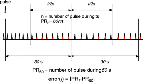
The error was simulated according to the following procedures. The number of pulses for ‘t’ seconds was assumed to be ‘n’; therefore, the pulse rate (PRt) was n × 60/t (bpm). For example, if 17 pulses were measured in 15 s, the rate (PR15) was 17 × 60/15 = 68 (bpm). The error (t) was thus defined as the absolute difference between PRt and PR60. Measurement duration (t) varied in the simulation between 6 and 60 s, and the average error at each duration was calculated.
HRV parameters
Interbeat interval signals were interpolated as 5 Hz equidistant data according to the instantaneous interbeat intervals method described by de Boer, Karemaker, and Strackee (Citation1985). Linear detrend and Hanning window were applied to the interpolated signals. Power spectra of HRV were calculated from 1024 points (204.8 s) of the interpolated interval sequences using fast Fourier transformation processing.
High-frequency (HF), low-frequency (LF), very low-frequency (VLF) and total-power (TP) components were obtained by integration of the power spectra at their respective ranges of 0.15–0.35, 0.04–0.15, 0.01–0.04 and 0.01–0.35 Hz. The natural logarithms of the HRV indexes (lnHF, lnLF, lnVLF and lnTP) were then calculated, because it has been reported that the raw HRV components indicate skewed distributions (Kobayashi, Park, and Miyazaki Citation2012). Characteristics of heartbeat recordings of the subjects in sitting position were summarised in Table .
Table 1 Summary of heartbeat recordings in sitting position (n = 70).
Results
Figure shows the continuously calculated error for durations from 6 to 60 s with subjects in the standing, sitting and supine positions. The error increased hyperbolically as the measurement duration decreased. The error was slightly lower in the supine position, although it was similar for the standing and sitting positions.
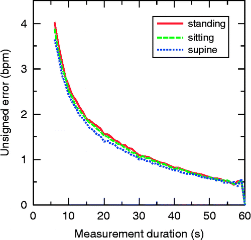
Table shows the average values and distribution characteristics of the error in standing, sitting and supine positions. For instance, the average error was 1.89 bpm with a measurement duration of 15 s in the sitting position. In this case, the probability of the error being greater than 4 and 7 bpm was 10% and 1%, respectively.
Table 2 Simulated errors in sitting, standing and supine positions (bpm).
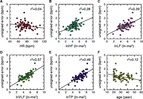
Correlations of error with age, heart rate and frequency components of HRV in the sitting position are shown in Figure . A negative correlation was observed between the error and subjects' age (r2 = 0.12). No relationship was found between the error and heart rate. Although a correlation was observed between the error and both LF and VLF components (r2 = 0.39 and 0.57, respectively), it was more apparent with VLF than with LF. Similar tendencies were observed in the standing and supine positions.
Discussion
Some studies have examined the relationship between measurement duration and the accuracy of pulse-counting. A representative study by Jones (Citation1970) quantified the errors of pulse-counting at 15, 30 and 60 s. In that study, 58 nurses and 30 nursing students counted human pulse rates by radial palpation, which were then compared with the rates simultaneously obtained using electrocardiography. The error was defined as the absolute difference between the two pulse rates. Because the measurement durations of these two counting methods were identical, the difference was attributed to human error. Hollerbach and Sneed (Citation1990) also conducted an experiment using similar procedures. In that study, the average absolute error was 3.372 bpm in pulse-counting for 15 s.
In the present study, the error was defined as the absolute difference between the simulated pulse rate counted for a given duration and the actual pulse rate counted for 60 s in healthy subjects. The definition of the error in pulse-counting was different between this study and the previous ones. In this study, the error was not a result of a human mistake but a direct result of the natural instability of human heartbeats. The errors presented in this study demonstrate the essential limit of accuracy due to limitation in data length.
The relation between HRV and measurement accuracy was also examined in this study. No correlation was found between error and heart rate, whereas a positive correlation was found between the error and the LF and VLF components of HRV. The correlation coefficient was higher in VLF than in LF. This result suggested that the error was the result of lower frequency fluctuation in heart rate.
In this study, greater accuracy was obtained with subjects in the supine position compared with the standing and sitting positions. This difference may be explained by the fact that in supine position, LF fluctuations are suppressed (Kobayashi Citation1996). Consequently, more constant and stable heart rates are observable in the supine position, resulting in better accuracy in pulse counting.
In this study, the error decreased with age, although the effect of this variable was small. This tendency may also be attributed to lower LF and VLF in elderly subjects. Smaller LF and VLF components have been reported in elderly people (Yeragani et al. Citation1997; Kuo et al. Citation1999; Ziegler et al. Citation1999; Agelink et al. Citation2001). Thus, we can expect errors to be smaller in elder people.
In this study, female subjects were not included. Previous studies on gender differences in LF fluctuations in HRV have been inconclusive. Some researchers have reported no gender differences in HRV (Kuo et al. Citation1999; Ziegler et al. Citation1999). In contrast, some researchers have reported larger LF (Liao et al. Citation1995; Agelink et al. Citation2001) or VLF (Jensen-Urstad et al. Citation1998) in males. Therefore, we can at least predict that the errors in females will not be significantly larger than the present results.
In this study, the mean value of the error was 1.88 bpm for a 15 s measurement in the sitting position. This relatively small value may seem acceptable; however, the percentile value of the error is more important in actual clinical situations. According to the results of this study, an error greater than 5 bpm will occur with a 5% probability when the pulse is counted for 15 s and multiplied by 4. Therefore, when a value of 70 bpm is obtained for a 15 s measurement, the 95% confidence interval is estimated to be 65–75 bpm. The SD of intra-individual variation of resting heart rate has been estimated to be 3% of the mean; thus, when the mean heart rate is 70 bpm, the SD is approximately 2 bpm (Kobayashi Citation2007). The author considers that the 15 s measurement is acceptable in clinical situations; however, it is open to discuss whether this is sufficiently accurate or not.
The present study examined the effect of HRV accuracy of pulse-counting. However, factors concerning human error were not taken into consideration. Medical practitioners should understand that the errors of pulse-counting in the actual situations will be larger than the results presented in this study. Future studies must examine the accuracy of pulse-counting considering both human factors and natural fluctuations in HRV.
References
- Agelink, M. W., T.Malessa, B.Baumann, T.Majewski, F.Akila, T.Zeit, and D.Ziegler. 2001. “Standardized Tests of Heart Rate Variability: Normal Ranges Obtained from 309 Healthy Humans, and Effects of Age, Gender, and Heart Rate.” Clinical Autonomic Research11: 99–108.
- de Boer, R. W., J. M.Karemaker, and J.Strackee. 1985. “Description of Heart-Rate Variability Data in Accordance with a Physiological Model for the Genesis of Heartbeats.” Psychophysiology22 (2): 147–154.
- Hollerbach, A. D., and N. V.Sneed. 1990. “Accuracy of Radial Pulse Assessment by Length of Counting Duration.” Heart & Lung19 (3): 258–264.
- Jensen-Urstad, M., K.Jensen-Urstad, M.Ericson, and J.Johansson. 1998. “Heart Rate Variability is Related to Leucocyte Count in Men and to Blood Lipoproteins in Women in a Healthy Population of 35-Year-Old Subjects.” Journal of Internal Medicine243 (1): 33–40.
- Jones, M. L.1970. “Accuracy of Pulse Rates Counted for Fifteen, Thirty and Sixty Seconds.” Military Medicine135 (12): 1127–1136.
- Kobayashi, H.1996. “Postural Effect on Respiratory Sinus Arrhythmia with Various Respiratory Frequencies.” Applied Human Science15 (2): 87–91.
- Kobayashi, H.2007. “Inter and Intra Individual Variations of Heart Rate Variability in Japanese Males.” Journal of Physiological Anthropology26 (2): 173–177.
- Kobayashi, H.2009. “Does Paced Breathing Improve the Reproducibility of Heart Rate Variability Measurements?” Journal of Physiological Anthropology28 (5): 225–230.
- Kobayashi, H., B. J.Park, and Y.Miyazaki. 2012. “Normative References of Heart Rate Variability and Salivary Alpha-Amylase in a Healthy Young Male Population.” Journal of Physiological Anthropology31: 9.
- Kuo, T. B., T.Lin, C. C.Yang, C. L.Li, C. F.Chen, and P.Chou. 1999. “Effect of Aging on Gender Differences in Neural Control of Heart Rate.” American Journal of Physiology Heart and Circulatory Physiology277 (6 Pt 2): H2233–H2239.
- Liao, D., R. W.Barnes, L. E.Chambless, R. J.Simpson, P.Sorlie, and G.Heiss. 1995. “Age, Race and Sex Differences in Autonomic Cardiac Function Measured by Spectral Analysis of Heart Rate Variability – The ARIC Study.” American Journal of Cardiology76: 906–912.
- Sayers, B. M.1973. “Analysis of Heart Rate Variability.” Ergonomics16 (1): 17–32.
- Smith, S. F., D. J.Duell, and B. C.Martin. 2003. Clinical Nursing Skills: Basic to Advanced Skills. 6th ed., 250–257. Upper Saddle River, NJ: Prentice Hall.
- Vanderlei, L. C. M., R. A.Silva, C. M.Pastre, F. M.Azevedo, and M. F.Godoy. 2008. “Comparison of the Polar S810i Monitor and the ECG for the Analysis of Heart Rate Variability in the Time and Frequency Domains.” Brazilian Journal of Medical and Biological Research41: 854–859.
- Yeragani, V. K., E.Sobolewski, J.Kay, V. C.Jampala, and G.Igel. 1997. “Effect of Age on Long-Term Heart Rate Variability.” Cardiovascular Research35: 35–42.
- Ziegler, D., K.Srasburger, H.Lambeck, and K.Dannehl. 1999. “Normal Ranges and Reproducibility of Statistical, Geometric, Frequency Domain, and Non-Linear Measures of 24-Hour Heart Rate Variability.” Hormone and Metabolic Research31: 672–679.