Abstract
Macro-autoradiographs of alpha emitters in environmental materials were obtained with solid-state nuclear track detector CR-39. This method is applicable only to alpha particles, and because their penetration ranges are very short, the obtained images are very clear. Since the method is simple and inexpensive, it can be used even at the high-school level. From the etch pit density obtained by microscopic observation, the radioactivity density of materials can be determined by using some theoretical calculations.
1. Introduction
A geometrical distribution image of radioactivity in a specimen is called a macro-autoradiograph. Such images of environmental materials are very helpful to understand natural radioactivity and natural radiations. With this intention, so far images of beta-ray emission from 40K, which is naturally present in vegetables, meat, and other environmental materials, have been obtained using an imaging plate [Citation1,Citation2]. These images have been widely used in public education. However, an expensive image reader is necessary to obtain such images.
A new method to obtain macro-autoradiographs of alpha emitters was developed by N. Ishigure [Citation3] who used a solid-state track detector CR-39 plate to shorten the exposure time compared to that required by an X-ray film, when he obtained plutonium distribution image in a mouse's body after the injection of plutonium in a vein. This method was considered to be adaptable to obtain natural alpha-particle emission images of environmental materials such as stone and ceramics [Citation4], although a very long exposure time would be necessary for these environmental materials contrary to N. Ishigure's intention to shorten the required exposure time by using artificial intense alpha-particle specimens. Such images would be very useful to understand natural radioactivity and natural radiations. Since this method is inexpensive and simple, it can easily be used even at the high-school level.
2. Characteristics and handling of the solid-state track detector CR-39
2.1. Procedure to obtain alpha-particle images with CR-39
An inexpensive solid-state track detector CR-39 plate, 280 × 280 × 0.9 mm in size, known as BARYOTRAK is commercially available through Nagase Landauar Ltd, Japan. For the present study, this plate was cut into 15 pieces. The detector is sensitive to alpha particles and insensitive to beta- and gamma-rays. Stone, ceramic ware, lead plate, and glass wares are appropriate specimens, because they emit natural alpha particles and the penetration ranges of alpha particles are very short to obtain very sharp images.
The exposure process is very simple and the specimen is simply placed on a CR-39 plate. It is not necessary to shield environmental radiations or light. However, the exposure time is as long as ∼2 or 3 months, because the alpha radioactivity of environmental specimens is very small. It is important to make contact the specimen surface with the plate surface to obtain a sharp image.
Alpha particle leaves a radiation damage trail called a latent track. The chemical etching enlarges it to be visible size under the microscope or naked eyes. In the present case, the CR-39 plate was immersed in caustic soda liquid (7.5N, 80°C) for 5 hours. Cone-shaped etch pits with diameter in the range of 10–100 μm appear on the plate surface. Since the exposure time is as long as 2–3 months, the surface density of the number of etch pits is large and the reflection of light by these dense etch pits allows observation of etch pits with the naked eyes. If the observation with an optical microscope is desired, the etch pit density must be small. Therefore, in this case, the exposure time was chosen to be as short as several days depending on the radioactivity of the specimen. Although there is a method to reduce the etch pit density by increasing the chemical etching time, this method involves large error in counting pit density.
2.2. Observation of the geometrical distribution images of alpha-particle emission
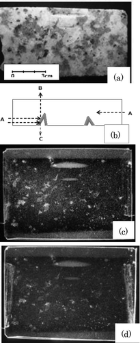
After chemical etching, the CR-39 plate was washed with water. The etch pit density image can be seen with the naked eyes. (a) shows a photograph of a piece of granite with a smooth surface. The granite was placed on the CR-39 plate for 3 months. After chemical etching, the plate was illuminated with light A (Figure (b)), and the photograph was taken. Figure (c) is obtained by observing scattered light B (Figure (b)) from the side opposite to the etch pit side (granite side) of the CR-39 plate. The white portions show areas having high density of alpha-particle etch pits. The image (d) is obtained by observing scattered light C (Figure (b)) from the same side as the etch pit side. Image (c) is clearer than (d). This difference is because of observation efficiency, i.e., the scattered light C shown in Figure (b) does not effectively appear owing to the multi-scattering by the rough surface of the etch pits.
Figure 1 (a) Photograph of a piece of granite with smooth surface. (b) Light illumination A and observation side B or C of CR-39. (c) Macro-autoradiograph of the granite alpha emitter obtained with CR-39 for three-month exposure. The image was observed from the side B, which is opposite to etch pit side in (b). White portions correspond to the large density of alpha-particle etch pits. (d) The image was observed from the etch pit side C in (b)
3. Obtained images
The radionuclides present in granite are thorium, uranium and their daughter nuclides. Therefore, the etch pits caused by alpha particles emitted from the radon gas emanated from granite and its daughter nuclides can be seen at outside the granite portion in Figure (c) and in the two fish-shape concaved portions on the granite surface. (a) is obtained, for comparison, with the imaging plate, which is sensitive to beta particles as well as alpha particles. The gray portions indicate beta-rays emitted from potash feldspar with the white color shown in Figure (a). The black points indicate alpha particles emitted from biotite with the black color shown in Figure (a). The position resolution of the imaging plate itself is around 100 μm and the beta-ray penetration range is much longer than the alpha-particle range; therefore, the position resolution of Figure (c) is far superior than that of Figure (a). Figure (b) shows an enlarged image of a part of Figure (c) demonstrating excellent position resolution.
Figure 2 (a) Alpha- and beta-ray image obtained with imaging plate. (b) Enlarged image of a part of Figure (c)
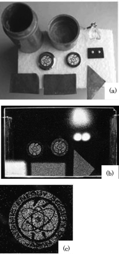
shows auto-radiographs of alpha emitters of various specimens. Figure (a) shows a photograph of specimens such as a lead container and cap, from upper left to right, uranium glass, two emblems of the Atomic Energy Society of Japan, two monazite spheres with a diameter of 2.5 mm, and three different lead plates. Figure (b) shows the auto-radiographs obtained by the exposure of specimens on CR-39 for 2 months. Since the surfaces of the lead container and cap were covered with white paint during the exposure time, there was no alpha-particle emission from them. The surfaces of uranium glass and monazite spheres were not flat, so that alpha particles emitted from a surface which did not directly contact with the CR-39 surface flew further and the images were not sharp. The monazite spheres contain thorium and uranium of 320 and 36 Bq/g, respectively [Citation5]. The alpha-particle emission of the lead plate on the left side was greater than that of other two lead plates on the right side depending on the elapsed time after refinery. Usually, 210Pb (RaD, half-life is 22.3 year) is included in lead and its daughter nuclide 210Po emits alpha particles. The emblem figure on the right side was enlarged as shown in Figure (c), and the letters “ATOMIC ENERGY SOCIETY OF JAPAN” can be read. The emblem is Cloisonné ware; thus, thorium, uranium, and their daughter nuclides are included.
Figure 3 (a) Photograph of various specimens: from the upper left, lead container and its cap, uranium glass, two emblems of the Atomic Energy Society of Japan, two monazite balls and three different lead plates. (b) Macro-autoradiograph of (a) obtained for two-month exposure. (c) Enlarged image of one of the emblem, in which letters can be read
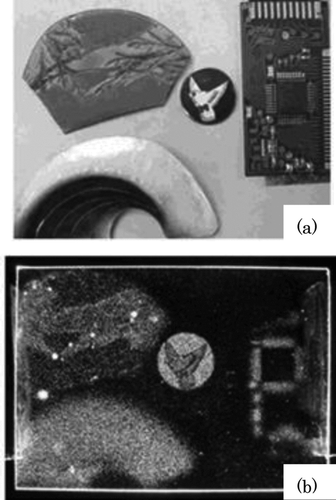
shows surrounding materials including two broaches, an emblem with a pigeon pattern, and an IC (integrated circuit) part of a broken electronic desk calculator. Figure (a) shows the photographs of these materials and Figure shows auto-radiographs of their alpha emitters. The wirings of the IC were attached with solder containing lead. Lead contains natural alpha emitters, as mentioned above, although recently manufactured solder does not contain lead. Pseudo-colored figures obtained by using “filter” software equipped in a personal computer can give beautiful images.
Figure 4 (a) Photograph of two personal ornaments, an emblem and soldered electronic IC parts. (b) Macro-autoradiograph of (a) obtained by the exposure for 2 months
4. Radioactivity determination of specimens
4.1. Relation between radioactivity and pit density
Determination of the radioactivity density (Bq/cm3) of one the specimen in Figure was made. However, the validity of the determination method itself was first confirmed as follows.
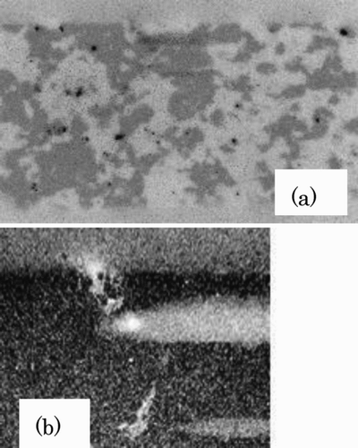
The radioactivity of an electro-deposit standard source, whose radioactivity was known and where the self-absorption of alpha particles could be neglected, was determined by measuring the etch pit density on the surface of CR-39. Then, the determined radioactivity was compared with the known radioactivity of the standard source. The standard source was RaDEF, which emits 210Po alpha particles with a radioactivity of 462 ± 2 Bq measured by Dr T. Yamada of the Japan Radioisotope Association.
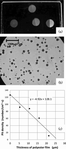
The half area of the source was covered with a polyester film of different thicknesses and the CR-39 surface was irradiated with alpha particles for different exposure times. A part of the result is shown in (a). The microscope image of a part of Figure (a) is shown in Figure (b), which is obtained by irradiating the CR-39 surface for 8-minute irradiation without the polyester film. The etch pit density N was 126.5 ± 6.3 (cm2 s)−1. The relation between the etch pit density and thickness of the polyester film is shown in Figure (c).
Figure 5 (a) Macro-autoradiographs of RaDEF alpha-particle standard source with known radioactivity, which were obtained with different exposure time. Half of the source surface was covered with polyester films with different thicknesses. (b) Microscopic observation of a part of (a). (c) Etch pit density against thickness of polyester film
Equation (Equation1 for determining the critical angle θc of the etch pit observation is obtained by transforming the equation shown by Price et al. [Citation6] with reference to other papers [Citation7,Citation8]
Then, the radioactivity S of the source with the area of 1.13 cm2 is determined from the etch pit density as follows:
This value is larger than the original radioactivity 462 ± 2 Bq by about 11%. The value S, however, contains around ±9.6% error, indicating that the validity of this method for determining the radioactivity of a plane source without self-absorption is confirmed.
4.2. Determination of radioactivity density of unknown specimens
As an example, the radioactivity density A (Bq/cm3) of the upper white part of the two wings of the pigeon emblem shown in Figure (b) is determined as follows.
The standard source mentioned in Section 4.1 is a plane source without the self-absorption of alpha particles. However, the unknown specimen is a bulk source, and thus it is necessary to determine the absorption of alpha particles in the specimen by calculating the energy loss of alpha particles.
The validity of using the Bethe equation [Citation9] for energy loss calculation was first examined. From Fig-ure (c), the polyester thickness at which the pit density is zero is experimentally determined to be 26.0 μm. On the other hand, from the Bethe equation (−dE/dx) the polyester thickness T at which alpha-particle energy E becomes 0.3 MeV can be determined by the following equation:
where E 0 is the incident initial energy of an alpha particle and (−dE/dx) P is the energy loss in polyester. It is assumed that an alpha particle with less than 0.3 MeV cannot make an etch pit on the CR-39 surface. The penetration range of an alpha particle with 0.3 MeV is only 2.2 μm in CR-39, and this seems to be a reasonable assumption with reference to Figure (b). The thickness T = 26.6 μm obtained by Equation (Equation3 is almost the same as the experimental value 26.0 μm, demonstrating that the calculation of energy loss using the Bethe equation is appropriate.
To determine the radioactivity A (Bq/cm3) of the upper white part of the two wings of the pigeon emblem shown in Figure (b), etch pit density N ((cm2 s)−1) must be determined experimentally. However, when Figure (b) was obtained with a two-month exposure, the etch pit density was very large and the number of etch pits was not countable separately. Therefore, the etch pit density N was obtained with a short exposure time (7 days) and the result was 0.083 (cm2 s)−1.
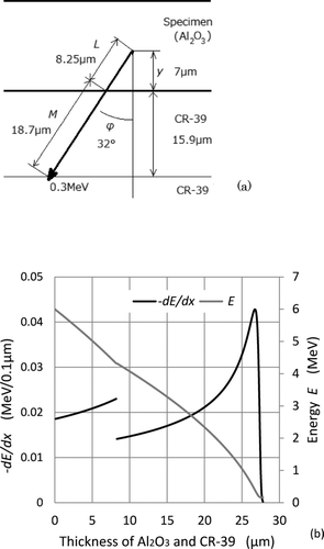
The relation between pit density N ((cm2 s)−1) and radioactivity A (Bq/cm3) was then theoretically determined to obtain A from N. The composition of the upper white part of the two wings of the pigeon emblem is almost the same as aluminum oxide (Al2O3). As shown in (a), an alpha particle emitted at depth y from the surface of the specimen with angle ϕ passes length L in Al2O3 and length M in CR-39. The angle ϕm (y) at which the energy Em becomes 0.3 MeV is determined whose procedure will be explained later. This energy is the threshold energy required to make an etch pit at a newly appeared surface of CR-39 after removing thickness 15.9 μm by chemical etching. This angle ϕm (y) is the maximum angle for the detection of an alpha particle emitted in 4π direction at depth y. Energy Em is determined by the following equation:
where (−dE/dx) U is the energy loss of alpha particle with incident energy E 0 in Al2O3 and (−dE/dx) C is the energy loss of alpha particle with incident energy EL in CR-39.
Figure 6 (a) Illustration of the calculation of maximum angle for the detection of an alpha particle emitted at depth y from the specimen surface. (b) Energy loss −dE/dx and energy against the thickness of Al 2 O3 of an alpha particle emitted at y = 7 μm with angle of ϕ = 32°
To obtain the maximum angle ϕm (y), an appropriate angle ϕ(y) is tentatively supposed. Then Em is calculated by Equation (Equation4. A few trial-and-error calculations yield Em = 0.3 MeV and ϕm (y). Finally, the detection efficiency η(y) for an alpha particle emitted in the 4π direction at depth y is obtained by the following equation:
As an example, when an alpha particle with 6 MeV emitted at depth y = 7 μm and Em = 0.3 MeV, the values of ϕ m (y), L, and M are determined to be 32°, 8.25 μm, and 18.7 μm, respectively, as shown in Figure (a) by the calculation mentioned above. In this case, energy loss (−dE/dx) and energy E are shown in Figure (b) for reference.
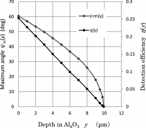
Such calculations give the relation between ϕ m (y), η(y), and y as shown in . The angle ϕ m (y) at y = 0 is 60.4° which gives the critical angle θc = 29.6°. The experimental value obtained by Equation (Equation1 is 26.2° which shows a fairly good agreement with the above calculated one.
Figure 7 Maximum angle ϕm (y) and detection efficiency η(y) against depth y in Al 2 O3
The relation between the etch pit density N ((cm2 s)−1) and the radioactivity A (Bq/cm3) of a specimen is given by
where r is the penetration range of alpha particle including the bulk etching length (= 15.9 μm) of CR-39. Calculations are performed using centimeter as the unit of measurement. The pit density is obtained using the same method as shown in Figure (b), and N is 0.083 (cm2 s)−1. Using the numerical calculation of η(y) in Figure , the value of the part of the integration in Equation (Equation6 is 0.13 × 10⁻³ cm. Then, A is determined to be 640 Bq/cm3.
Such calculation includes various errors: the error (around ±10%) mentioned in Section 4.1, the error (around ±20%) estimated for the deviation from the assumption that the specimen composition is Al2O3, and the error (around ±10%) estimated by the consideration that alpha particles have the energy of not only 6 MeV but also other energies. The total error would be around ±25%. Usually, radioactive equilibrium is maintained and the daughter nuclides of 232Th and 238U contribute in making the etch pits. The radioactivity of 232Th including 238U in the glaze of the upper white part of the wings of the pigeon emblem shown in Figure (b) is therefore considered to be around 1/5–1/6 of 640 Bq/cm3, which is more than 10 times of the average content of alpha emitter in ordinary granite.
4.3. Simplified equation to determine radioactivity in specimens
For a plane source of alpha particle in which self-absorption can be neglected, the relation between pit density N ((cm2 s)−1) and radioactivity density A (Bq/cm2) is shown by the following equation (with reference to Equation (Equation2):
For a bulk source, the relation between N and A (Bq/cm3) is shown by Equation (Equation8 which is obtained by referring to Equation (Equation6 and the following discussion:
There is almost a linear relation between detection efficiency η(y) and depth y, as shown in Figure . Therefore, the average detection efficiency is half of the detection efficiency (= (1 − sinθc ) at y = 0. Finally, radioactive density A ((cm2 s)−1) can be determined from etch pit density N ((cm2 s)−1) and critical angle θc . If angle 20° is chosen as a representative value [Citation10] for the critical angle, then the value of (1−sinθ c ) is nearly equal to 0.6, and we obtain the following equation:
5. Conclusions
Using solid-state track detector CR-39, we can obtain very sharp alpha-particle macro-autoradiographs that are very useful for understanding natural radioactivity and natural radiations. Since this method is very simple and inexpensive, it can be adapted in extra-curricular activities under the supervision of a teacher at a high-school or junior high-school level. At universities or research institutes, this method can be used for studying petrology. Simple equations are deduced to obtain radioactivity density in a specimen from the pit density obtained by microscopic observation.
Acknowledgements
This research was initiated as a joint effort between the Super Science High School project promoted by Mr Makoto Mishima of Okayama prefectural Ichinomiya High School and Mr Jun Kawai of the Mitsubishi Research Institute. Prof. Nobuhito Ishigure of Nagoya University provided the author with valuable comments on this study. Dr Takahiro Yamada of Japan Radioisotope Association determined the radioactivity of a RaDEF alpha-particle standard source and provided the author with an opportunity to use it. Prof. Takao Iida and Assoc. Prof. Jun Moriizumi of Nagoya University gave valuable advice on chemical etching of CR-39. The author would like to express his sincere thanks to each of them.
References
- Mori , C . 1999 . Measurement of extremely low level radioactivity distributions with imaging plate . Radioisotopes , 48 : 589 – 599 . Japanese
- Natural radiations through naked eyes . 1996 . Brochure of Chubu Atomic Power Conference . Chubu Atomic Power Conference . 1996 , Japan. pp. 8 – 9 . Chubu Genshiryoku Kondankai .
- Ishigure , N . 1985 . Application of CR-39 plastic to rapid and quantitative macro-autoradiography of α-emitters in a whole body section of an experimental animal . Radioisotopes , 34 : 101 – 102 .
- Mori , C . 2011 . Obtaining alpha-ray image of surrounding material with CR-39 . Paper presented at: Proceeding of 2011 Annual Meeting of the Atomic Energy Society of Japan . Mar 28–30 2011 , Japan. Vol. M20 , pp. 617 Fukui University . ISBN 978-4-89047-151-5 [CD-ROM]
- Mori , C . 2012 . [Theoretical calculation of the number of α-ray track in a Petri dish-type cloud chamber with monazite source] . Radioisotopes , 61 : 399 – 406 . Japanese
- Price , P B and Fleischer , R L . 1971 . Identification of energetic heavy nuclei with solid dielectric track detector: Application to astrophysical and planetary studies . Ann Rev Nucl Sci , 21 : 295 – 334 .
- Henke , R H , Ogura , K and Benton , E V . 1986 . Standard method for measurement of bulk etch in CR-39 . Nucl Tracks , 12 : 307 – 310 .
- Enomoto , H iroko and Ishigure , N obuhito . 1998 . [Aging and fading effects on registration properties for nuclear tracks in CR-39] . Hoken Butsuri , 33 : 407 – 413 . Japanese
- Evans , R D . 1955 . The atomic nucleus , 637 – 643 . New York : McGraw-Hill Book Company .
- Tsuruta , T . 1987 . [Investigation of interaction between acetic anhydride and aniline or its derivatives through continuous measurement of specific dielectric constant] . Ann. Rep. Kinki Univ At Energy Res Inst , 24 : 7 – 15 . Japanese