
Abstract
Boron neutron capture therapy (BNCT) is a promising cancer therapy. Epi-thermal neutron (0.5 eV < En < 10 keV) flux intensity is one of the basic characteristics for modern BNCT. In this work, based on the 71Ga(n,γ)72Ga reaction, a new simple monitor with gallium nitride (GaN) wafer as activation material was designed by Monte Carlo simulations to precisely measure the absolute integral flux intensity of epi-thermal neutrons especially for practical BNCT. In the monitor, a GaN wafer was positioned in the center of a polyethylene sphere as neutron moderator covered with cadmium (Cd) layer as thermal neutron absorber outside. The simulation results and related analysis indicated that the epi-thermal neutron flux intensity could be precisely measured by the presently designed monitor.
1. Introduction
Boron neutron capture therapy (BNCT) is a promising cancer therapy which kills tumor cells while suppressing exposure dose to normal tissues [Citation1]. In the early phase of BNCT, thermal neutrons (En < 0.5 eV) were primarily used for clinical studies in Japan to treat patients with brain tumors [Citation2] and cutaneous melanomas [Citation3]. However, since the kinetic energy of thermal neutrons is low, thermal neutrons could not reach sufficiently deep places in a human body. Operation thus became quite necessary for treating deep-seated tumors. In order to avoid operation risk, epi-thermal neutrons (0.5 eV < En < 10 keV) have been used in BNCT in recent years [Citation4–7] since they can penetrate deep inside tissues.
For modern BNCT, epi-thermal neutron flux intensity is one of the basic characteristics. However, it is quite difficult to measure it directly and accurately taking into account the neutron spectrum shape, because there are no suitable spectrometers to measure the neutron spectrum and no activation materials covering the epi-thermal neutron energy range appropriately. The objective of this work is hence to design a monitor using a suitable activation material to precisely measure the absolute integral flux intensity of epi-thermal neutrons especially for practical BNCT.
2. Design of monitor
2.1. Design requirements
Neutron source for BNCT produced in a nuclear reactor or in an accelerator-based neutron sources facility has energies normally distributing over a wide range, including thermal, epi-thermal and fast (En > 10 keV) neutrons, simultaneously. Thus, in order to precisely measure the absolute integral flux intensity of only epi-thermal neutrons, an ideal monitor to be designed in the present study should have a flat sensitivity to epi-thermal neutrons and no sensitivities to thermal and fast neutrons. On the other hand, the size of the monitor should be designed to be as small as possible so that it can be used in the neutron fields for practical BNCT, and hopefully, it would be expected to be used even on the surface of a patient. In addition, since practical BNCT needs very high neutron flux intensity, i.e., >109 n/sec/cm2, the monitor is required to be used in such high intensity neutron fields.
2.2. Materials
The principle of the epi-thermal neutron flux intensity monitor to be designed for BNCT in this work is based on the activation method with an appropriate activation material which is sensitive to epi-thermal neutrons. However, most activation materials have large sensitivity to thermal neutrons. Thus, it is necessary to reduce the contribution of thermal neutrons incident to the monitor. Cadmium (Cd) was selected in this work as the thermal neutron absorber, due to its availability, fewer activated products and lower cost. On the other hand, in order to precisely measure the absolute integral flux intensity of epi-thermal neutrons, effective neutron moderator is also required to obtain a flat sensitivity in the epi-thermal neutron energy range. For this purpose, polyethylene (CH2) was selected because it has high atomic density of hydrogen (H) and good productivity.
Then we had to choose a suitable activation material. After considering the characteristics of various nuclides, such as the amount of radioactivity, half-life and emitted main γ-rays’ energy, etc., we selected candidates including 197Au, 151Eu, 127I, 115In, 71Ga, 55Mn, 37Cl and 23Na owing to their higher feasibility to be used as activation materials for the monitor to be designed in this work. listed the information of the considered nuclides and corresponding activation reactions.
Table 1. Information of the considered nuclides and corresponding activation reactions.
2.3. Monte Carlo simulations
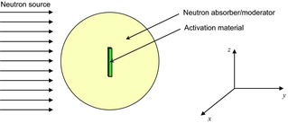
In order to estimate the amount of activities for the considered activation materials by neutrons, a Monte Carlo transport code, MCNP5 [Citation8], was employed to carry out design calculations of the monitor. The calculation model was shown in . A spherical shape was used as a monitor body to avoid dependence of neutron incidence direction to the monitor. It was assumed that the considered activation materials with conventional dimensions of 1.0 cm×1.0 cm and 0.01 cm in thickness were positioned in the center of a spherical neutron absorber/moderator, i.e., Cd/polyethylene in this work.
Figure 1. Calculation model for MCNP5.
In the Monte Carlo simulations, mono-energetic broad parallel neutron beams were used as neutron sources to obtain the monitor sensitivity curve as a function of neutron energy in the range from 0.01 eV to 10 MeV, in which each energy decade was divided into two bins so as to have an equal lethargy. Track length neutron tally was employed to estimate the average neutron flux in each cell of the monitor and the activation reaction rates were calculated directly with JENDL-4.0 [Citation9] and JENDL/A-96 [Citation10] as the activation cross section libraries. The number of source histories (more than 3.0×10⁷) was determined to satisfy the statistical error of the reaction rate for each energy bin below 5.0%.
3. Results and discussion
3.1. Determination of monitor configuration
Based on the MCNP5 calculation model shown in , two simple configurations can be used for the monitor to be designed in this work: (1) a considered activation material was positioned in the center of a polyethylene sphere covered with a Cd layer outside and (2) a considered activation material covered with a Cd layer was positioned in the center of a polyethylene sphere.
For the first configuration, thermal neutrons can be absorbed by the outside Cd layer, while epi-thermal neutrons can pass through it and be moderated by the polyethylene sphere. Thus it becomes possible to obtain a flat sensitivity to epi-thermal neutrons by adjusting the size of the polyethylene sphere (see section 3.3 in detail). However, in the case of the second one, epi-thermal neutrons can be moderated by the polyethylene sphere to become thermal neutrons and captured by the inside Cd layer, which is disadvantageous to an epi-thermal neutron flux intensity monitor. Thus, the first configuration of the monitor was finally selected in this work.
3.2. Selection of activation material
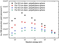
According to the simulation results of the considered nuclides listed in , it was found that the size of the neutron moderator (polyethylene sphere) was strongly dependent on the shape of the activation cross section curve, especially in the epi-thermal neutron energy range. Practically, if the activation cross section has an inverse velocity curve and drops faster with the neutron energy, a larger size polyethylene sphere is required to moderate neutrons with higher energy and makes the monitor more sensitive to higher energy neutrons. However, if a larger polyethylene sphere is employed, some of the lower energy neutrons will be over-moderated so that they cannot reach the activation material. It means the lower the neutron energy is, the lower the efficiency become. For instance, the cross section curve of the 197Au(n,γ)198Au reaction drawn based on the data taken from JENDL-4.0 [Citation9] was shown as the dashed line in , and the calculated sensitivities (in the form of 198Au production yields) in the epi-thermal neutron energy range for an Au foil monitor with different sizes of polyethylene sphere were shown in .
Figure 2. Cross section curves of the 71Ga(n,γ)72Ga and 197Au(n,γ)198Au reactions in the epi-thermal neutron energy range.
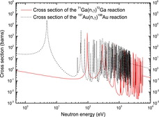
Figure 3. Calculated sensitivities (in the form of 198Au production yields) in the epi-thermal neutron energy range for an Au foil monitor with different sizes of polyethylene sphere. Here, the 198Au production yield is the number of 198Au atoms/cm3 produced from the 197Au(n,γ)198Au reaction.
Among the considered nuclides listed in , 71Ga shows the most promising overall trend of the activation cross section curve. The solid line in showed the cross section curve of the 71Ga(n,γ)72Ga reaction drawn based on the data taken from JENDL-4.0 [Citation9]. Compared to other considered nuclides, e.g., 197Au, the cross section curve of the 71Ga(n,γ)72Ga reaction drops more slowly with the neutron energy in the epi-thermal neutron energy range. This characteristic indicates that 71Ga has potential advantages over the other considered nuclides for finally designing an epi-thermal neutron flux intensity monitor for practical BNCT.
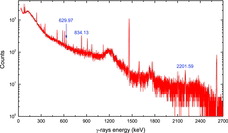
There are two stable isotopes of Ga: 69Ga (60.1%) and 71Ga (39.9%). Since the half-life of 72Ga (14.10 h) is sufficiently longer than that of 70Ga (21.14 min), the effect of 69Ga can be avoided in the measurement by waiting for the decay of 70Ga. Experimentally, it was found out that the main γ-rays emitted via the decay of 72Ga at 629.97, 834.13 and 2201.59 keV could be measured separately from background γ-rays with a high purity germanium (HPGe) detector, as shown in . However, since pure Ga metal cannot be flattened into a foil because of its poor malleability and since the melting point of Ga metal is 29.76 °C, just slightly above room temperature, gallium nitride (GaN) wafer, of which the melting point is greater than 2500 °C [Citation11], was finally selected as activation material in the present study due to its availability and very few activated products resulted from its nitrogen (N) content.
Figure 4. γ-ray spectrum measured by an HPGe detector after irradiating a Ga metal sample with an 241Am-Be neutron source. The main γ-rays emitted via the decay of 72Ga at 629.97, 834.13 and 2201.59 keV could be measured separately from background γ-rays.
3.3. Design results
After the configuration and activation material of the monitor were determined, a series of design calculations were carried out by MCNP5 to optimize the monitor configuration in size by varying the diameter of the polyethylene sphere and the thickness of the Cd layer, simultaneously.
The schematic view of the optimized configuration of the presently designed epi-thermal neutron flux intensity monitor for BNCT was illustrated in . Correspondingly, the calculated monitor sensitivities, in the form of 72Ga production yields, as a function of neutron energy in the range from 0.01 eV to 10 MeV were shown in . summarized the specification of the materials used for the monitor in this work.
Table 2. Specification of the materials used for the monitor in this work.
Figure 5. Schematic view of the optimized configuration of the presently designed epi-thermal neutron flux intensity monitor for BNCT. The monitor has a spherical body with a diameter of 7.12 cm. The left-hand side is the schematic view in y–z plane, and the right-hand side is the schematic view in x–z plane. Neutrons are incident along y-axis.
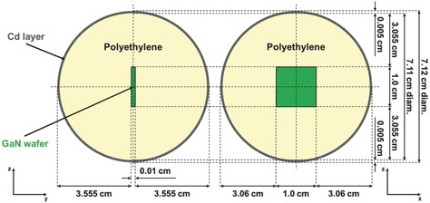
Figure 6. Calculated monitor sensitivities, in the form of 72Ga production yields, as a function of neutron energy in the range from 0.01 eV to 10 MeV. The fluctuation of 72Ga production yields in the epi-thermal neutron energy range was suppressed to be within ±4.6% relative to the average value. Here, the 72Ga production yield is the number of 72Ga atoms/cm3 produced from the 71Ga(n,γ)72Ga reaction.
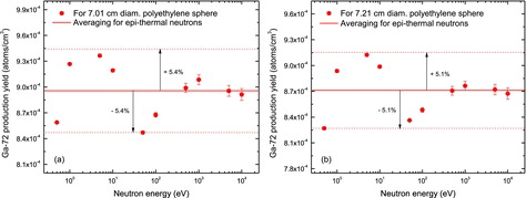
As shown in , the presently designed monitor with 7.11 cm diameter polyethylene sphere as neutron moderator had a flat sensitivity to epi-thermal neutrons, i.e., the fluctuation of 72Ga production yields in the epi-thermal neutron energy range was suppressed to be sufficiently small, within ±4.6% relative to the average value. If the diameter of the polyethylene sphere was changed to be thinner or thicker than 7.11 cm, such flat sensitivity would be destroyed (see section 3.2 for details). As examples, the calculated results in the epi-thermal neutron energy range for the GaN wafer monitor with 7.01 and 7.21 cm diameter polyethylene spheres were shown in (a) and (b), respectively.
Figure 7. Calculated results in the epi-thermal neutron energy range for the GaN wafer monitor with (a) 7.01 cm and (b) 7.21 cm diameter polyethylene spheres. The fluctuations of 72Ga production yields relative to the average values were within ±5.4% and ±5.1%, respectively. Here, the 72Ga production yield is the number of 72Ga atoms/cm3 produced from the 71Ga(n,γ)72Ga reaction.
On the other hand, it was demonstrated by the simulation results that the thickness of the Cd layer covering the monitor was a key factor for reducing the contribution of thermal neutron incident to the monitor. A thicker Cd layer could remove thermal neutrons more effectively. However, if the employed Cd layer was too thick, the epi-thermal neutrons between 0.5 and 1 eV would also be considerably removed since the neutron capture cross section of Cd in this energy range is not small. The simulation results shown in suggested that 0.005 cm was the most suitable thickness for the Cd layer covering the presently designed monitor.
From the above results and discussion, it was confirmed that the presently designed monitor had an optimized configuration and a flat sensitivity to epi-thermal neutrons. Moreover, the size of the monitor was small, i.e., suppressed to be around 7 cm in diameter. It was found that the absolute integral flux intensity of epi-thermal neutrons could be measured within 4.6% by the presently designed monitor.
However, strictly speaking, it was made clear that it was difficult to completely remove fast neutrons especially in the neutron energy range between 10 keV and 1 MeV for the presently designed monitor, as shown in . Thus, we have to investigate to find an effective way to reduce the contribution of fast neutron incident to the monitor in the next step. In addition, experimental activation tests with a prototype monitor will be carried out in an epi-thermal neutron field [Citation12] produced at the intense 14 MeV neutron source facility OKTAVIAN of Osaka University, Japan.
4. Conclusions
In the present work, based on the activation reaction of 71Ga(n,γ)72Ga, a new simple monitor with GaN wafer as activation material was designed by Monte Carlo simulations to precisely measure the absolute integral flux intensity of epi-thermal neutrons especially for practical BNCT. In the monitor, a GaN wafer with a dimension of 1.0 cm×1.0 cm and 0.01 cm in thickness was positioned in the center of a polyethylene sphere (7.11 cm in diameter) as neutron moderator covered with a 0.005-cm Cd layer as thermal neutron absorber outside. The simulation results and related analysis suggested that the monitor had a flat sensitivity to epi-thermal neutrons. In addition, the size of the monitor was small, i.e., around 7 cm in diameter. It was concluded that the epi-thermal neutron flux intensity could be measured within 4.6% by the presently designed monitor.
In the next step, we have to investigate an effective way to completely reduce the contribution of fast neutron incident to the monitor. Meanwhile, activation tests with a prototype monitor will be carried out experimentally.
Acknowledgements
This work is partly supported by Mitsubishi Heavy Industries Mechatronics Systems, Ltd. One of the authors, Xingcai Guan, is very grateful to the China Scholarship Council (CSC) for the financial support.
References
- Locher GL. Biological effects and therapeutic possibilities of neutrons. Am J Roentgenol. 1936; 36:1–13.
- Hatanaka H, Kamano S, Amano K, Hojo S, Sano K, Egawa S, Yasukochi H. Clinical experience of boron-neutron capture therapy for gliomas: a comparison with convectional chemo-immuno-radio-therapy. Niigata: Nishimura Co., Ltd.; 1986. p. 349–378.
- Mishima Y, Ichihashi M, Tsuji M, Hatta S, Ueda M, Honda C, Susuki T. Treatment of malignant melanoma by single thermal neutron capture therapy with melanoma-seeking 10B-compound. Lancet. 1989;2:388–389.
- Diaz AZ. Assessment of the results from the phase I/II boron neutron capture therapy trials at the Brookhaven National Laboratory from a clinician's point of view. J Neurooncol. 2003;62:101–109.
- Miyatake S, Kawabata S, Yokoyama K, Kuroiwa T, Michiue H, Sakurai Y, Kumada H, Suzuki M, Maruhashi A, Kirihata M, Ono K. Survival benefit of boron neutron capture therapy for recurrent malignant gliomas. J Neurooncol. 2009;91:199–206.
- Kawabata S, Miyatake S, Kuroiwa T, Yokoyama K, Doi A, Iida K, Miyata S, Nonoguchi N, Michiue H, Takahashi M, Inomata T, Imahori Y, Kirihata M, Sakurai Y, Maruhashi A, Kumada H, Ono K. Boron neutron capture therapy for newly diagnosed glioblastoma. J Radiat Res. 2009;50:51–60.
- Kankaanranta L, Seppälä T, Koivunoro H, Saarilahti K, Atula T, Collan J, Salli E, Kortesniemi M, Uusi-Simola J, Välimäki P, Mäkitie A, Seppänen M, Minn H, Revitzer H, Kouri M, Kotiluoto P, Seren T, Auterinen I, Savolainen S, Joensuu H. Boron neutron capture therapy in the treatment of locally recurred head-and-neck cancer: final analysis of a phase I/II trial. Int J Radiat Oncol Biol Phys. 2012;82:e67–75.
- X-5 Monte Carlo Team. MCNP – a general Monte Carlo n-particle transport code, Version 5 (LA-UR-03-1987). Los Alamos, NM: Los Alamos National Laboratory; 2003.
- Shibata K, Iwamoto O, Nakagawa T, Iwamoto N, Ichihara A, Kunieda S, Chiba S, Furutaka K, Otuka N, Ohsawa T, Murata T, Matsunobu H, Zukeran A, Kamada S, Katakura J. JENDL-4.0: a new library for nuclear science and engineering. J Nucl Sci Technol. 2001;48(1):1–30.
- Nakajima Y, JNDC WG on activation cross section data. JENDL activation cross section file. Proceedings of the 1990 Symposium on Nuclear Data; 1990 Nov. 29–30; Tokai (Japan); JAERI-M 91-032. Tokyo: Japan Atomic Energy Research Inst.; 1991. p. 43–57.
- Harafuji K, Tsuchiya T, Kawamura K. Molecular dynamics simulation for evaluating melting point of wurtzite-type GaN crystal. J Appl Phys. 2004;96:2501–2512.
- Tsubouchi K, Obata T, Murata I. Design calculation of epi-thermal neutron field with DT neutron source for low energy neutron spectrometer developed for BNCT. Proceedings of the 2013 Symposium on Nuclear Data; 2013 Nov. 14–15; Tsuruga (Japan). (to be published as a JAEA-Conference report).