
 ?Mathematical formulae have been encoded as MathML and are displayed in this HTML version using MathJax in order to improve their display. Uncheck the box to turn MathJax off. This feature requires Javascript. Click on a formula to zoom.
Abstract
Hastelloy-N alloys were irradiated by Xe+ with energies of 7 and 2 MeV at room temperature in order to investigate the effects of dose and dose rate on the change of micro-structure and nanohardness. Hardness was measured by nanoindentation, and micro-structure evolution and irradiation defects were identified by transmission electron microscopy (TEM) and positron annihilation coincidence Doppler broadening (CDB), respectively. The nanoindentation results showed that the hardness of alloys increased after irradiation, especially at a lower dose rate. At higher dose rate, TEM results indicated that irradiation defects were observed at first in this experiment, and the size of the defects increased, while their number density decreased as the dose increased. However, for a same total dose, the defects formed at a lower dose rate were larger, and their number density was also higher than that at a higher dose rate. Meanwhile, CDB analysis verified the growth of vacancy-type defects with increasing dose.
1. Introduction
As one of the six most promising Generation IV reactors, the thorium molten salt reactor (TMSR) has been attracting increasing attention. Recently, numerous materials have been qualified for use in the TMSR; however, since materials to be used in future commercial TMSRs need to exhibit corrosion and irradiation resistance, many researchers have focused efforts on developing such materials [Citation1–3].
Hastelloy-N alloy is selected as a candidate material for structural and loop materials in the TMSR because of several favorable properties such as high irradiation and corrosion resistance [Citation4–8] compared to austenitic and ferritic–martensitic steels [Citation9–12]. Many studies have reported the effect of corrosion and irradiation on Fe- and Zr-based alloys [Citation13–15]. However, studies dealing with the correlation between the micro-structure and mechanics of alloys after irradiation are rare. Allen et al. [Citation16] analyzed the effect of dose rate on radiation-induced segregation and observed that the segregation profiles strongly depend on the dose rate. By considering dose rate changes with the different radial distances from the reactor center, it is important to study the effect of the dose rate on the change of material properties at a given total dose. In this paper, the effect of irradiation dose and dose rate on the change in the micro-structure and nanohardness in Hastelloy-N alloy were studied, and presumably, some relationship was observed to exist between the micro-structure and mechanics.
2. Experimental
2.1. Material and sample preparation
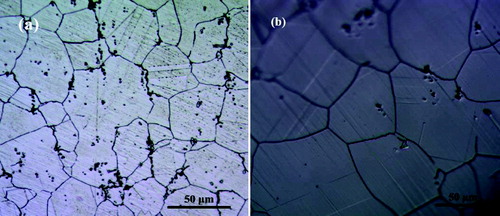
The raw Hastelloy-N alloy rod (2 cm diameter) was provided by the Institute of Metal Research, Chinese Academy of Sciences. lists its chemical composition. To homogenize the micro-structure of the alloy, solution heat treatment was conducted at the temperature of 1450 K for 30 min, after which the alloy was rapidly cooled in air. shows the difference in the metallographic structure before and after heat treatment. The strengthening phase around the grain boundary was fully dissolved after solution heat treatment.
Table 1. Chemical composition of the Hastelloy-N alloy.
Figure 1. Metallography of Hastelloy-N alloy: (a) before heat treatment and (b) after heat treatment.
After solution heat treatment, the alloy was cut into wafers of 1 mm thickness. Each wafer was wet-polished with 2000-grit silicon carbide paper, and then successively polished with 0.5 and 0.05 μm diamond paste. Finally, the polished alloys were sonicated with deionizer water for 15 min.
2.2. Heavy-ion irradiation
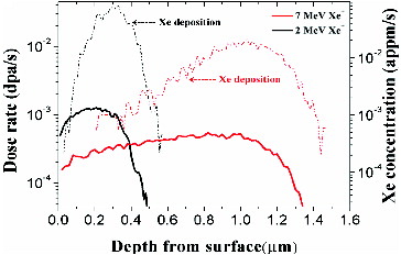
Ion irradiation was performed at the 320 kV highly charged ions comprehensive research platform of the Institute of Modern Physics (IMP), Chinese Academy of Sciences. Irradiation was conducted using 7.0 and 2.0 MeV Xe+ with average dose rates of approximately 3.0×10⁻⁴ and 1.3×10⁻³ dpa/s, respectively. Samples to be irradiated were fixed onto a large stainless steel target frame to rapidly transfer heat, and the temperature of the chamber was maintained at 300 K. Considering the increase in temperature caused by beam heating, the temperature of the samples did not exceed 400 K during irradiation. summarizes the details of irradiation. The vacuum was maintained below 2×10⁻⁵ Pa during implantation, and no visible change was observed in the sample surfaces after implantation. The doses and dose rates were calculated using the stopping and range of ions in the material (SRIM) 2010 program [Citation17], and a displacement energy of 40 eV was chosen for the calculation of dpa. The damage rate (dpa/s) at different depths of the alloy is shown in .
Table 2. Irradiation conditions at the Institute of Modern Physics.
Figure 2. Damage rate and Xe deposition with Xe+ implantation depth calculated by SRIM: black line represents the damage rate for high dose rate (1.3×10⁻³ dpa/s) and red line represents the damage rate for low dose rate (3.0×10⁻⁴ dpa/s).
2.3. Nanoindentation with continuous stiffness measurement
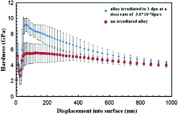
The nanohardness of the as-prepared and post-irradiated Hastelloy-N alloys was measured using a NanoIndenter G200 instrument. The continuous stiffness measurement mode was used to acquire hardness (H) as a function of depth. Nanoindentation nanohardness is defined as the indentation load divided by the projected contact area of indentation [Citation18]. It is a measure of the mean pressure by which a material can support under a given load. In our test, samples were mounted on aluminum stubs with hot wax, and the indents were produced in a direction normal to the sample surface. The load–displacement data for each preparation condition are obtained from arrays of six indents with a gap of 100 μm between the indents. All load–displacement data induced by a strain rate of 0.05/s were analyzed according to the method developed by Oliver and Pharr [Citation19]. As seen from , since the maximum implantation depth obtained by Xe-ion irradiation with energies of 7 and 2 MeV was about 1400 and 550 nm, respectively, the maximum indentation depth was controlled at 1000 nm, which can be observed in . Hardness data at an indentation depth of less than 50 nm were discarded because of the roundness of the indenter tip and oxidation of the surface [Citation18,Citation20]. Thus, the actual irradiation depth range was only selected from 50 to 150 nm to exclude the effects of the un-irradiated matrix below the damaged layer [Citation21].
Figure 3. Hardness versus indentation depth for Hastelloy-N alloy.
2.4. Coincidence Doppler broadening
Positron annihilation coincidence Doppler broadening (CDB) was conducted at the Institute of High Energy Physics, Chinese Academy of Sciences. 22Na was selected as the positron source, and the energy of the positron beam was varied from 0.18 to 20 keV. Positron Doppler broadening spectroscopy was employed to investigate the changes in the overall vacancy-type defect density induced by the heavy ion-irradiated Ni-based alloy. For CDB spectroscopy, the energies of annihilation γ rays shift because the momentum distribution of different electrons can be measured by high-purity Ge-detectors. The S-parameter is defined as the intensity of the γ rays energy around 511 keV and reflects the annihilation with low-momentum electrons [Citation22]. For a high vacancy concentration, the high relative probability of annihilation with low-momentum electrons brings about an increase in the S-parameter [Citation23,24].
2.5. Transmission electron microscopy
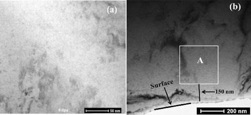
Micro-structures of the as-prepared and post-irradiated Hastelloy-N alloys were measured using FEI Tecnai G2F20 S-TWIN transmission electron microscopy (TEM) instrument. The preparation of the specimens was as follows. After irradiation, the alloy was cleaved into strips with a section of 2 mm×1 mm and the irradiated sides of two strips were glued to each other. Then, the glued sample was placed in a brass rod with an outer diameter of 3 mm. The sample with a thickness of about 1000 μm can be obtained by a wire cutting device, then the cleaved sample was reduced to a thickness of 20 μm using a SiC abrasive paper. Finally, a thickness of 200 nm was carried out by Ar+ bombardment before TEM measurement. (a) shows the TEM micrograph of the Hastelloy-N alloy before Xe+ implantation. Nearly no defects were observed in the observation field. The region A of (b) is a part of the irradiation zone at a depth of 150 nm under the surface, and the method of determining the observation area is the same for all irradiated alloys discussed later.
Figure 4. TEM micrograph of Hastelloy-N alloy: (a) before implantation and (b) irradiation with 7 MeV Xe+ at 0.5 dpa.
3. Results
3.1. Hardness measurements
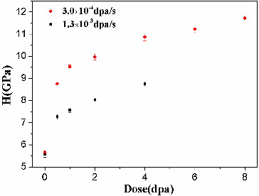
shows the difference of the average hardness for un-irradiated and irradiated alloys during indentation tests. The variation of hardness with dose was characterized by mean hardness. As shown in the figure, irradiation hardening was clearly observed after the irradiation dose increased from 0 to 0.5 dpa, while at a given total dose, greater hardening was observed in the Hastelloy-N alloys irradiated at a low dose rate.
Figure 5. Hardness of alloy as a function of irradiation dose at different dose rates.
3.2. CDB measurements
shows the changes of the S-parameter with the positron penetration depth at different doses. The mean depth of the annihilation positrons from the surface is calculated from the established Equation (1), where R is the implantation depth of the positron (nm), E0 is the incident energy of the positron (keV), and ρ is the density of the alloy (g/m3). As shown in the figure, the S-parameters of all the irradiated alloys were higher than that of the un-irradiated alloy, and the value increased with irradiation dose. This result indicates that high concentrations of vacancies were produced with high dose.
(1)
(1)
Figure 6. S-parameter as a function of depth in the alloys irradiated with different dose.
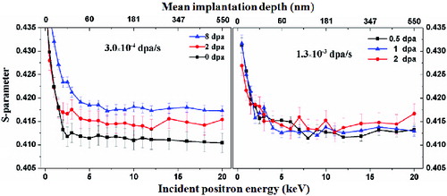
The S-parameter was high at an incident positron energy of lower than 3 keV, mainly caused by the annihilation of positrons on the surface [Citation25,26]. When the incident energy increases, the probability of the diffusion of positrons to the surface sharply reduces; thus, all the positrons will annihilate in the bulk of the alloy.
3.3. TEM observations
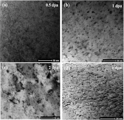
and show TEM micrographs of Hastelloy-N alloy under high and low dose rates, respectively. As shown in the micrographs, Xe+ irradiation significantly affected the micro-structure of the Hastelloy-N alloy. The density and diameter of defects caused by radiation damage at different irradiation conditions were estimated, they are summarized in .
Table 3. Number density and mean size of defects at the different irradiation conditions.
Figure 7. TEM micrograph of Hastelloy-N alloy irradiated by different Xe ion doses with a low dose rate. (a) Magnified image of region A of (b).
Figure 8. TEM micrograph of Hastelloy-N alloy irradiated by different Xe ion doses with a high dose rate.
For the low dose-rate irradiation, no visible defects were observed when the dose is 0.5 dpa ((a)). However, when the dose reached 1 dpa, many black dots with a density of about 3.3×10¹¹/cm2 and some defects with sizes varying from 2 nm appeared. On the other hand, when the total dose increased, radiation damage was quite different; that is, increase in size and decrease in density of defects with doses. Furthermore, as the dose increased to 4 dpa, the upper limit diameter of the defects increased to 12 nm ((d)), which is probably caused by the aggregation of vacancies or interstitials. The two main processes that occur during irradiation are generation and recovery of the defects. The irradiation temperature significantly affects the migration and recovery of the defects in particular, and as the temperature increased, the defects were more prone to migration and annihilation with each other. At an irradiation temperature of below 400 K, the dominant evolution processes of defects are recombination and migration of interstitial atoms. Considering the lower migration energy and higher diffusivity of interstitials at the irradiation temperature, the observed defects are conformed to be interstitial defects [Citation27,28].
Compared with that at low dose rates, radiation damages caused by high dose rate were visible when the dose reached 0.5 dpa ((a)). Similar to the size of the defects at low dose rates, the size of defects at high dose rates increased with increasing dose. In contrast to the density of defects at low dose rates, the density of the defects at high dose rates decreases with increasing dose; moreover, with the same dose, the density and size of defects were lower than those at low dose rates.
4. Discussion
The nanoindentation results revealed that the hardness of all alloys increased after irradiation and the greater hardening was observed for the alloy irradiated at a lower dose rate. The change in the mechanical properties of the alloy was determined by its micro-structural evolution. The vacancy and interstitial defects produced by irradiation and their migration were the primary factors for the growth of the defects, which finally caused irradiation hardening in alloys. The total dose would change the micro-structure of the Hastelloy-N alloy. As observed in and , more irradiation defects were produced when the total dose increased. Besides the total dose, the dose rate was also an important factor that affected the change in the micro-structure.
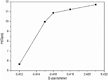
Both vacancy- and interstitial-type defects could affect the mechanical properties of Hastelloy-N alloy. To explore the effect of vacancy-type defects on mechanical properties, the change in hardness was studied as a function of the S-parameter. As shown in , hardness increased with the increase in the S-parameter, and the S-parameter for the un-irradiated sample was 0.412. However, the S-parameter for the irradiated sample only increased marginally from 0.412 to 0.421. The ΔS/S parameter (calculated from the established Equation (2)) for the most heavily irradiated sample was just 2.2%, suggesting that a few large-vacancy clusters exist after irradiation. According to the view of Makin, the region containing poverty atoms contributing to irradiation hardening is only confined to include above 10 vacancies [Citation29], the existence of vacancy defects could also harden the alloys; however, vacancy defects are not the main contributors in our experiments.
(2)
(2)
Figure 9. Hardness of alloy (irradiation with 7 MeV Xe+) as a function of the average S-parameter (in depth from 50 nm to 150 nm).
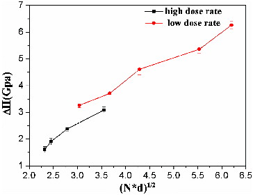
Besides the contribution of vacancy-type defects to irradiation hardening, dislocation loops, voids, and precipitates produced by irradiation also could promote irradiation hardening in Hastelloy-N alloy. These defects often act as barriers to dislocation glide and result in increased slip stress [Citation30,31]. As reviewed elsewhere [Citation32], dispersed hardening models predict that the change in the yield strength (σ) is proportional to the average obstacle spacing (N×d)1/2, where d is the average diameter and N is the number density of irradiation defects. Tabor's expression [Citation33] can be used to relate the nanohardness (H) to yield strength as follows: H ≈ 3σ. As shown in , a low dose rate would bring about more number of surviving defects and defects nucleated to clusters with larger size at the end of a cascade. According to the collision theory, the implantation of Xe ions into the alloy could displace atoms from crystal lattice (primary knocked atom), cause collision cascades, and create vacancies and interstitials. However, radiation damage generates point defects from a single cascade event, which are too small to be observed by TEM until such point defects interact with additional cascades, suggesting that the agglomeration of defects into visible clusters does not occur until doses at which cascade volumes begin to overlap. For a low irradiation dose rate, the timescale of the average duration for cascade overlap was much longer than that of defect migration and subsequent micro-structural evolution, suggesting that defect migration was stopped before the end of cascade overlap; in contrast, for a high dose rate, the timescale of the average duration for cascade overlap was shorter than that of the defect migration and subsequent micro-structural evolution, and the defects produced by the high dose rate have a priority to grow larger and hence are observed. A similar phenomenon was observed in the study by Hardie and Williams et al. with Fe-based alloys [Citation34]. shows the relationship between increased hardening ΔH (calculated from the established Equation (3)) and average obstacle spacing by considering the fact that the impact of the matrix is different at different irradiation conditions. The trend of lines agreed well with the prediction by the analytical model. The size of clusters at a high dose rate was smaller than that at a low dose rate, large size, and high number density of defects in the sample aggravate hardening.
(3)
(3)
Figure 10. Hardness as a function of average obstacle spacing.
5. Conclusions
The strengthening phase around the grain boundary of Hastelloy-N was fully dissolved after solution heat treatment. Nanoindentation results showed that all irradiated samples of Hastelloy-N alloy exhibited hardening, especially for alloys irradiated with a low dose rate. Irradiation caused the formation of vacancy and interstitial-type defects, which contribute to hardening. The S-parameter obtained from CDB measurement indicated that the concentration of vacancy-type defects increased as the dose increased, while a small increase of the S-parameter after irradiation suggests that the existence of interstitial-type defects, and not vacancy, was the main contributor to alloy hardening. TEM results showed that these sizes of the defects caused by irradiation increased, while their number density decreased, with increasing doses for all irradiated Hastelloy-N alloys. Experimental results agreed well with the prediction by the analytical model of radiation hardening, where yield strength is proportional to (N×d)1/2. Compared to the size of the defects produced at a high irradiation dose rate, that of the defects produced at a low irradiation dose rate was larger and their number density was higher. Large size and high number density of defects in the sample aggravate hardening.
Acknowledgements
The authors gratefully acknowledge the Institute of Modern Physics (IMP) for assistance in conducting Xe+ irradiation experiments. The authors also acknowledge the staff of the Key Laboratory of Nuclear Analysis Techniques, Institute of High Energy Physics, Chinese Academy of Sciences, for their assistance in conducting positron annihilation spectroscopy experiments.
Additional information
Funding
References
- Allen TR, Tan L, Was GS, Kenik EA. Thermal and radiation-induced segregation in model Ni-base alloys. J Nucl Mater. 2007;361:174–183.
- Azhazha VM, Bakai AS, Lavrinenko SD, Bobrov YP, V’yugov PN, Kovtun KV, Pylypenko MM, Savchenko VI, Solopikhin AD, Stetsenko SP, Malykhin DG. Alloys for molten-salt reactors. Radiat Damage Phys Radiat Mater Sci. 2005;87:40–47.
- Yamakawa K, Shimomura Y. Damage structures in fission-neutron irradiated Ni-based alloys at high temperatures. J Nucl Mater. 1999;264:319–326.
- Rowcliffe AF, Mansur LK, Hoelzer DT, Nanstad RK. Perspectives on radiation effects in nickel-base alloys for applications in advanced reactors. J Nucl Mater. 2009;392:341–352.
- Bakai AS. Combined effect of molten fluoride salt and irradiation on Ni-based alloys. Conference of the NATO Advanced Study Institute on Materials Issues for Generation IV Systems; 2007 Sep 24–Oct 06; France.
- Azhazha VM, Bakai AS, Bobrov Y, Virich VD, Yemlyaninova TG, Kapustin VL, Kovtun KV, Lavrinenko SD, Malykhin DG, Petel’guzov IA, Pylypenko MM, Savchenko VI, Semyonov NA, Solopikhin AD, Shirokov BM. The corrosion resistance of heatproof nickel alloy in molten fluoride salts. Radiat Damage Phys Radiat Mater Sci. 2005;87:74–81.
- Olson LC, Ambrosek JW, Sridharan K, Anderson MH, Allen TR. Materials corrosion in molten LiF–NaF–KF salt. J Fluor Chem. 2009;130:67–73.
- Jin SX, Guo LP, Yang Z, Fu DJ, Liu CS, Xiao W, Tang R, Liu FH, Qiao YX. Microstructural evolution in nickel alloy C-276 after Ar+ ion irradiation. Nucl Instrum Methods Phys Res B. 2011;269:209–215.
- Kondo M, Nagasaka T, Xu Q, Muroga T, Sagara A, Noda N, Ninomiya D, Nagura M, Suzuki A, Terai T, Fujii N. Corrosion characteristics of reduced activation ferritic steel, JLF-1 (8.92Cr–2W) in molten salts Flibe and Flinak. Fusion Eng Des. 2009;84:1081–1085.
- Meslin E, Radiguet B, Pareige P, Toffolon C, Barbu A. Irradiation-induced solute clustering in a low nickel FeMnNi ferritic alloy. Exp Mech. 2011;51:1453–1458.
- Dai Y, Henry J, Tong Z, Averty X, Malaplate J, Long B. Neutron/proton irradiation and He effects on the microstructure and mechanical properties of ferritic/martensitic steels T91 and EM10. J Nucl Mater. 2011;415:306–310.
- Jiao Z, Was GS. Segregation behavior in proton- and heavy-ion-irradiated ferritic–martensitic alloys. Acta Mater. 2011;59:4467–4481.
- Hengstler-Eger RM, Baldo P, Beck L, Dorner J, Ertl K, Hoffmann PB, Hugenschmidt C, Kirk MA, Petry W, Pikart P, Rempel A. Heavy ion irradiation induced dislocation loops in AREVA's M5 alloy. J Nucl Mater. 2012;423:170–182.
- Jiao ZJ, Shankar V, Was GS. Phase stability in proton and heavy ion irradiated ferritic-martensitic alloys. J Nucl Mater. 2011;419:52–62.
- Allen TR, Was GS, Kenik EA. The effect of alloy composition on radiation-induced segregation in Fe-Cr-Ni alloys. J Nucl Mater. 1997;244:278–294.
- Allen TR, Cole JI, Kenik EA, Was GS. Analyzing the effect of displacement rate on radiation-induced segregation in 304 and 316 stainless steels by examining irradiated EBR-II components and samples irradiated with protons. J Nucl Mater. 2008;376:169–173.
- Ziegler JF, Ziegler MD, Biersack JP. SRIM – the stopping and range of ions in matter. Nucl Instrum Methods Phys Res B. 2010;268:1818–1823.
- Li XD, Bhushan B. A review of nanoindentation continuous stiffness measurement technique and its applications. Mater Charact. 2002;48:11–36.
- Oliver WC, Pharr GM. Measurement of hardness and elastic modulus by instrumented indentation: advances in understanding and refinements to methodology. J Mater Res. 2004;19:3–20.
- Egeland GW, Valdez JA, Maloy SA, McClellan KJ, Sickafus KE, Bond GM. Heavy-ion irradiation defect accumulation in ZrN characterized by TEM, GIXRD, nanoindentation, and helium desorption. J Nucl Mater. 2013;435:77–87.
- Hardie CD, Roberts SG. Nanoindentation of model Fe–Cr alloys with self-ion irradiation. J Nucl Mater. 2013;433:174–179.
- Wan QM, Shu GG, Wang RS, Ding H, Peng X, Zhang Q, Lei J. Characterization of proton irradiation-induced defect in the A508-3 steel by slow positron beam. Nucl Instrum Methods Phys Res B. 2012;287:148–152.
- Lambrecht M, Malerba L. Positron annihilation spectroscopy on binary Fe–Cr alloys and ferritic/martensitic steels after neutron irradiation. Acta Mater. 2011;59:6547–6555.
- Yabuuchi A, Maekawa M, Kawasuso A. Vacancy defects in a stress-corrosion-cracked Type 304 stainless steel investigated by positron annihilation spectroscopy. J Nucl Mater. 2011;419:9–14.
- Jean YC, Zhang RW, Cao H, Yuan JP, Huang CM. Glass transition of polystyrene near the surface studied by slow-positron-annihilation spectroscopy. Phys Rev B. 1997;56:R8459–R8462.
- Uedono A, Tanigawa S, Sugiura J, Ogasawara M. A study of vacancy-type defects in B+-implanted SiO2/Si by a slow positron beam. Jpn J Appl Phys. 1989;28:1293–1297.
- Wharry JP, Jiao ZJ, Was GS. Application of the inverse Kirkendall model of radiation-induced segregation to ferritic–martensitic alloys. J Nucl Mater. 2012;425:117–124.
- Ehrhart P, Averback RS. Diffuse X-ray scattering studies of neutron- and electron-irradiated Ni, Cu and dilute alloys. Philos Mag A. 1989;60:283–306.
- Makin MJ, Minter FJ. Irradiation hardening in copper and nickel. Acta Met. 1960;8:691–699.
- Onimus F, Dupuy L, Mompiou F. In situ TEM observation of interactions between gliding dislocations and prismatic loops in Zr-ion irradiated zirconium alloys. Prog Nucl Energy. 2012;57:77–85.
- Gupta G, Jiao Z, Ham AN, Busby JT, Was GS. Microstructural evolution of proton irradiated T91. J Nucl Mater. 2006;351:162–173.
- Lucas GE. The evolution of mechanical property change in irradiated austenitic stainless steels. J Nucl Mater. 1993;206:287–305.
- Rodriguez R, Gutierrez I. Correlation between nanoindentation and tensile properties: influence of the indentation size effect. Mater Sci Eng A. 2003;361:377–384.
- Hardie CD, Williams CA, Xu S, Roberts SG. Effects of irradiation temperature and dose rate on the mechanical properties of self-ion implanted Fe and Fe–Cr alloys. J Nucl Mater. 2013;439:33–40.