
ABSTRACT
Two- and three-dimensional images were obtained by X-ray CT in the reaction product between zircaloy-2 cladding tube and MOX fuel. The gamma-ray intensity distributions in the same specimen were also obtained by gamma-ray measurements of two fission products (Cs-137 and Eu-154) and one neutron-activated nuclide (Co-60). The average values of the fuel density (about 10.5 g/cm3) and the cladding density (about 6.55 g/cm3) were obtained in the metallic phase region by evaluation of the density distributions on two-dimensional X-ray CT images. The distributions of the crushed fuel pellet and the pores were also clearly observed in the three-dimensional X-ray CT images. The following results were found from the gamma-ray measurement. First, Cs-137 was observed in the unreacted fuel region and the pore region in the metallic phase region. Second, Eu-154 was widely distributed to all regions. Finally, Co-60 was confirmed only in the metallic phase region.
1. Introduction
It has been predicted that core melting took place in reactor cores during the severe accident of the Fukushima Daiichi Nuclear Power Plant. Two cases are considered in the core melting; in case 1, both fuel and cladding melted, and, in case 2, only cladding melted and fuel did not melt. A study simulating the latter case was performed in a facility of JAEA, and the release behavior of fission products was investigated. The details of the experimental procedure and the results have been published elsewhere [Citation1].
In the present study, two- and three-dimensional images were obtained by X-ray CT in the reaction product formed in the previous study. In addition, the distributions of density and fission products in the specimen were evaluated by X-ray CT and gamma-ray measurement of the fission products Cs-137 and Eu-154. In addition, the distribution of the neutron-activated nuclide, Co-60, was measured.
2. Experimental
In the previous study [Citation1], pieces of zircaloy-2 cladding tube were melted on the fuel irradiated to about 40,000 MWd/t in the heavy water reactor, FUGEN. Details of the experimental procedure have been given in the previous paper [Citation1].
In the present study, X-ray CT and gamma-ray measurement were applied to the photographing of two- and three-dimensional images and the analyses of distributions of density and fission products in the reaction product formed in the previous study.
A process flow sheet from the preparation of the specimen to measurement by X-ray CT is shown in . Additionally, the main specifications of the X-ray CT apparatus are shown in .
Figure 1. Process flow sheet showing steps from specimen preparation to X-ray CT measurement.
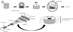
Table 1. Main specifications of the X-ray CT apparatus.
The X-ray CT measurements were made at intervals of 1 mm along the axial direction of the metal holder containing the reaction product specimen. The X-ray source was obtained from a linear electron accelerator having an electron energy of 9 MeV, and silicon semiconductor detectors with 100 channels were used for the measurement of X-ray intensities. The X-ray CT image was obtained in a cross section by rotating the X-ray CT apparatus around the specimen and subsequently moving in the parallel direction. In the X-ray CT image, the density increases with the increase of brightness. The density distribution in the specimen was evaluated from the relationship between brightness value (CT value) and densities published in other paper [Citation2].
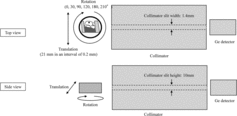
schematically illustrates the gamma-ray measurement of Cs-137, Eu-154, and Co-60. The gamma-rays were measured using a high-purity germanium detector (Canberra). In this measurement, the detector window was 10 mm in width and 14 mm in length, which was larger than the specimen thickness. The distance between the specimen and the detector was 2209.3 mm. The specimen was rotated in 6 angles (0°, 30°, 90°, 120°, 180°, 210°) and was axially moved along the slit for a distance of 21 mm at intervals of 0.2 mm for each rotated position. The radial distributions of nuclides were analyzed using the cross section nuclide analysis program arranged based on the MART [Citation3].
Figure 2. Schematic views of the gamma-ray measurement.
3. Results and discussion
In order to understand the obtained X-ray CT image deeply, the optical image was taken by a low-magnification camera. shows the comparison between X-ray CT image and the optical image. From the observation of the specimen in the previous study [Citation1], the gray region in the upper side of the specimen enclosed by dotted-line corresponds to the metallic phase region, while the dark region in the lower side of it corresponds to the unreacted fuel region.
Figure 3. Optical image and X-ray CT image.
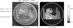
In the X-ray CT image, the high-density regions are observed on the unreacted fuel region. The density of these regions is evaluated about 10 g/cm3. It can be assumed from this result that these regions correspond to the roughly crushed fuel pellets, although this dispersion cannot be confirmed by the optical image. The density of the region, excluding the high-density regions in the unreacted fuel region, is about 8 g/cm3. It is assumed that these regions correspond to the accumulation of finely crushed fuel pellets.
In the metallic phase region of molten zircaloy, many pores were observed on both the X-ray CT image and the optical image. The density in the metallic phase region is about 8 g/cm3 and higher than that of the zircaloy-2 cladding tube (about 6.55 g/cm3). It is considered from this result that the molten zircaloy was mixed with the fuel having high density. In regions other than the upper and right regions, the connection of large pores and an accumulation of relatively large pores were observed.
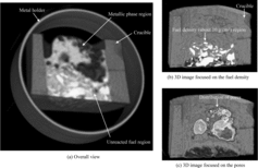
In order to confirm the occurrence of the above behavior, three-dimensional X-ray CT images were taken. shows the three-dimensional X-ray CT image (3D image) of the holder containing the reaction product specimen. This 3D image was composed of many X-ray CT images obtained on a number of transverse cross sections along the axial direction at the intervals of 1 mm. The overall 3D image that includes the metal holder is shown in (a). The X-ray CT image focused on the fuel density (about 10 g/cm3) in the unreacted fuel region is shown in (b). From this image, it is found that several crushed pellets were dispersed in the specimen. (c) shows the X-ray CT image for the metallic phase region above the unreacted fuel region. This image clearly showed three-dimensional distributions of the pores.
Figure 4. 3D images of the specimen.
shows the distributions of Cs-137, Eu-154, and Co-60 in the internal region of the specimen obtained by gamma-ray measurement with the X-ray CT images. The fission product nuclides, Cs-137 and Eu-154, were observed mainly in the unreacted fuel region, while the neutron-activated nuclide, Co-60, was located mainly in the metallic phase region. In addition, Cs-137 was accumulated in the pores of the metallic phase region, and Eu-154 was distributed widely in the metallic region.
Figure 5. X-ray CT image and gamma-ray intensity distributions.
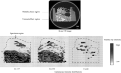
High release rates of cesium from the fuel have been reported at high temperatures in several studies [Citation4–7]. It is presumed from this reason that the cesium released from fuel is condensed in the pores when the molten zircaloy was solidified.
Eu-154 is produced in the fuel by fission and dissolved into it as oxide. Europium oxide is the stable oxide [Citation8–10], and the melting and boiling points are reported as 2050 ± 30 °C and 3790 °C, respectively [Citation11–14]. The chemical reaction between fuel and cladding takes place beyond about 1800 °C [Citation15–17], and then the europium oxide contained in the irradiated fuel partially moves into the molten zircaloy. Therefore, it seems that Eu-154 of fission product was dispersed in the metallic phase region as shown in .
4. Summary
Two- and three-dimensional X-ray CT images were obtained for the reaction product formed between several pieces of cut zircaloy-2 cladding tube and MOX fuel irradiated to 40,000 MWd/t. In addition, the distributions of Cs-137, Eu-154, and Co-60 were measured in this reaction product by gamma-ray measurement.
It was found from the results of two-dimensional X-ray CT images how the density, pores, molten cladding, and crushed pellets were distributed on the surface of the reaction product. From three-dimensional X-ray CT images, the detailed shapes of crushed pellets and pores can be observed with reasonable clarity. In addition, the distributions of Cs-137, Eu-154, and Co-60 in the reaction product could be measured. Eu-154 produced into the fuel is found in the metallic phase region composed chiefly of the molten zircaloy.
The elucidation of the process and these results are expected to assist in the analyses of debris taken from the molten cores of the Fukushima Daiichi reactors.
Acknowledgments
This work is supported by Alpha-Gamma Facility of Japan Atomic Energy Agency, E&E Techno Service Company Ltd. and Inspection Development Company Ltd.
Disclosure statement
No potential conflict of interest was reported by the authors.
References
- Tanaka K, Miwa S, Sato I, et al. Effects of interaction between molten zircaloy and irradiated MOX fuel on the fission product release behavior. J Nucl Sci Technol. 2014;51:876–885.
- Ishimi A, Katsuyama K, Maeda K, et al. Upgrading of X-ray CT technology for analyses of irradiated FBR MOX fuel. J Nucl Sci Technol. 2012;49:1144–1155.
- Minerbo GN, Sanderson JG. Reconstruction of a source from a few (2 or 3) projections [ART, CART, and MART, based on algebraic reconstruction technique; in FORTRAN]. Los Alamos (NM): Los Alamos Scientific Laboratory; 1977. ( DOE publication; no. LA-6747-MS).
- Walker CT, Bagger C, Mogensen M. Observations on the release of cesium from UO2 fuel. J Nucl Mater. 1996;240:32–42.
- Iglesias FC, Lewis BL, Reid PJ, et al. Fission product release mechanisms during reactor accident conditions. J Nucl Mater. 1999;270:21–38.
- Lewis BJ, Dickson R, Iglesias FC, et al. Overview of experimental programs on core melt progression and fission product release behavior. J Nucl Mater. 2008;380:126–143.
- Hidaka A. Outcome of VEGA program on radionuclide release from irradiated fuel under severe accident conditions. J Nucl Sci Technol. 2011;48(1):85–102.
- Johansson B. The boiling points of the rare earth metals. J Phys F Metal Phys. 1975;5:1241–1247.
- Grossman LN, Lewis JE, Rooney DM. The system UO2–EU2O3 at high temperatures. J Nucl Mater. 1967;21:302–309.
- Kleykamp H. The chemical state of the fission products in oxide fuels. J Nucl Mater. 1985;131:221–246.
- Krishnan RV, Panneerselvam G, Antony MP, et al. Solubility studies and thermal expansion coefficient of uranium–lanthanum mixed oxide system. J Nucl Mater. 2010;403:25–31.
- Ploetz GL, Krystyniak CW, Dumas HE. Sintering characteristics of rare-earth oxides. J Am Ceram Soc. 1958;41(12):551–554.
- Curtis CE, Tharp AG. Ceramic properties of europium oxide. J Am Ceram Soc. 1959;42(3):151–156.
- Adachi G, Imanaka N. The binary rare earth oxides. Chem Rev. 1998;98(4):1479–1514.
- Hofmann P, Kerwin-peck D, Nikolopoulos P. Explanation of the UO2/zircaloy-4 reaction layer sequence in terms of the total interfacial energy of the system. J Nucl Mater. 1984;124:114–119.
- Wilhelm AN, Garcia EA. Simulation of the kinetics of the UO2 dissolution by molten zircaloy-4 between 1950 and 2250°C. J Nucl Mater. 1988;158:143–158.
- Hofmann P. Current knowledge on core degradation phenomena, a review. J Nucl Mater. 1999;270:194–211.