
ABSTRACT
Tungsten (W) has been proposed as a plasma-facing material in fusion reactors due to its outstanding properties. Degradation of the material properties is expected to occur as a result of hydrogen (H) isotope permeation and trapping in W. In this study, two polycrystalline W plates were implanted with 80 keV H2+ ions to a fluence of 2 × 10²¹ H+/m2 at room temperature (RT). Time-of-flight secondary ion mass spectrometry (ToF-SIMS), focused ion beam (FIB), and scanning electron microscopy (SEM) were used for sample characterization. The SIMS data shows that H atoms are distributed well beyond the ion projected range. Isochronal annealing appears to suggest two H release stages that might be associated with the reported activation energies. H release at RT was observed between days 10 and 70 following ion implantation, and the level was maintained over the next 60 days. In addition, FIB/SEM results exhibit H2 blister formation near the surface of the as-implanted W. The blister distribution remains unchanged after thermal annealing up to 600 ˚C.
1. Introduction
Tungsten (W) is considered as a leading candidate for the plasma-facing material of fusion reactors due to its outstanding properties, such as high melting point and low erosion rate. The material will be subject to high-flux D+, T+, and He+ ion irradiation and implantation. The implanted hydrogen (H) isotopes can permeate into bulk and be trapped at defects and impurities, which can lead to degradation of W properties over the time. Extensive research on H retention and trapping in W has been reported [Citation1–4], where ion-induced defects are one of the important contributors. Low energy (10 eV to 10 keV) and high fluence (1021–1027ions/m2) H isotope ions [Citation5,Citation6] have been applied to simulate the irradiation conditions in fusion reactors. In addition, He ions, neutrons, and high-energy H ions have also been used in the experimental studies [Citation7,Citation8]. The results from the surface topography and depth profile in H implanted tungsten [Citation9] as well as the damage effects on H retention in self-ion irradiated W [Citation8,Citation9] showed that a high level of lattice damage can enhance H retention in W and modify the W surface.
A number of techniques have been applied for characterizing the H distribution and retention in ion-implanted samples. Nuclear reaction analysis (NRA) and elastic recoil detection analysis can analyze the H depth profiles, but their depth resolution and detection sensitivity are limited. Thermal desorption spectroscopy (TDS) is regarded as a useful tool to obtain desorption behavior of H molecules, but the technique does not measure H distribution in samples. A method capable of providing H distribution with both high depth resolution and high detection sensitivity is needed for a detailed study of H diffusion and trapping. Secondary ion mass spectrometry (SIMS) has been applied to analyze H in W in previous studies [Citation1]. More recent efforts [Citation10,Citation11] have demonstrated that time-of-flight secondary ion mass spectrometry (ToF-SIMS) is a suitable tool for H analysis with high sensitivity (10–100 ppm detection limit) and decent depth resolution (several nanometers). In this study, in situ ToF-SIMS was used to measure H depth profiles in a polycrystalline W during thermal annealing up to 600 °C. Focused ion beam combined with scanning electron microscopy (FIB/SEM) were also used to study H2 blistering.
2. Experimental procedures
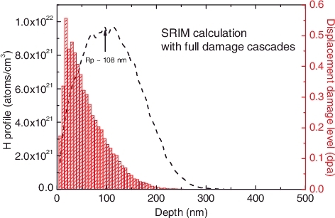
The sample in this study was a hot-rolled polycrystalline tungsten plate with a purity of 99.98%. One side of the plate was mechanically polished to a mirror-like surface. The plate with a dimension of 10 mm × 15 mm × 0.85 mm was then cut into two samples of equal areas. SEM was used to examine the as-polished samples, and clear grains with an approximate size of ∼10 µm were observed. A pre-check was applied using ToF-SIMS and showed very low contaminations on the surface. Ion implantation was performed with 80 keV H2+ beam to a fluence of 2 × 10²¹ H+/m2 at an ion flux of 2.4 × 10¹⁷ H+/(m2 s). The molecular ions, equivalent to 40 keV H+, were chosen to maximize the beam current for ion implantation. The incident angle was 60° and the implantation temperature was nominally room temperature (RT). The implantation was performed over a half of each of the two plates with the unimplanted area as a reference. A simulation based on SRIM code [Citation12] was performed with the displacement threshold energy of 90 eV [Citation13] and the incident angle of 60°. The results indicate that under the irradiation conditions, a shallow damage peak with the maximum dose of 0.5 displacements per atom (dpa) at 20 nm and a broader H profiles peaked at ∼108 nm are resulted in W, as shown in .
Figure 1. H depth distribution (black curve) and displacement per atom (red bars) simulated with SRIM-2012 for tungsten irradiated with 40 keV H+ ions to a fluence of 2 × 10²¹ H+/m2, assuming a displacement threshold energy of 90 eV.
ToF-SIMS depth profiling was performed within the Environmental Molecular Sciences Laboratory at the Pacific Northwest National Laboratory using a TOF-SIMS 5 spectrometer (IONTOF GmbH, Müster, Germany). The W sample was mounted on a heating/cooling sample holder during depth profiling analysis. The sample temperature could be controlled from −150 °C to + 600 °C with a precision of +/− 1 °C. A thermocouple was clamped on the surface of the W sample to monitor the temperature during the measurement. A 2.0 keV Cs+ beam of about 60 nA was used as the sputtering beam due to its great enhancement of negative ion yields. The incident angle was 45° and the sputtering area was set to be 200 × 200 µm2. A 25 keV pulsed Bi+ beam was used as the analytical beam, which was rastered over a 50 × 50 µm2 area at the center of the Cs+ sputtered crater. Negative secondary ions were collected because H− is more sensitive than H+ [Citation10,Citation11].
One of the samples was isochronally annealed at 100, 150, 200, 300, 400, 500, and 600 °C for 60 min each. The sample was cooled down to 100 °C after each annealing step before H− depth profile was measured by ToF-SIMS. The heating/cooling rate was controlled to be ∼1 °C per second. It should be noted that the in situ annealing SIMS in this study is preferred because the H− ion yield could be affected by experimental conditions, including base vacuum and sample temperature. For the other sample, H− depth profiles were performed at RT in 10, 70, and 130 days, respectively. The same annealing experiment was subsequently performed, except that the analysis temperature was set to 25 °C. At RT, the base vacuum of the ToF-SIMS analysis chamber was better than 3 × 10⁻⁹ mbar. During depth profiling measurement, the vacuum slightly increased but was still better than 8 × 10⁻⁹ mbar. Surface topography and microstructure of the cross section on the samples were characterized by FIB/SEM at Lanzhou University. The FIB/SEM (LYRA3, TESCAN) instrument provides a dual beam system and combines a high-resolution FE-SEM column with a versatile high-performance gallium (Ga) ion source FIB. The lateral resolution was better than 100 nm. Prior to the FIB cutting, a 0.2 μm thick platinum was deposited for surface protection. SEM was performed at a tilting angle of 55°.
3. Results and discussion
3.1. H diffusion and retention in W at RT
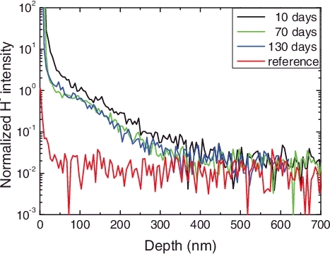
The H depth profiles in a H2+ implanted sample after 10, 70, and 130 days of storage at RT are shown in . The H intensity was very high near the surface region (0–30 nm), partly because of the surface roughness and absorption. No apparent H peak was found at the ion projected range of ∼108 nm (), but the H intensity in the sample gradually decreases over the depths from 30 to 450 nm. This behavior is consistent with a recent ion-channeling study [Citation14] that did not show an interstitial peak in tungsten after self-ion irradiation at RT. A previous study [Citation15] based on NRA indicated that H atoms implanted in tungsten to fluences higher than 5 × 10²³ ions/m2 could diffuse into the bulk for several micrometers. ToF-SIMS has a higher H detection sensitivity (∼10−4 at. % [Citation16]) than NRA (∼10−3 at. % in [Citation15]), but such a long H tail is not observed in this study due to a much lower H concentration. From , about 31% ± 2% of the retained H atoms in the implanted W were released spontaneously at RT from days 10 to 70 after implantation. The release rate in the near-surface region was slightly higher. H profile remains nearly unchanged in the next 60 days, indicating that the rest of the H atoms could hardly be de-trapped at RT. The released H could originate mainly from interstitials or weakly trapped atoms in W. Based on a reported diffusion coefficient D = 3 × 10⁻⁷ exp (−0.39 eV/kT) m2/s at temperatures between 25 and 80 °C [Citation17], more than 32% of the interstitial H atoms are expected to release within 1 h after implantation. The diffusion coefficients of both interstitials and trapped H atoms can vary significantly with defect type and concentration. It was reported [Citation1] that the concentration of deuterium atoms in single-crystal tungsten implanted with 6 keV D+ ions at RT decreased considerably beyond the implantation range from 30 min to 64 h after implantation. The saturated concentration of 42 at.% D implanted into tungsten was found to decrease to 3 at.% D after one-year storage at RT [Citation18]. The data in suggest that there is little change in the H depth profiles after sample storage between 70 and 130 days at RT.
Figure 2. H depth profile in as-implanted W after 10, 70, and 130 days at RT. H− Intensity was normalized to 186W−. The un-implanted H profile is shown in red as a reference.
3.2. Isochronal annealing behavior
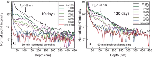
The H depth profiles in the samples stored at RT for 10 and 130 days as a function of annealing temperature are shown in for comparison. There is a faster H release at temperatures from 200 to 300 °C and from 400 to 500 °C in both samples during one-hour isochronal annealing. A very low H concentration was still detected after annealing at 600 °C. The general H behavior is similar for the two samples in spite of their different initial H concentrations. However, a rapid H decrease occurs from 25 to 100 °C in the 10-day stored sample. In contrast, the H profile in the 130-day-stored sample remains nearly unchanged after the same thermal annealing. It also appears that H diffused from the implanted to unimplanted area (reference sample) after 130 days, leading to a higher H concentration in the latter sample.
Figure 3. H distribution as a function of temperature in the 60-min isochronal annealing of the samples stored at RT for (a) 10 and (b) 130 days after ion implantation. H− Intensity was normalized to 186W−. Rp is the projected range from SRIM simulation.
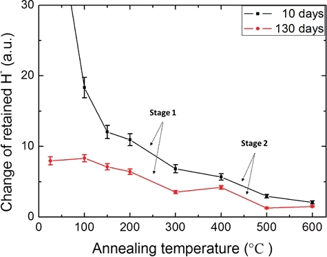
shows the integrated H intensities in the region of depths from 30 to 450 nm. A rapid decrease of H is observed below 150 °C for the 10-day stored sample and a much more gradual decrease for the 130-day stored one. For both samples, there appeared to be two release stages from 200 to 300 °C and from 400 to 500 °C. At other temperatures, the H retention changed less significantly. Moreover, additional information of different release rates during the annealing is indicated. The H retention in this study is generally low but is still detectable by ToF-SIMS. The possible release stages may be associated with two of the reported trapping energies [Citation19,Citation20] (discussed below). The results in are generally consistent with previous TDS studies [Citation20,Citation21], but the locations of the release peaks cannot be accurately determined due to the large temperature steps used in this study. TDS usually provides a much higher heating rate (from 1 to 10 K/s) and is well suited to analyze H desorption peaks. However, the analysis is likely a dynamic process and two factors may affect the TDS results. One is the heating rate that can lead to deviation of trapping peaks [Citation22] and the other is the physical depth of the trapping sites that can affect the peak location. A reference data from other measurements, such as ToF-SIMS, may be needed for calibration.
Figure 4. H retention changes as a function of temperature in the 60-min isochronal annealing of the samples stored at RT for 10 (black) and 130 days (red).
Hydrogen annealing behavior involves a complex process of trapping, de-trapping and diffusion of H atoms, accompanied by defect clustering and recovery. Various types of trapping sites (ion-induced and intrinsic defects) in tungsten and corresponding trapping energies from the literature are summarized in . Due to an extremely low solubility of H in W, most of the H atoms trapped at interstitial sites were able to release after 10 days. The retained H atoms in chemisorption sites also decreased after annealing at 150 °C. The H release from the 130-day stored sample shows a similar but slower release behavior. The major trapping sites in both cases should be identical, but the temperature effect on the de-trapping rate could exist. The corresponding activation energy of these trapping sites is 0.4–0.9 eV. During annealing at higher temperatures, the first possible release stage from 200 to 300 °C is assumed to be associated with dislocations, and the second one from 400 to 500 °C with vacancies and small vacancy clusters. The corresponding trapping energies are ∼0.85 and ∼1.45 eV for the first and second stages, respectively. H+ ion implantation produces point defects and defect interactions lead to formation of dislocation loops and vacancy clusters in W [Citation21]. In addition, the H intensities in the surface layer (0–30 nm) are hardly changed during thermal annealing up to 600 °C. One assumption is that some larger vacancy cluster microstructures could be formed at higher defect concentrations. They are thermally stable at 600 °C and could serve as a H trapping center with higher trapping energies [Citation23].
Table 1. Trapping energies of H in defects and related temperatures
3.3. H2 blisters in tungsten
Surface topography of the as-implanted sample is shown in , where H2 blisters are observed. Most of the blisters are about 0.5 ± 0.2 μm in size ((a)). Some large blisters have sizes around 1.4 ± 0.4 µm ((b)). After annealing of the sample at 600 °C, the blister shape and size distribution do not change visibly. Blisters are observed only in some of the grains ((c)). It may be possible that H atoms diffuse faster along some specific orientations in a grain and accumulate at some specific sites along the diffusion paths to an oversaturated concentration, leading to formation of the H2 blisters.
Figure 5. SEM images for the surface areas of the implanted polycrystalline tungsten.
Extensive studies have been performed for the H2 blistering behavior as a function of H ion energy, flux, fluence, and implantation temperature [Citation21,Citation24,Citation25]. However, the different experiments cannot be compared directly due to the different implantation conditions. Obviously, blisters are formed due to the diffusion of the implanted H atoms, which can be trapped at vacancy-type defects and grain boundaries along the pathway leading to the nucleation and growth of H2 gas filled blisters at the trapping sites. Our experiment indicates that 40 keV implanted H atoms are retained in a shallow region (depth <400 nm). The observed depth profiles and blistering behavior agree well with Ogorodnikova's assumptions on the ion energy effects [Citation21].
Cross-sectional view of the blisters with a tilting angle of 55° is shown in . The blisters have a flat shape located at the depth of ∼160 nm beneath the surface, as indicated in (a). This depth has a relatively small vacancy concentration compared to vacancy peak at ∼ 20 nm (). Although self-interstitials have been observed to be extremely mobile in tungsten even at RT [Citation14], vacancies are not expected to migrate for a long distance under the irradiation conditions of this study. The observed behavior might be associated with that H2 blisters of those sizes can survive only beyond a certain depth because shallower blisters with a thinner wall at smaller depths could burst. In addition, an overestimation of the electronic stopping powers in the SRIM database for slow heavy ions in many materials has been reported, but it is unknown whether H+ ions in tungsten at energies below 40 keV would also have smaller stopping powers, which would shift the vacancy peak to a larger depth. Further studies are needed to better understand the observation. It should be noted that some larger blisters are also observed, as shown in (b), which have an irregular crack-like shape. The microstructure could be formed as a result of blister nucleation and growth into a deeper region.
Figure 6. Cross-sectional view of H2 blisters near the surface region of the implanted polycrystalline tungsten.
4. Conclusions
In situ thermal annealing behavior and surface topography of polycrystalline tungsten are investigated by ToF-SIMS and FIB/SEM. ToF-SIMS has been proved a useful technique for H depth profiling with a high spatial resolution and high detection sensitivity. Two possible H release stages from 200 to 300 °C and from 400 to 500 °C are observed and their associated activation energies are discussed. A ∼ 31% ± 2% release of the H atoms occurred during RT storage from days 10 to 70 after implantation and no further release was observed for the next 60 days. In addition, H2 blisters of 0.5–1.4 μm in size and the extended microstructures are formed at the depth of ∼160 nm beneath the surface.
Acknowledgments
Ion implantation was performed at Texas A&M University. ToF-SIMS analysis was performed at the Environmental Molecular Sciences Laboratory, a national scientific user facility located at the Pacific Northwest National Laboratory (PNNL), and sponsored by the Department of Energy's (DOE) Office of Biological and Environmental Research. The authors would also like to thank Dr Tamas Varga (PNNL), Dr Limin Zhang, (LZU) and Dr Hang Zang (Xi'an Jiaotong University) for their valuable support and discussion.
This study was supported by National Natural Science Foundation of China [grant number 11405079], [grant number 11275085].
Disclosure statement
No potential conflict of interest was reported by the authors.
Additional information
Funding
Related Research Data
References
- Alimov VK, Ertl K, Roth J. Deuterium retention and lattice damage in tungsten irradiated with D ions. J Nucl Mater. 2001;s 290–293(2001):389–393.
- Causey RA, Doerner R, Fraser H, et al. Defects in tungsten responsible for molecular hydrogen isotope retention after exposure to low energy plasmas. J Nucl Mater. 2009;390–391:717–720.
- Hatano Y, Shimada M, Alimov VK, et al. Trapping of hydrogen isotopes in radiation defects formed in tungsten by neutron and ion irradiations. J Nucl Mater. 2013;438:S114–S119.
- Markelj S, Ogorodnikova OV, Pelicon P, et al. In situ nuclear reaction analysis of D retention in undamaged and self-damaged tungsten under atomic D exposure. Phys Scripta. 2014;T159:014047.
- Roth J, Schmid K. Hydrogen in tungsten as plasma-facing material. Phys Scripta. 2011;T145:014031.
- Tanabe T. Review of hydrogen retention in tungsten. Phys Scripta. 2014;T159:014044.
- Tokunaga K, Doerner RP, Seraydarian R, et al. Surface morphology and helium retention on tungsten exposed to low energy and high flux helium plasma. J Nucl Mater. 2003;s 313–316(2):92–96.
- Alimov VK, Hatano Y, Tyburska-Püschel B, et al. Deuterium retention in tungsten damaged with W ions to various damage levels. J Nucl Mater. 2013;441(1–3):280–285.
- Fukumoto M, Kashiwagi H, Ohtsuka Y, et al. Hydrogen behavior in damaged tungsten by high-energy ion irradiation. J Nucl Mater. 2009;386(2):768–771.
- Zhu Z, Shutthanandan V. Are cluster ion analysis beams good choices for hydrogen depth profiling using time-of-flight secondary ion mass spectrometry ? Surf Interface Anal. 2012;44(1):89–93.
- Zhu Z, Shutthanandan V, Engelhard M. An investigation of hydrogen depth profiling using ToF-SIMS. Surf Interface Anal. 2012;44(2):232–237.
- Zeigler JF, Biersack JP, Littmark U. The stopping and ranges of ion in solids. Vol.1. New York (NY): Pergamon Press; 1985.
- ASTM E521-16. Standard practice for investigating the effects of neutron radiation damage using charged-particle irradiation. West Conshohocken (PA): ASTM International; 2016.
- Jiang W, Edwards D, Nandipati G, et al. Characterization of self-Ion irradiated tungsten. Fusion Materials Semiannual Progress Report for the Period Ending June 30, 2017. Oak Ridge (TN). 2017. ( DOE-ER-0313/62).
- Alimov VK, Roth J, Mayer M. Depth distribution of deuterium in single- and polycrystalline tungsten up to depths of several micrometers. J Nucl Mater. 2005;337–339:619–623.
- Wilson RG. SIMS quantification in Si, GaAs, and diamond - an update. Int J Mass Spectrom Ion Process. 1995;143(94):43–49.
- Ikeda T, Otsuka T, Tanabe T. Application of tritium tracer technique to determination of hydrogen diffusion coefficients and permeation rate near room temperature for tungsten. Fusion Sci Technol. 2011;60(4):1463–1466.
- Zhao JT, Wang Q, Liu DD, et al. Dynamical concentration and static retention of deuterium in tungsten foil studied by low energy D(d,p)T reaction and elastic recoil detection. Nucl Instrum Methods Phys Res B. 2015;360:139–144.
- Eleveld H, Veen AV. Deuterium interaction with impurities in tungsten studied with TDS. J Nucl Mater. 1992;s 191–194(5):433–438.
- Franzen P, Plank H, Alimov VK, et al. Hydrogen trapping in and release from tungsten: modeling and comparison with graphite with regard to its use as fusion reactor material. J Nucl Mater. 1997;241–243(1):1082–1086.
- Ogorodnikova OV, Roth J, Mayer M. Ion-driven deuterium retention in tungsten. J Appl Phys. 2008;103(3):034902.
- Poon M, Haasz AA, Davis JW. Modelling deuterium release during thermal desorption of D+-irradiated tungsten. J Nucl Mater. 2008;374(3):390–402.
- Tyburska B, Alimov VK, Ogorodnikova OV, et al. Deuterium retention in self-damaged tungsten. J Nucl Mater. 2009;395(1–3):150–155.
- Wang W, Roth J, Lindig S, et al. Blister formation of tungsten due to ion bombardment. J Nucl Mater. 2001;299(2):124–131.
- Alimov VK, Shu WM, Roth J, et al. Temperature dependence of surface topography and deuterium retention in tungsten exposed to low-energy, high-flux D plasma. J Nucl Mater. 2011;417(1–3):572–575.