

ABSTRACT
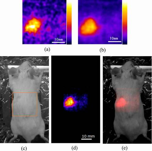
Imaging of 14C outside of the subject is considered to be difficult because it is a radionuclide that emits only low-energy beta particles. However, we found that bremsstrahlung X-rays form 14C could be imaged from outside of subjects and is thus applicable to in vivo small animal imaging. We developed a high-resolution low-energy X-ray imaging system using a (Gd, La)2Si2O7:Ce(La-GPS) plate combined with a flat panel photomultiplier tube (FP-PMT) for in vivo imaging of a mouse to detect the X-rays from a 14C solution administered. Without using a parallel hole collimator, accumulated 14C in the mouse’s abdomen was imaged in 1 min and dynamic in vivo imaging was possible although the spatial resolution was moderate. With a parallel hole collimator, 14C in the abdomen was obtained with a higher spatial resolution with a 60-min acquisition time. We conclude that in vivo imaging of 14C is possible by using the developed high-resolution La-GPS imaging system and may be promising for molecular imaging research.
GRAPHICAL ABSTRACT
Acknowledgments
This work was partly supported by JSPS KAKENHI Grant Number 19H00672. The authors thank Drs. T. Fukuchi and J. Kataoka for providing and processing the pixelated La-GPS scintillator plate.
Disclosure statement
No potential conflict of interest was reported by the author(s).