Abstract
There are 21 species of Verbena L. in Argentina, six of which are found in the Buenos Aires province: Verbena gracilescens (Cham.) Verter; V. bonariensis L.; V. intermedia Gillies and Hook; V. litoralis Kunth; V. montevidensis Spreng. and V. rigida Spreng. These species are used in popular medicine for their hepatoprotective, digestive, anti-inflammatory, antidiarrheal and healing properties. The stems and leaves of these species were fixed and transverse sections were cut for observation with a light microscope in order to differentiate their anatomical features. Likewise a scanning electron microscope and an environmental scanning electron microscope were used to study the leaf surface of these species. The type and density of stomata and glandular and nonglandular trichomes were analysed in all species, as well as the internal structure of the leaf and stem. The Verbena species of Buenos Aires province have been characterized using these features to help in their identification.
Introduction
In recent years, the return to using natural products in therapeutics has been facilitated by factors such as the serious side effects of synthetic drugs, greater chemical and pharmaceutical knowledge of plant-based drugs and their derivatives and the development of analytical methods that guarantee better quality control (Cañigueral et al. Citation2003). This resurgence raises the need for correct identification and appropriate selection of medicinal plants.
Even though medicinal plants of the native flora of Argentina have become very important for use in popular medicine for various treatments, as well as in the formulation of herbal medicines, there is only a partial understanding of their characterization, either as mono herbs or commercially available mixtures (Barboza et al. Citation2001). Only a small group of species has been included in the Farmacopea Nacional Argentina VI ed. (1978) (Leonardi et al. Citation2002).
In Argentina there are 21 species of Verbena, six of which are native in the province of Buenos Aires: Verbena gracilescens (Cham.) Verter; V. bonariensis L.; V. intermedia Gillies and Hook; V. litoralis Kunth; V. montevidensis Spreng. and V. rigida Spreng. (Troncoso Citation1974; Múlgura Citation1999; O’Leary 2007). Numerous authors report the medicinal properties of V. litoralis, V. bonariensis, V. intermedia and V. gracilescens. However, these properties have not been cited for V. montevidensis and V. rigida. The four species mentioned are used for their hepatoprotective, digestive, emmenagogue, healing, anti-inflammatory, antineuralgic, antidiarrheic, anti-gangrene and antimicrobial properties, among other uses (Arenas & Moreno Azorero Citation1977; Ratera & Ratera Citation1980; Martínez Crovetto Citation1981; Alonso Citation1998; Cáceres Citation1999; Alonso & Desmarchelier Citation2005; Barboza et al. Citation2006).
Most anatomical studies of the Verbenaceae are of genera such as Lippia (Nunes et al. Citation2000; Barboza et al. Citation2001; Bonzani et al. Citation2003), Aloysia (Matesevach Becerra et al. Citation2000; Barboza et al. Citation2001; Bonzani et al. Citation2003), Acantholippia (Carmona & Ancibor Citation1995; Barboza et al. Citation2001; Bonzani et al. Citation2003), Glandularia (Botta Citation1993) and Junellia (Botta Citation1989). Metcalfe and Chalk (Citation1979) performed comprehensive anatomical revisions of the Verbenaceae. Inamdar (Citation1969) described the epidermis and the ontogenesis of stomata of different genera of the Verbenaceae. However, few anatomical studies have been carried out on Verbena. Bhoj (Citation1983) determined the pollen characteristics of the genus. Souza et al. (Citation2005) studied the stem and leaf anatomy of V. litoralis to find diagnostic features for its detection in pharmaceutical products. Munsif et al. (Citation2007) analysed the epidermal surfaces of various Verbena species from the Middle East. Mathew and Shah (Citation1983) studied taxonomic and structural aspects of the trichomes present on nine species of Verbena and also their development. In Argentina O’Leary (Citation2007) and O’Leary et al. (Citation2007) presented a taxonomic revision of the genus on the basis of morphological characters. However, no information was included for using their anatomical features to differentiate the Verbena species, especially the natives of Buenos Aires province. The study of anatomical characters is a useful tool for the identification and systematic revision of taxa (Metcalfe & Chalk Citation1979). Moreover, it allows proper identification of herbal medicines used in popular medicine (Barboza et al. Citation2001).
The objective of this study is to describe the anatomical features of leaves and stems of Verbena native species of Buenos Aires province, as a contribution to future taxonomical studies of the genus and for its characterization as a crude drug.
Materials and methods
Plant material
Whole specimens of each species were prepared and deposited in the herbarium of the Instituto de Botánica Darwinion (SI), Buenos Aires, Argentina ().
Table 1 List of specimens of each species collected in Buenos Aires province, Argentina.
Light microscopy
Transverse section
Pieces of fresh stem were cut at the height of the fourth internode from the apex from each species under study. This material was cut in 20 µm transverse sections with a MICROM HM 400 R sliding microtome (Microm, Germany), stained with safranin-fastgreen and mounted in synthetic balsam. Also stems and leaves were fixed in FAA (ethanol 96%:distilled water:formaldehyde:acetic acid, 10:7:2:1), embedded in paraffin, cut in 15–20 µm transverse sections with a MICROM HM 325 rotary microtome (Microm, Germany), stained with saffranin-fastgreen and mounted in synthetic balsam. Likewise, small pieces of the leaves of these specimens were fixed in 4% glutaraldehyde, post-fixed in 1.5% osmium tetroxide in 0.1 M phosphate buffer, embedded in epoxy resin and cut with an ultramicrotome in transverse sections of 1–2 µm. Subsequently, these sections were stained with 1% toluidine blue and mounted in synthetic balsam (PMYR, Instrumental Pasteur, Argentina).
Clearing
Expanded leaves from the fourth node from the apex were cleared using Dizeo de Strittmater’s method (D’Ambrogio Citation1986). This technique involves: removal of chlorophyll with boiling ethanol for 30 s; the dissolution of protoplasm using boiling 5% NaOH and ethanol (1:1) for 1 min and washing three times with distilled water; removal of brown coloration by bleaching with 50% sodium hypochlorite and washing three times with water; clearing with 2.5 g cm−3 chloral hydrate 2–3 min; and staining with diluted safranine and then mounting with glycerin-gelatin.
Microchemical tests
Fresh transversal sections of leaves were used to determine cellular contents. Thus, cresyl blue was used for mucilages and 2M HCl for calcium carbonate (cystoliths) (Martínez et al. Citation2008).
Observations, drawings and photographs were made using an UNICO light microscope with a computerized system for the capture of images (Intellicam; Matrox Electronic Systems, Canada) and an OPTIMA 6.5 image processor of the Luján National University (Buenos Aires, Argentina).
Scanning electron microscopy
Pieces of leaves (0.5 cm×0.5 cm) of the different species under study were fixed in FAA, dehydrated in an ascending series of alcohol and were dried to the critical point. Subsequently they were coated with a gold-palladium alloy so that they could be observed and photographed with a Phillips XL-30 SEM microscope (Microscopy Service, Bernardino Rivadavia Natural Sciences Museum, Buenos Aires, Argentina).
Environmental scanning electron microscopy
Fresh leaves of the species under study were observed and photographed using an ESEM Electroscan Phillips 2010 microscope (Microscopy Service, CITEFA, Buenos Aires, Argentina).
Statistical analysis
Stomatal and glandular trichomes density was measured in 30 randomly chosen optical microscope fields on 10 slides from each species. The results for each parameter were analysed with ANOVA and compared with Tukey's multiple comparison test, using SPSS v.12.0 software (SPSS Inc., Chicago, Illinois, USA).
Results
Leaf
Surface view
The leaves of the Verbena species under study show isodiametric epidermal cells with wavy borders on both leaf surfaces. The stomata are anisocytic and anomocytic in all species, rarely diacytic in V. montevidensis. The stomatal density calculated shows significant differences between the adaxial and abaxial surfaces (A–1E; ). The highest values were recorded for V. litoralis, 318.7/mm2 on the abaxial surface and 139.7/mm2 on the adaxial surface ().
Figure 1 Leaf surface of Verbena native species of Buenos Aires province. A, Adaxial epidermis of V. bonariensis (SEM). B, Abaxial epidermis of V. bonariensis (SEM). C, Anomocytic stomatal apparatus, adaxial epidermis of V. bonariensis (SEM). D, Adaxial epidermis of V. bonariensis (SEM). E, Adaxial epidermis of V. montevidensis (LM). F, Vascularization, abaxial epidermis of V. rigida (LM). Abbreviations: g, glandular trichomes; LM, Light Microscopy; ng, nonglandular trichomes; s, stomata; SEM, scanning electron microscopy. Bars: A, B, F, 200 µm. C, D, E, 50 µm.
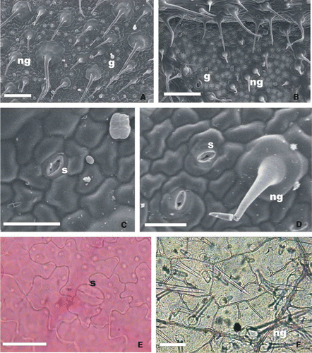
Table 2 Density of stomata and glandular trichomes in the leaves of Verbena native species of Buenos Aires province, Argentina.
The cuticle on both the adaxial and abaxial epidermis is slightly striated, the striations being more marked over the mid-vein (F).
Figure 2 Glandular and nonglandular trichomes of Verbena native species of Buenos Aires province, leaf surface view. A, General view of abaxial epidermis of V. litoralis (ESEM). B, Detail of appressed nonglandular trichome, adaxial epidermis of V. gracilescens (ESEM). C, Long-stalked glandular trichome and nonglandular patent trichome, abaxial epidermis of V. bonariensis (SEM). D, Long-stalked glandular trichome adaxial epidermis of V. rigida (SEM). E, Long-stalked glandular trichome, adaxial epidermis of V. intermedia (ESEM). F, Four-celled head of short-stalked glandular trichome, abaxial epidermis of V. rigida (SEM). G, Multicellular head of short-stalked glandular trichome, abaxial epidermis of V. montevidensis (ESEM). Abbreviations: ESEM, environmental scanning electron microscopy; g, glandular trichome; LM, light microscopy; ng, nonglandular trichome; SEM, scanning electron microscopy. Bars: A, 250 µm. B, 150 µm. C–E, 50 µm. F, G, 10 µm.
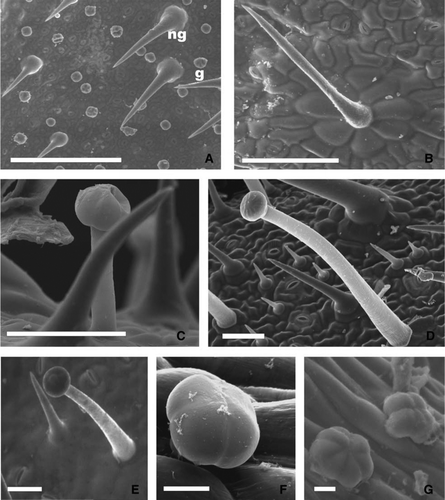
All species have nonglandular and glandular trichomes in both epidermis (A, 1B, 1F; A; ).
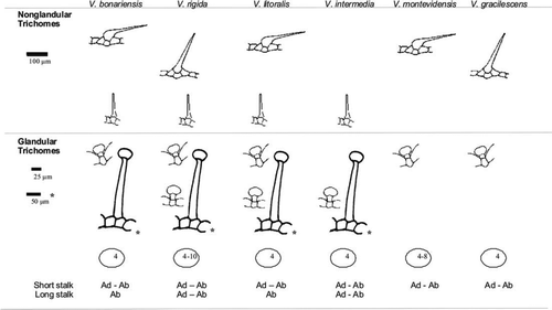
Figure 3 Anatomical features that differentiate the leaves of Verbena native species of Buenos Aires province, Argentina. Abbreviations: Ab, Trichomes in the abaxial epidermis; Ad, Trichomes in the adaxial epidermis. The number inside the oval indicates number of cells in head of glandular trichomes.
The nonglandular trichomes have unicellular body and two multicellular and radially arranged basal cells. The body of these trichomes is elongated, with a pointed tip and smooth or verrucose walls. They are distributed over both epidermal surfaces of the leaves and may be appressed or slightly appressed. Moreover, there are many unicellular patent trichomes (A, 1B, 1D, 1F; A–2E).
The glandular trichomes have a multicellular head and a stalk of varying length and they are distributed on both epidermal surfaces (A, C–2G).
All the leaves of these species have cladodromous venation, with incomplete vascularization on the leaf blade and margin, and pentagonal to polygonal areoles, generally with ramified veins (F).
Transverse section
The mesophyll is dorsiventral in all species studied, with three layers of palisade chlorenchyma. The epidermis has stomata on both surfaces (amphistomatic leaf) (A–4D). The cuticle is thin and slightly striated.
Figure 4 Leaf transverse section of Verbena native species of Buenos Aires province (LM). A, General view of V. litoralis. B, Detail mid-vein of V. montevidensis. C, Detail of abaxial epidermis and spongy chlorenchyma of V. bonariensis. D, Detail of adaxial epidermis and palisade chlorenchyma of V. bonariensis. E, Patent nonglandular trichome in V. rigida. F, G, Nonglandular trichome with cystoliths in the basal cells of V. bonariensis. H, Short-stalked glandular trichome of V. litoralis. I, Sunken short-stalked glandular trichome of V. litoralis. J, Long-stalked glandular trichome and nonglandular trichomes of V. intermedia. K, Long-stalked glandular trichomes of V. bonariensis. L, Adaxial epidermis of V. bonariensis. Abbreviations: lg, long-stalked glandular trichome; LM, light microscopy; ps, parenchymatous sheath; s, stomata; sg, short-stalked glandular trichome; black arrow, cystoliths. Bars: A, B, F, J, L, 100 µm. C–E, K, 50 µm. G, H, I, 25 µm.
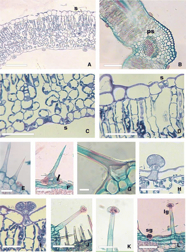
The nonglandular trichomes have a multicellular base with cystoliths detected by microchemical test. Mucilages were not detected in these cells. The wall of the body cell is lignified (F–4G). The glandular trichomes are formed of a basal cell, a stalk cell of varying length and a bi- to multicellular head. If the stalk cell of glandular trichomes is short it may be sunken (H–4L). Each species has a particular type, density and distribution of trichomes (; A, 4B, 4E, 4F, 4H–4L).
Vascular bundles surrounded by a parenchymatous sheath were seen in all species studied (B).
Stem
Transverse section
The stems of all species have tetragonal to quadrangular transverse sections (; A). Nonglandular and glandular trichomes were observed in the unistratified epidermis as described for the leaf. The type of glandular trichome present in the stem is the same as that described for the leaf of the same species.
Figure 5 Stem transverse section of Verbena native species of Buenos Aires province. A, V. gracilescens. B, V. montevidensis. C, V. bonariensis. D, V. litoralis. E, V. intermedia. F, V. rigida. Bars: 500 µm.
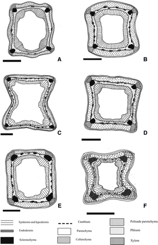
Figure 6 Stem transverse section of Verbena native species of Buenos Aires province. A, General aspect of V. montevidensis (LM). B, Detail of stem angle in V. gracilescens (LM). C, Detail of the stem side in V. bonariensis (LM). D, Epidermis and hypodermis in V. litoralis (LM). E, Endodermis with Casparian bands in V. gracilescens (LM). F, Vascular bundle and cap of fibres in V. intermedia (LM). Abbreviations: c, collenchyma; e, endodermis; f, fibres; h, hypodermis; LM, light microscopy; s, stomata; black arrow, Casparian bands. Bars: A, 250 µm. B, C, 100 µm. D–F, 25 µm.
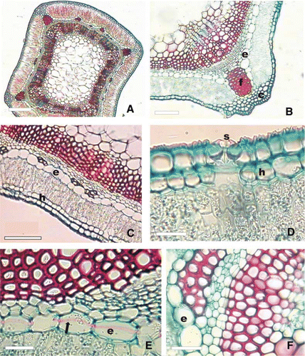
There is a layer of hypodermic cells underneath the epidermis (C–6D). Caps of collenchyma and sclerenchyma are found in the stem angles (; A–6B) and the chlorenchyma of cortex is interrupted in these areas (A–6C). All species studied have a continuous endodermis surrounding the parenchymatous cortex and vascular system (A–6C, 6E). Casparian bands have been observed in the endodermis in V. gracilescens and V. montevidensis (E), but no thickening of this type was seen in the rest of the species studied (C).
The vascular bundles are collateral and have caps of phloem fibres (A, 6B, 6F). All species studied show incipient secondary growth at the height of the fourth node from the apex (; A, 6C).
Discussion
The stems of the species studied show the characteristics of the Verbenaceae, such as the square transverse section and the presence of sclerenchyma and collenchyma caps at the angles, coinciding with that observed by Metcalfe and Chalk (Citation1950), O’Leary et al. (Citation2007) and O’Leary (Citation2007).
According to Schnack and Covas (Citation1944) and Botta (Citation1989), the stem of Verbena has discontinuous cortical parenchyma. On the other hand, O’Leary et al. (Citation2007) and O’Leary (Citation2007) described two types of cauline anatomy for the genus: Type A: with continuous cortical parenchyma (V. rigida and V. gracilescens); Type B: with discontinuous cortical parenchyma (V. bonariensis, V. litoralis, V. montevidensis and V. intermedia). However, in our study all species including V. rigida and V. gracilescens show stems with discontinuous parenchyma (Type B), whereas both types of parenchyma distribution are observed in V. montevidensis and V. bonariensis (Types A and B).
Caps of angular collenchyma are seen in the angles of all stems. However, Souza et al. (Citation2005) reported the presence of lamellar collenchyma in the stem angles of V. litoralis. The presence of a hypodermis in the stem of Verbena was also reported by Metcalfe and Chalk (Citation1950) and Souza et al. (Citation2005). On the other hand, some authors indicate that there is continuous collenchyma around the perimeter of the Verbena stem (Botta Citation1989; O’Leary Citation2007; O’Leary et al. Citation2007) rather than a hypodermis, but this has not been seen in the species considered in this study.
The endodermis is seen to be continuous around the perimeter of the stem in all species and Casparian bands are present in the endodermal cells of V. gracilescens and V. montevidensis. These features are cited for the Verbenaceae and Verbena by Metcalfe and Chalk (Citation1950, Citation1979), O’Leary et al. (Citation2007) and O’Leary (Citation2007). However, Souza et al. (Citation2005) considered that this layer of cells was a starch parenchymatic sheath.
The sclerenchyma, represented by caps of fibres in the stem angles and phloem fibres, of Verbena species studied is similar to that described for other genera of the family (Barboza et al. Citation2001; Souza et al. Citation2005; O’Leary et al. Citation2007).
The leaves of Verbena species considered in this study present the general characteristics of the Verbenaceae family and specifically those of the Verbena genus (Metcalfe & Chalk Citation1950, Citation1979; Inamdar Citation1969; Souza et al. Citation2005; O’Leary et al. Citation2007): nonglandular and glandular trichomes of different types, stomata on both epidermal surfaces and heterogeneous mesophyll.
The presence of anisocytic and anomocytic stomata has also been reported for other genera of the Verbenaceae (Metcalfe & Chalk Citation1979), in Acantholippia, Aloysia and Lippia (Barboza et al. Citation2001) and in Glandularia (Botta Citation1993). However, Souza et al. (Citation2005) reported paracytic stomata in V. litoralis and Inamdar (Citation1969) found diacytic ones in V. venosa (Syn. V. rigida Spreng.). The high stomatal density seen in V. litoralis shows a ratio of 3:1 stomata (abaxial surface:adaxial surface), as cited by Souza et al. (Citation2005) for Verbena species.
The species in this study show nonglandular unicellular trichomes with multicellular bases with cystoliths as in other representatives of the family (Inamdar Citation1969; Metcalfe & Chalk Citation1979; Botta Citation1993; Barboza et al. Citation2001). This type of trichome has been previously cited for the Verbena genus (Inamdar Citation1969; Mathew & Shah Citation1983; Cañigueral et al. Citation1998; Souza et al. Citation2005; O’Leary et al. Citation2007).
The different types of glandular trichomes observed were previously cited for representatives of the family (Metcalfe & Chalk Citation1950, Citation1979; Inamdar Citation1969; Mathew & Shah Citation1983; Botta Citation1993; Barboza et al. Citation2001). Cañigueral et al. (Citation1998) report the presence of long-stalked glandular trichomes and others with a short stalk and tetracellular head in V. officinalis. On the other hand the presence of long-stalked glandular trichomes is refuted by Souza et al. (Citation2005).
Although the general anatomical features of all Verbena species considered in this study coincide with those reported for the Verbenaceae, some anatomical parameters and characteristics have been found that are valid for differentiating between the species of Buenos Aires province. These distinctive anatomical characters can be applied to taxonomic revisions of the genus and also aid the identification of Verbena native species of Buenos Aires province as crude drugs.
References
- Alonso J 1998 . Tratado de Fitomedicina. Bases clínicas y farmacológicas . Buenos Aires, Issis SRL . 1040 p.
- Alonso J , Desmarchelier C 2005 . Plantas Medicinales Autóctonas de la Argentina . Buenos Aires, L.O.L.A . 664 p.
- Arenas , P and Moreno Azorero , R . 1977 . Plants of common use in Paraguayan folk medicine for regulating fertility . Economic Botany , 31 : 298 – 301 .
- Barboza G , Bonzani N , Filippa E , Luján M , Morero R , Bugatti M , Decolatti N , Ariza Espinar L 2001 . Atlas histo-morfológico de plantas de interés medicinal de uso corriente en Argentina. Serie Especial I . Córdoba, Museo Botánico de Córdoba . 212 p.
- Barboza G , Cantero J , Nuñez C , Ariza Espinar L 2006 . Flora Medicinal de la Provincia de Córdoba (Argentina) . Córdoba, Museo Botánico de Córdoba . 1251 p.
- Bhoj , R . 1983 . A contribution to the pollen morphology of Verbenaceae . Review of Palaeobotany and Palynology , 39 : 343 – 422 .
- Bonzani , N , Filippa , E and Barboza , G . 2003 . Estudio anatómico comparativo de tallo en algunas especies de Verbenaceae . Anales del Instituto de Biología Serie Botánica , 74 : 31 – 45 .
- Botta , SM . 1989 . Estudios en el género sudamericano Junellia (Verbenaceae, Verbenoideae) I . Delimitación y tratamiento infragenérico. Darwiniana , 29 : 371 – 396 .
- Botta , SM . 1993 . Notas sobre el género Glandularia (Verbenaceae-Verbenoideae) III . Parodiana , 8 : 9 – 36 .
- Cáceres A 1999 . Plantas de uso medicinal en Guatemala . Colección Monografías . Guatemala , Universitaria . 402 p.
- Cañigueral S , Vila R , Witchl M 1998 . Plantas Medicinales y Drogas Vegetales para infusión y tisana. Manual para Farmacéuticos y Médicos . Barcelona , OEMF International . 606 p.
- Cañigueral , S , Dellacassa , E and Bandoni , A . 2003 . Plantas Medicinales y Fitoterapia: ¿Indicadores de Dependencia o Factores de Desarrollo? . Acta Farmacéutica Bonaerense , 22 : 265 – 278 .
- Carmona , CS and Ancibor , E . 1995 . Anatomía ecológica foliar de las especies de Acantholippia (Verbenaceae) . Boletín de la Sociedad Argentina de Botánica , 31 : 3 – 12 .
- D'Ambrogio A 1986 . Técnicas en histología vegetal . Buenos Aires , Hemisferio Sur . 83 p.
- Inamdar , JA . 1969 . Epidermal structure and ontogeny of stomata in some Verbenaceae . Annals of Botany , 33 : 55 – 66 .
- Leonardi , D , Di Sapio , O , Gattuso , MA and Gattuso , S . 2002 . Caracteres morfoanatómicos de la corteza y hojas de Tabebuia impetiginosa y T. heptaphylla (Bignoniaceae) . Boletín de la Sociedad Argentina de Botánica , 37 : 51 – 61 .
- Martínez Crovetto , R . 1981 . Las plantas utilizadas en medicina popular en el NO de Corrientes . Miscelánea , 69 : 89 – 92 .
- Martínez A , Valencia GA , Jiménez N , Mesa M , Galeano E 2008 . Manual de prácticas de laboratorio de farmacognosia y fitoquímica 2008 . Medellín, Colombia, Facultad de Química Farmacéutica, Universidad de Antioquía . 95 p.
- Matesevach Becerra , AM , Dottori , N and Cosa , MT . 2000 . Desarrollo de la semilla y del fruto de Aloysia polystachya (Griseb.) Moldenke (Verbenaceae) . Kurtziana , 28 : 239 – 250 .
- Mathew , L and Shah , G . 1983 . Structure, development, organographic distribution and taxonomic significance of trichomes in nine species of Verbena . Feddes Repert , 94 : 123 – 333 .
- Metcalfe CR , Chalk L 1950 . Anatomy of Dicotyledons. Leaves, stem, and wood in relation to taxonomy with notes on economic uses . Vol. 2 . Oxford , Clarendon Press . 706 p.
- Metcalfe CR , Chalk L 1979 . Anatomy of Dicotyledons. Systematic anatomy of leaf and stem, with a brief history of the subject . Vol. 1 . Oxford , Clarendon Press . 794 p.
- Múlgura ME 1999 . Verbenaceae . In : Zuloaga FO , Morrone O Catálogo de las Plantas Vasculares de la República Argentina. Monographs in Systematic Botany from the Missouri Botanical Garden 74 . St. Louis , MO , Missouri Botanical Garden Press . Pp. 1136 – 1169 .
- Munsif , S , Khan , M , Ahmad , M , Zafar , M , Shah , G and Shaheen , N . 2007 . Leaf epidermal anatomy as an aid to the identification of Genera Lantana, Verbena and Vitex of family Verbenaceae from Pakistan . Journal of Agricultural and Social Sciences , 3 : 43 – 46 .
- Nunes , R , Xavier , HS , Rolim Neto , P , Santana , D and Albuquerque , U . 2000 . Padronizaçao Botanica de Lippia sidoides Cham. (Verbenaceae) . Acta Farmacéutica Bonaerense , 19 : 115 – 118 .
- O’Leary N 2007 . Estudios sistemáticos y filogenéticos en el género Verbena L. (Verbenaceae) . Unpublished PhD thesis, Facultad de Ciencias Exactas y Naturales, Universidad Nacional de Buenos Aires, Argentina . 259 p.
- O’Leary , N , Múlgura , ME and Morrone , O . 2007 . Revisión Taxonómica de las especies del género Verbena (Verbenaceae): serie Pachystachyae . Annals of the Missouri Botanical Garden , 94 : 571 – 621 .
- Ratera L , Ratera M 1980 . Plantas de la flora argentina empleadas en medicina popular. Buenos Aires, Hemisferio Sur . 192 p.
- Schnack , B and Covas , G . 1944 . Nota sobre la validez del género Glandularia . Darwiniana , 6 : 469 – 476 .
- Souza TJ , Manfron MP , Zanetti GD , Hoelzel SC , VPagliarin VP 2005 . Análise Morfo-Histológica e Fitoquímica de Verbena litoralis Kunth . Acta Farmacéutica Bonaerense 24 : 209 – 214 .
- Troncoso , NS . 1974 . Los géneros de Verbenáceas de Sudamérica extratropical . Darwiniana , 18 : 295 – 412 .