The paper by MR Alley, RA Fairley, DG Martin, L Howe and T Atkinson published in the New Zealand Veterinary Journal 56, 247–251, 2008, entitled, “An outbreak of avian malaria in captive yellowheads/mohua (Mohoua ochrocephala)”, contained errors with regard to Figure 2, Figure 3, and Figure 4a and 4b. In Figures 2 and 4a, the arrows were omitted, and in Figures 3 and 4a and 4b, the labels on the fi gures were omitted. The correct fi gures, with their captions, are reproduced here.
Figure 2. High-power light photomicrograph of a section of lung from a mohua (Bird 4) which died due to infection with Plasmodium spp., showing swollen endothelial cells containing numerous intracytoplasmic protozoal organisms (arrows) (H&E, bar=10 µm).
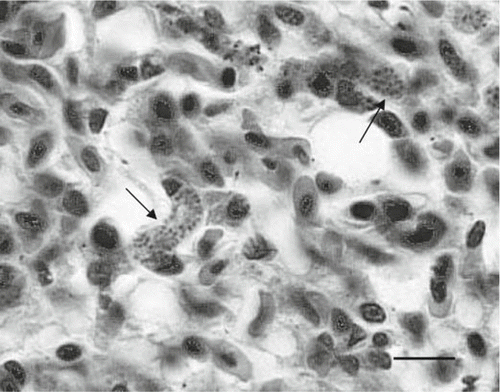
Figure 3. Transmission electron micrograph of a section of lung from a mohua (Bird 5) which died due to infection with Plasmodium spp., showing a protozoal schizont containing numerous merozoites within a parasitophorus vacuole in the cytoplasm of an endothelial cell (E). The adjacent capillary (C) contains a nucleated erythrocyte (bar=5 µm).
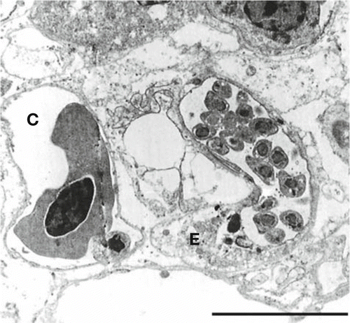
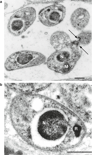
Figure 4. Higher-power transmission electron micrographs of Plasmodium spp.-like merozoites within pulmonary endothelial cells of a mohua (Bird 5) which died due to infection with Plasmodium spp., demonstrating (a) a clump of merozoites showing polar rings (arrows) and a single mitochondria (M) behind the nucleus (N), and (b) a single merozoite with a pear-shaped rhoptry (R) and broken-down pellicular complex (P) (bars=0.5 µm).