Abstract
AIM: To determine whether working dogs in New Zealand with carpal injuries and treated with unilateral pancarpal arthrodesis (PCA), using a dorsal hybrid-plating method, are able to return to satisfactory working ability.
METHODS: Fourteen working dogs presented to the Veterinary Specialist Group (VSG) and the Massey University Veterinary Teaching Hospital (MUVTH) with carpal injuries were prospectively treated using dorsal hybrid plating. Dogs were eligible if actively involved in farm, hunting or police work. Dogs had a standardised PCA surgical procedure performed, and similar instructions for post-operative care were provided. Dogs were re-evaluated clinically and radiographically at 6 weeks, 6 months, and 12 months after surgery. A questionnaire was completed by 12 owners, to assess each dog's working ability.
RESULTS: Twelve months following arthrodesis, 10/12 (83%) dogs could perform most or all duties normally. Eleven owners (92%) reported that the result of the surgery met their expectations, and nine owners (75%) were very satisfied with the outcome of the surgery. No owners were disappointed or very disappointed with the surgical outcome. Post-operative complications requiring surgical removal of the implant occurred in three (25%) dogs.
CONCLUSIONS: Unilateral PCA using a standardised surgical procedure and dorsal hybrid plating of carpal injuries has a good prognosis for working dogs in New Zealand to return to work.
CLINICAL RELEVANCE: These results may allow veterinarians to provide a more accurate prognosis to owners of working dogs that have debilitating carpal injury
| CAP | = |
Hybrid carpal arthrodesis bone plate |
| ESF | = |
External skeletal fixator |
| MUVTH | = |
Massey University Veterinary Teaching Hospital |
| PCA | = |
Pancarpal arthrodesis |
| VSG | = |
Veterinary Specialist Group |
Introduction
The canine carpus is a complex anatomical structure that is occasionally injured in large-breed dogs during athletic activities such as running, jumping and climbing. The carpus has the greatest range of motion of all appendicular joints in the dog during the trot gait, and is subjected to high-impact forces, especially during deceleration (Eward et al. Citation2003). This biomechanical effect predisposes the canine carpus to injury. Common carpal injuries include fractures or ligamentous injuries, including hyperextension with resultant luxation or subluxation of the carpus. Such injuries can cause pain, inflammation and instability of the joint, leading to debilitating lameness. The condition and surgical treatment were reviewed by Buote et al. (Citation2009).
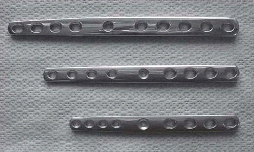
Working dogs are an important component of farming, hunting, and police activities in New Zealand. In our opinion, due to the athletic nature of the working activity expected, New Zealand's working breeds of dogs are probably more prone to carpal injury than more sedentary breeds. The success in conservatively treating fractures and ligamentous injuries of the canine carpus can be expected to be poor, particularly in working dogs. The use of objective prognostic data is helpful when making treatment decisions for working dogs with carpal injuries, as the ability for these dogs to return to full work is generally a requirement of the owners. Recently, a retrospective study of unilateral pancarpal arthrodesis in working dogs in New Zealand concluded that this procedure carried a good prognosis for return to work (Worth and Bruce Citation2008). Post-operative complications were high (50%) but this did not affect the eventual outcome for most dogs. The inclusion of eight surgeons and the use of two different surgical implant systems involved in managing these cases indicated further prospective studies were necessary. With the recent availability of a commercial hybrid carpal arthrodesis bone plate (CAP) (Veterinary Instrumentation, Sheffield, UK) (Figure ), we wanted to determine whether using the CAP to treat working dogs in New Zealand with carpal injuries would provide secure fixation, and allow the dogs to return to work following PCA. The CAP has been shown experimentally to be significantly stronger than the more traditional dynamic compression bone plates (Wininger et al. Citation2007). The aim of this study was to determine whether use of the CAP would allow working dogs with carpal injury to return to work following PCA.
Materials and methods
From July 2006 to December 2008 bonafide working dogs referred to the small animal surgical services at the VSG and MUVTH for diagnosis and treatment of unilateral carpal injury were evaluated. Dogs determined to be candidates for PCA were entered into the study. Dogs with concurrent orthopaedic or neurological disease in another limb were not enrolled. Whether previous ipsilateral carpal surgery had been performed was recorded but not considered as a reason for exclusion from the study. Each owner signed an informed consent form prior to inclusion of their dog in the study, and a financial incentive (discount) was offered to encourage the owners to return their dogs for post-operative evaluations.
Pre-operative assessment
A full orthopaedic and neurological examination was performed on each dog. Previous orthopaedic problems or surgery were recorded, and an assessment of soft-tissue swelling, joint effusion, pain, crepitus and carpal instability was made on both thoracic limbs.
Radiographs were taken of both antebrachia, to include the distal radius, ulna and manus. Further radiographs, with application of medial, lateral, dorsal and flexion stress across the carpal joints, were taken of both thoracic limbs.
Anaesthesia and analgesia
Each dog received the standard anaesthetic protocol for orthopaedic surgery patients at the institution where the surgery was performed (VSG or MUVTH). Pre-operatively, each dog received an injection of 2 mg/kg bupivacaine (Marcain; AstraZenca, Auckland, NZ) in the brachial plexus, for pre-emptive analgesia. Postoperative pain relief included opioid medication, as required, for 24–48 hours post-operatively. Non-steroidal anti-inflammatory medication was prescribed for 7–10 days following surgery.
Surgical procedure
All surgeries were performed by a registered specialist small animal surgeon or a surgical resident. The nature of any intra-operative complications was recorded.
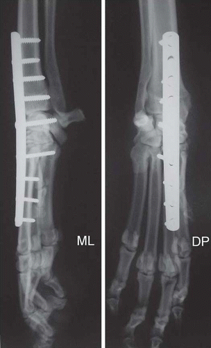
All dogs were clipped and prepared for surgery, using standard techniques. Surgical draping of the limb followed standard orthopaedic protocols, including sterile stockinette bandage, adhesive plastic shield, and suturing of the S/C tissue to the drape. A skin incision was made from the mid-diaphysis of the radius to the distal end of the metacarpal bones. The S/C tissues were transected, and underlying extensor tendons elevated. At the surgeon's discretion, two or three holes were drilled in the distal radial epiphysis entering the medullary canal, to aid vascularisation of the fusion. The cartilage on all joint surfaces was debrided, using a pneumatic bone bur. Cancellous bone graft was obtained from the ipsilateral proximal humerus, and packed into the joint spaces. Arthrodesis was performed by placing a 3.5/2.7-mm tapered CAP and appropriate bone screws. The size of plate chosen was dependent on the bodyweight of the dog. Screws were placed in the distal radius, the radial carpal bone, and the proximal aspect of the third metacarpal bone. The tendons were sutured over the implants, using monofilament absorbable suture material, and closure of S/C tissue and the skin was routine.
Post-operatively, radiographs were taken of the operated limb, to confirm appropriate positioning of the orthopaedic implants, with bicortical purchase of the screws in the distal radius and third metacarpal bones as well as a single screw positioned in the radial carpal bone. A split-cast bandage (3M Scotchcast Plus; 3M Health Care, Borken, Germany) was applied to the limb, and the dog was recovered from anaesthesia. All dogs were discharged, with instructions for changing of the cast weekly, suture removal 10–14 days post-operatively, and strict confinement for 6 weeks. Instructions were given for pain relief, using non-steroidal antiinflammatory medication, and passive physical therapy, including heat therapy and massage.
Post-operative assessment
Dogs were returned to the VSG, MUVTH or the referring veterinary clinic 6 weeks, 6 months, and 12 months following surgery. Dogs were evaluated for lameness and pain. Any postsurgical complications were recorded. Radiographs were taken of the operated carpus at each re-evaluation. The radiographs were assessed by a specialist veterinary radiologist, and the stability of surgical implants and level of bony healing of the arthrodesis were recorded. Sedation was required for radiography. The dogs were recommended for a gradual return to normal activity, with working activity beginning approximately 4 months following surgery. The owners were asked to complete a questionnaire (adapted from Worth and Bruce 2008; Supplementary Table Footnote 1 ) 12 months after surgery, concerning the dog's willingness to, ability to, and level of work. Assessment of the success of surgery from the owner's perspective was recorded.
Statistical analysis was not performed on the data from the questionnaire due to the low number of cases in the study.
Results
Signalment
Fourteen dogs were entered into the study; the results are summarised in Table 1. There were six Huntaway dogs, with a median bodyweight of 31 (range 26–38) kg, four Heading dogs, with a median bodyweight of 22 (range 20–26) kg, three mixed-breed farm dogs, with a median bodyweight of 28 (range 23–36) kg, and a single German Shepherd Police dog weighing 41 kg. Eleven of the dogs were male, and the average age of all the dogs was 4.4 (range 1–9) years. All dogs were injured whilst jumping, falling, or by a kick from cattle during active work. The desired outcome following surgery was a return to work.
Two dogs were lost to follow-up prior to completion of the study. Of the remaining 12 dogs with a complete questionnaire, seven were working on flat farmland or rolling hill country, and four were working on hill or hard hill-country properties; the Police dog was required to perform duties in a variety of urban environments. Eight respondents described their property as extensive sheep/beef, two dogs were working on an intensive beef property, one dog worked on a dairy farm, and the Police dog worked on urban terrain.
Table 1. The age, sex, breed, and weight at treatment of 14 working dogs treated using pancarpal arthrodesis, with cause of injury, and the responses of the owners to a questionnaire on the results of the surgery.
Pre-operative assessment
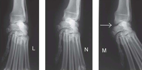
There was radiographic evidence of instability and hyper-extension injury of the antebrachiocarpal joint in 12/14 dogs, varying from instability evident on stress-view radiographs to complete luxation of the carpus (Figure ). In addition to carpal instability, two dogs had fractured the ipsilateral fifth metacarpal bone. One dog had a chronic fracture of the radial carpal bone, and another dog presented with resolving septic arthritis of the antebrachial carpal joint. Three dogs had had previous carpal surgery, with attempted stabilisation of a medial carpal ligament injury using wire; attempted partial carpal arthrodesis using crossed pins; and an external skeletal fixator (ESF) used in the case of septic arthritis of the carpus.
Surgical complications
Eleven dogs were treated using a 2.7/3.5-mm CAP, and three using a 3.5/3.5-mm CAP (Figure ). No intra-operative complications were recorded.
Post-operative assessment and complications
During the first 6 weeks following surgery, no complications were detected in 12/14 dogs. One dog developed a decubital ulcer over the accessory carpal bone, that was assumed to be due to pressure from the cast. The cast was changed to a spoon splint applied laterally, and the ulcer healed with regular management of the wound. Another dog developed interdigital dermatitis, requiring changing the bandage to a half-cast, applied in a palmar aspect.
Radiographic assessments at 6 weeks, 6 months, and 12 months demonstrated satisfactory progression of PCA, with healing and remodelling seen in all dogs that completed the study. There was evidence of loosening of the implant in four dogs. Radiographic evidence of bony union was present in all dogs at the 6-month assessment, except one dog that had problems associated with the implant, and subsequent placement of an ESF (see below). Failure of bony arthrodesis was not recorded in any dog during the study period.
Figure 2. Dorsopalmar radiographs of a working dog with instability of the medial side of the carpus and subluxation of the antebrachioradial joint (arrow). The image on the left (L) has a stress force applied laterally, the image in the middle (N) has no stress applied, and the image on the right (M) has a stress force applied medially.

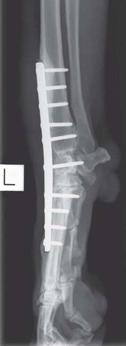
Three months following surgery two dogs developed a discharging sinus over the distal end of the plate. In one of these dogs, serial radiographs over the next 7 months showed continued lucency around the plate and several of the screws. The CAP was removed, and a transarticular ESF was placed 11 months following the original procedure; this device remained in place for 8 weeks. Thirteen months from the original surgical procedure, the ESF was removed, and a further CAP applied. This dog was subsequently lost to follow-up. The other dog showed radiographic evidence of lucency at the distal end of the plate (Figure ). The dog was allowed to continue light work duties, and was treated with injectable cefovecin sodium (Convenia; Pfizer, Auckland, NZ) at 2-weekly intervals, until the plate and screws were removed at 6 months following surgery.
Figure 3. Post-operative mediolateral (ML) and dorsopalmar (DP) radiographs of a working dog, with application of a nine-hole 2.7/3.5-mm carpal arthrodesis bone plate.

Seven months after surgery another dog developed an acute onset of non-weight-bearing lameness affecting the operated limb, with swelling and heat around the carpus. Radiographs showed evidence of breakage of several of the distal screws, and lucency around the implants that had not been present at the radiographic assessment one month earlier. The surgical implants were removed by the referring veterinarian. Bacterial culture and sensitivity were not performed. The dog was then returned to the VSG, and was treated with hyperbaric oxygen (10 sessions @ 1 atmosphere), and empirical antibiotic therapy comprising 25 mg/kg metronidazole (Trichozole; Pacific Pharmaceuticals, Auckland, NZ), 10mg/kg amoxicillin/clavulanic acid (Vetamox; Ethical Agents, South Auckland, NZ), and 10 mg/kg ciprofloxacin (Ciproxin; Pacific Pharmaceuticals, Auckland, NZ) for 6 weeks. The dog recovered well, and was able to return to work before dying from gastric dilatation/volvulus at 10 months following the PCA procedure.
Figure 4. Mediolateral radiograph of a working dog taken approximately 6 months following surgery for pancarpal arthrodesis, demonstrating evidence of lucency around the proximal and distal ends of the carpal arthrodesis bone plate.

Radiographs of one dog taken 12 months after surgery showed evidence of loosening of three of the four screws in the metacarpal bone, despite complete healing of the arthrodesis site. This dog was showing signs of intermittent lameness, and removal of the surgical implants was recommended but not performed.
Owner assessment
Of the 12 owners who completed the questionnaire three (25%) felt that their dog could perform normal duties, and a further seven (58%) indicated that their dog could perform most duties. Two owners (17%) felt that their dog could perform some duties but was of limited usefulness.
Three owners (25%) indicated that the surgical procedure did not have any adverse effect on their dog's gait, while seven (58%) reported that their dog had a constant gait abnormality and mild intermittent lameness. One owner felt that their dog had a constant gait abnormality, with mild persistent lameness, and the final owner felt that their dog had a constant gait abnormality, with moderate persistent lameness that was intermittently nonweight- bearing.
Eleven owners (92%) reported that the result of the surgery met their expectations, and eight owners were very satisfied with the outcome of the surgery. No owners were disappointed or very disappointed with the surgical outcome.
Discussion
This study provided further clinical evidence that most working dogs will return to good or excellent working ability post-operatively following unilateral PCA using a CAP. Twelve months following arthrodesis 10/12 (83%) dogs could perform most or all duties normally as before injury. The findings concur with those from a previous retrospective study of working dogs in New Zealand providing veterinarians and owners some prognostic information to assist with treatment decision-making in these cases (Worth and Bruce Citation2008).
Conservative supportive treatment of carpal injuries in largebreed dogs is generally unrewarding, therefore carpal arthrodesis is recommended as the treatment of choice for most dogs with carpal instability (Buote et al. Citation2009). Partial carpal arthrodesis is recommended for injuries of the intercarpal and carpometacarpal joints, while PCA is recommended for injuries that include instability of the antebrachiocarpal joint (Willer et al. Citation1990; Haburjak et al. Citation2003; Buote et al. Citation2009). In two studies of partial carpal arthrodesis, it was reported that 45–50% of dogs developed degenerative joint disease or further carpal instability post-operatively, which would probably limit this technique to selected cases and preclude the technique from being appropriate in working dogs (Willer et al. Citation1990; Haburjak et al. Citation2003). PCA is most commonly performed by the dorsal application of a bone plate from the distal radius across all three carpal joints, to include at least 50% of the length of the third metacarpal bone (Whitelock et al. Citation1999). Articular cartilage is debrided from the joints, and an autogenous cancellous bone graft is packed into the joint spaces prior to closure of the surgical site. The clinical ability of dogs to return to normal activity has been reported to be >70%, although no specific distinction of working dogs was made in those studies (Parker et al. Citation1981; Denny and Barr Citation1991; Maarschalkerweerd et al. Citation1996).
The majority of studies describing PCA are defined by the use of a standard dynamic compression plate typically used in fracture repair. The dynamic compression plate has a fixed width along its length, fixed screw-hole sizing, and holes are spaced evenly. These characteristics mean that the plate is generally wider than the third metacarpal bone distally, that screws may be difficult to accurately place in the small intercarpal bones, and that large bone screws >50% of the diameter of the third metacarpal bone are necessary. The CAP has been designed to avoid these problems; it allows the placement of larger bone screws, e.g. 3.5 mm, to secure the proximal section of the plate to the radius, and smaller bone screws, e.g. 2.7 mm, to secure the distal section of the plate to the third metacarpal bone. A single screw hole to accommodate either-sized screws is situated in the centre of the plate, for fixation to the radial carpal bone. These plates have been reported previously for successful PCA in dogs (Li et al. Citation1999). A mechanical study demonstrated that the CAP failed at higher bending loads than a dynamic compression plate and, therefore, the CAP was concluded to be more desirable for PCA (Wininger et al. Citation2007).
Post-operative complications are common after PCA, and most of those reported related to the bandage or cast placed over the limb during the first 6 weeks after surgery (Worth and Bruce Citation2008; Buote et al. Citation2009). Only two dogs had evidence of bandage-related problems in the current study, that resolved with more intensive management. Loosening of the surgical implants and presumed infection is demonstrated by radiographic evidence of soft-tissue swelling over the plate and lucency around the plate and screws. In this study, three dogs required removal of the implant, and in a further dog, removal of the implant was recommended. This finding is similar to previous reports of post-operative complications following PCA (Denny and Barr Citation1991; Li et al. Citation1999; Worth and Bruce Citation2008). Despite removal of the implant the overall outcome and owner satisfaction did not appear to be affected in these cases. For the dog in which the owner reported persistent lameness and only a fair overall outcome, removal of the implant was recommended but not performed. It is feasible that this dog may have had a superior outcome if the implant had been removed. The final dog to undergo removal of the implant was the Police dog, the largest and oldest dog entered into the study. Removal of the implant coincided with the conclusion of the study yet at that time the dog's handler assessed the outcome as fair. It is possible that this dog improved following removal of the implant. In addition, it may be that the expectations of a Police dog handler are different from those of farmers with respect to evaluating working soundness of their respective dogs, given the differences in work requirements between Police and farm dogs.
The mechanism of injury to the carpal joint in these working dogs, while not witnessed in all cases, was similar to that previously reported for working dogs in New Zealand, with most injuries occurring by a fall or jumping down during work activities (Worth and Bruce Citation2008). The final function of the affected limbs in the cases in the study presented here was assessed by the owners as having a gait abnormality in 75% of cases. This is not unexpected, as PCA eliminates flexion and extension of the carpus during the stride, requiring the dog to laterally circumduct the limb during the swing phase of the gait, to allow for correct placement of the foot at the beginning of the stance phase of the gait. Force-plate analysis of dogs following PCA has shown a prolonged stance phase, with decreased craniocaudal peak forces compared with normal dogs (Maarschalkerweerd et al. Citation1996). The maximal vertical loading rate of the limbs is not significantly different from normal dogs, indicating that the gait abnormality observed is a mechanical change in the gait rather than a paininduced lameness (Maarschalkerweerd et al. Citation1996; Worth and Bruce Citation2008).
Using an owner's assessment as the primary means of evaluation in this study created some limitations as the assessment was subjective and had the potential for bias due to the owner's possible desire to satisfy the expectations of the study. As it was our intention to assess the dogs' ability to return to work it was deemed critical to use the owners' observations, as veterinary assessment or objective measures such as force-plate analysis were unlikely to accurately mimic the working conditions of the dogs. Future studies could include force-plate data, to provide some more objective information of dogs following PCA. A greater number of dogs would have added more strength to this study but time limitations precluded any more dogs from being enrolled. All dogs had a veterinary orthopaedic examination prior to the final radiographic assessment at the completion of the study. With the exception of the complications with the implants previously described in two dogs no significant abnormalities were reported.
Three of the dogs had had previous unsuccessful surgery on the ipsilateral carpal joint prior to PCA. These dogs were included in this study as PCA is a salvage procedure where total bony fusion with no joint movement is the expected endpoint, therefore whether previous surgery has been performed is unlikely to influence the long-term outcome of the PCA procedure. In addition, working dogs are frequently presented to the VSG and MUVTH following unsuccessful carpal surgery for PCA, and assessment of the outcome in these dogs is valuable. In this study, the expectations of the owner were met in all three dogs that had had previous carpal surgery.
In summary, the results of this prospective study show that working dogs undergoing a standardised surgical procedure for unilateral PCA using a CAP applied dorsally have a good prognosis for return to work. These results may allow veterinarians to provide a more accurate prognosis to owners of working dogs with debilitating carpal injury.
60927supplementarytable1.pdf
Download PDF (90.6 KB)Acknowledgements
We would like to thank Angela Hartman, Mark Owen, and Chris Warman for assessing the radiographs of these cases. We would also like to acknowledge the support of the Companion Animal Society of the New Zealand Veterinary Association, whose granting of the Companion Animal Health Foundation Annual Study Grant made this study possible.
Notes
References
- Buote , NJ , McDonald , D and Radasch , R . 2009 . Pancarpal and partial carpal arthrodesis . Compendium on Continuing Education for the Practicing Veterinarian , 31 : 180 – 192 .
- Denny , HR and Barr , ARS . 1991 . Partial carpal and pancarpal arthrodesis in the dog: a review of 50 cases . Journal of Small Animal Practice , 32 : 329 – 334 .
- Eward , C , Gillette , R and Eward , W . 2003 . Effects of unilaterally restricted carpal range of motion on kinematic gait analysis of the dog . Veterinary and Comparative Orthopaedics and Traumatology , 16 : 158 – 163 .
- Haburjak , JJ , Lenehan , TM , Davidson , CD , Tarvin , GB , Carlson , KR and Hayes , A . 2003 . Treatment of carpometacarpal and middle carpal joint hyperextension injuries with partial carpal arthrodesis using a cross-pin technique: 21 cases . Veterinary and Comparative Orthopaedics and Traumatology , 16 : 105 – 111 .
- Li , A , Gibson , N , Carmichael , S and Bennett , D . 1999 . Thirteen pancarpal arthrodesis using 2.7/3.5 mm hybrid dynamic compression plates . Veterinary and Comparative Orthopaedics and Traumatology , 12 : 102 – 107 .
- Maarschalkerweerd , RJ , Hazewinkel , HAW , Mey , BP , Theyse , LTH and ven , der Brom WE . 1996 . Carpal arthrodesis in dogs, a retrospective study with force-plate analysis . Veterinary Quarterly , 18 ( Supplement 1 ) : 22 – 23 .
- Parker , RB , Brown , SG and Wind , AP . 1981 . Pancarpal arthrodesis in the dog: A review of forty five cases . Veterinary Surgery , 10 : 35 – 43 .
- Whitelock , RG , Dyce , J and Houlton , JEF . 1999 . Metacarpal fractures associated with pancarpal arthrodesis in dogs . Veterinary Surgery , 28 : 25 – 30 .
- Willer , RL , Johnson , KA , Turner , TM and Piermattei , DL . 1990 . Partial carpal arthrodesis for third-degree carpal sprains. A review of 45 carpi . Veterinary Surgery , 19 : 334 – 340 .
- Wininger , FA , Kapatkin , AS , Radin , A , Shofer , FS and Smith , GK . 2007 . Failure mode and bending moment of canine pancarpal arthrodesis constructs stabilized with two different implant systems . Veterinary Surgery , 36 : 724 – 728 .
- Worth , AJ and Bruce , WJ . 2008 . Long-term assessment of pancarpal arthrodesis performed on working dogs in New Zealand . New Zealand Veterinary Journal , 56 : 78 – 84 .
- http://www.sciquest.org.nz/node/60927
