
Article title: Phase II enzyme induction by α-lipoic acid through phosphatidylinositol 3-kinase-dependent C/EBPs activation
Authors: Ki, SH & Kim, SG
Journal: Xenobiotica
Bibliometrics: Volume 38, Number 6, pages 587–604
DOI: http://dx.doi.org/10.1080/00498250802126920
When the above article was published, was incorrect.
Figure 3B. Immunocytochemistry. C/EBPα, C/EBPβ, and C/EBPδ were immunochemically localized using their specific antibodies. The same fields were counter-stained with DAPI to verify the location and integrity of nuclei.
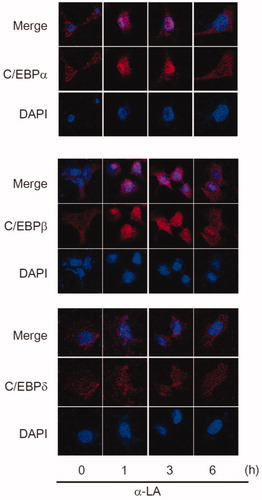
The correct figure is shown here:
Confocal microscopy
Briefly, H4IIE cells were grown on a coverslip and fixed in a 4% paraformaldehyde solution, followed by permeabilization with 0.1% Triton X-100. The cell samples were immunostained with antibodies directed against C/EBPα (sc-61), C/EBPβ (sc-150), and C/EBPδ (sc-515028) overnight, followed by incubation with either Alexa Fluor® 555 goat anti-rabbit or anti-mouse IgG (Invitrogen). After incubation, the samples were cover-slipped with mounting media. The samples were examined using a laser-scanning confocal microscope (A1, Nikon instruments Inc., NY, USA).