

Abstract
Here we carried out a comparative study on two cost and time effective methods of encapsulating silver nanoparticles (AgNP) in dipalmitoyl-phosphatidyl choline (DPPC)/cholesterol-based liposome to enhance nanoparticle cytotoxicity, and evaluated the effect of this on a blood cell line (THP1 monocytes) often involved in uptake of nanoparticles during human exposure. DLS and Zeta potential analyses over a 6-months period showed the extruded Lipo-AgNP (Ex-Lipo-AgNP) exhibited more stable characteristics when compared with the probe-sonicated Lipo-AgNP (PB-Lipo-AgNP). SEM microscopy indicated agglomeration of the PB-Lipo-AgNP which was not observed in Ex-Lipo-AgNP. Ex-Lipo-AgNP also exhibited higher temperature-dependent stability with 35.3% reduction in size from 20 °C to 37 °C while PB-Lipo-AgNP was less stable exhibiting 55% size reduction over same temperature range and 6 h period. Load release study over 24 h showed controlled release from Ex-Lipo-AgNP while the PB-Lipo-AgNP exhibited burst release at pH 4 and 6.5. Interestingly, it was found that Ex-Lipo-AgNP induced significantly higher cytotoxicity on THP1 cell line after 24 h exposure compared with control unexposed cells, uncoated AgNP and PB-Lipo-AgNP exposed cells at the same concentration. Thus, we report here that liposomal encapsulation of AgNP by extrusion produces a stable nanocapsule with enhanced cytotoxicity, thus preventing overreliance on high AgNP concentration to achieve desired toxicity for in vitro and possible in vivo applications.
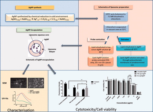
Graphical Abstract
Disclosure statement
No potential conflict of interest was reported by the authors.