

Abstract
In this study, we aimed to improve the hydrophilicity, cellular activities and osteogenesis of polycaprolactone (PCL) scaffolds by adding gelatin (Gel) and nano-hydroxyapatite (n-HA). We fabricated biocomposite scaffolds using the extrusion deposition 3 D printing method. The physical and biological properties of the composite scaffold were evaluated. Scanning electron microscopy (SEM) of the fabricated composite scaffolds revealed that the Gel and n-HA particles were uniformly embedded in the internal PCL. The composite scaffold (PCL + Gel + n-HA) showed dramatically improved mechanical properties. The porosity, hydrophilicity and hydrophobicity and ability to promote the attachment and proliferation of osteoblasts were then compared between the pure PCL and composite scaffolds. The PCL + Gel + n-HA scaffold was implanted in the rabbits for 5 weeks, and the results of H&E and Masson staining analyses demonstrated that the scaffold had good biocompatibility and good osteogenesis. The strategy developed in this study has promising prospects for future clinical applications of enhancing bone defect repair.
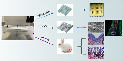
Graphical Abstract
Acknowledgements
We would like to thank Yi He at the Analytical and Testing Center of Sichuan University for the help with SEM analysis.
Disclosure statement
The authors declare that they have no known competing financial interests or personal relationships that could have appeared to influence the work reported in this paper.
Data availability statement
The data that supports this study are available within this article.