

Abstract
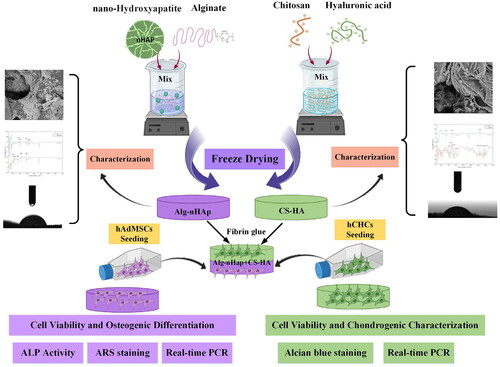
Cartilage joint lesions have created a significant challenge in the field of durable reconstruction and recovery due to their limited long-term repair and reconstruction. In this study, a biphasic scaffold was obtained using chitosan (CS)-hyaluronic acid (HA) and alginate-nanohydroxyapatite (nHAP) for osteoarthritis diseases. The scaffolds revealed porous structures and hydrophilic chemical groups by scanning electron microscopy (SEM) and Fourier-transform infrared spectroscopy (FTIR) methods, respectively. The resulting three-dimensional hydrogels were assembled by using fibrin glue and seeded with human chondrocytes cells (hCHCs) in the chondral layer and human adipose-derived mesenchymal stem cells (hAdMSCs) in the subchondral one. Then, the assessments including 3-(4, 5-dimethylthiazol-2-yl)-2, 5-diphenyltetrazolium bromide (MTT), SEM, compressive assay, weight loss, alizarin red staining, alkaline phosphatase (ALP) activity, alcian blue staining and Real-Time PCR were performed. The associated cell viability confirmed that there were no interventions between the cell types and SEM illustrated cell attachment and spreading. The subchondral and chondral layers were investigated separately to detect their ability for cellular commitment. Finally, the Real-time PCR was carried out on the bilayer scaffold and the related observations approved the osteogenic/chondrogenic differentiation of both cell types. Taken together, the Alg-nHAP/CS-HA scaffold provided an appropriate environment for cartilage and bone regenerations and could be recommended for all aspects of osteochondral tissue engineering.
Graphical Abstract
Acknowledgements
We are grateful to those who have helped us in this and other related projects.
Disclosure statement
The authors declare that there are no academic or financial conflicts between the authors of this article.