
ABSTRACT
A highly efficient in vitro micropropagation protocol, using newly emerged sprouts coming from greenhouse-grown plants, was developed to produce Actinidia melanandra Franch. and Actinidia rubricaulis Dunn. This is the first report of in vitro culture of A. rubricaulis Dunn. Axenic culture was possible using one- or two-node explants or terminal buds, surface-sterilised with commercial bleach tablets. Nearly 90% success rate in establishment was observed. The best axillary shoot proliferation and maximum adventitious shoots elongation were achieved on Quoirin-Lepoivre medium supplemented with 1.5 mg L−1 6-benzyladenine and 30 g L−1 sucrose. Although used as an inert support, agar brand has a significant effect. Kobe agar at 8 g L−1 gave the best response. 100% of in vitro rooting was observed. Roots length and their quality were highly improved by the use of vermiculite in transfer medium. Over 99% of rooted plants survived after acclimatisation.
Introduction
Actinidia melanandra Franch. and Actinidia rubricaulis Dunn. are dioecious species belonging to the Actinidiaceae family, originating from China. Although they produce edible fruit, they are not commercially cultivated. A. melanandra Franch. is a mid-sized, deciduous and climbing perennial vine. With its reddish flesh fruits, it is also known as purple ‘kiwi’. A. rubricaulis Dunn. is a semi-evergreen shrub with dark green and hairless fruits.
Since the first publication of Harada in 1975 on kiwifruit (Actinidia spp.), several protocols have been published on the two edible species, Actinidia chinensis Planch. and Actinidia deliciosa (Chev.) Liang and Ferguson (Revilla et al. Citation1992; Kumar and Sharma Citation2002; Xu et al. Citation2003; Sharma and Shirkot Citation2004; Ferguson and Seal Citation2008; Wang and Gleave Citation2012; Bourrain Citation2015). Micropropagation was also reported for Actinidia kolomikta (Kovac Citation1993), Actinidia polygama Miq. (Takahashi et al. Citation2004) and A. melanandra Franch., cited for the first and only time by Debenham et al. (Citation2016). To the best of our knowledge, no publication has been found on in vitro culture of A. rubricaulis Dunn.
Actinidia spp. have mostly been regenerated by adventitious organogenesis from callus using different types of explants: stamen, endosperm, mature embryo, root, stem segment and mature seed (Wiyaporn et al. Citation1990; Rey et al. Citation1992; Kim et al. Citation2007; Akbas et al. Citation2009). Plants produced by this technique can be variable in characters and genetically aberrant due to the callus phase. Where the objective is to maintain true-to-type material, adventitious organogenesis or somatic embryogenesis cannot be used to effectively produce plants. Micropropagation of axillary shoots from organised tissue, without a callus phase, is preferred. Shoot tips, buds, meristems or nodal segments from Actinidia spp. mature vines are highly desirable to maintain stable traits, however, few reports of such approaches are available (Standardi Citation1981; Marino and Bertazza Citation1990; Shen et al. Citation1990; Kovac Citation1993; Moncalean et al. Citation2001; Prado et al. Citation2005; Akbas et al. Citation2007; Debenham et al. Citation2016).
MS (Murashige and Skoog Citation1962) solid or liquid medium was successfully used by many authors for callus formation as well as for shoot proliferation (Harada Citation1975; Monette Citation1986; Marino and Bertazza Citation1990; Shen et al. Citation1990; Wiyaporn et al. Citation1990; Moncalean et al. Citation2001; Kim et al. Citation2007). Barbieri and Morini (Citation1988) grew bark explants on Gamborg B5 medium (Gamborg et al. Citation1968). LP medium (Quoirin and Lepoivre Citation1977) is mentioned by Piagnani et al. (Citation1986). Takahashi et al. (Citation2004) and Dimassi-Theriou and Bosabalidis (Citation1997) used WPM (Lloyd McCown Citation1980) for A. polygama Miq. and A. deliciosa, respectively. Induction of shoots was achieved by Lin et al. (Citation1994) using N6 medium (Chu Citation1978). Debenham et al. in (Citation2016) investigated, with A. chinensis and A. polygama, macro salt formulations of LP, WPM and B5 as alternatives to MS. 6-benzyladenine (BA) is the most widely used plant growth regulator (PGR) for the multiplication phase, sometimes in combination with other PGRs, typically used in the range of 0.5–2 mg L−1.
Rooting is achieved using MS or half strength MS media supplemented with 0.5–1 mg L−1 indole-3-butyric acid (IBA), depending on the process of rooting: agar medium or quick dip in IBA solution (Monette Citation1986; Pedroso et al. Citation1992; Kovac Citation1993; Moncalean et al. Citation2001; Prado et al. Citation2005; Nasib et al. Citation2008; Wu et al. Citation2011; Wang and Gleave Citation2012). Shoots were rooted by Akbas et al. (Citation2007) on MS medium with 1 mg L−1 α-naphthalene acetic acid (NAA). Shen et al. (Citation1990) found that IBA gave the best percentage of rooting and higher mean root number than indole-3-acetic acid (IAA) in A. chinensis. Debenham et al. (Citation2016) rooted shoots on a modified MS with 3 mg L−1 IBA.
This study was undertaken during the implementation of a varietal sensitivity test on Actinidia spp. against Pseudomonas syringae pv. actinidiae (Psa). A. melanandra Franch. and A. rubricaulis Dunn. probably carry genes resistant to Psa. This resistance could make them very useful as tolerant controls in the varietal sensitivity tests and could be used in a breeding program. To validate the results of sensitivity test on excised shoots, whole plants are needed. In vitro micropropagation was developed on these species in order to rapidly satisfy the demand of true-to-type, homogeneous, healthy and high-quality plants. Irrespective of seasons, in vitro plants can be produced and be available for tests in large numbers and in shorter periods than cuttings. The main objective of this study was to optimise and standardise an efficient and reliable micropropagation protocol for rapid regeneration without callus formation.
Materials and Methods
Plant material
One-year-old cuttings from male plants grown in pots, under insect-proof shelter conditions, were used as starting plant material. In February–March, when the chilling requirements were fulfilled, the pots were transferred to a heated greenhouse to accelerate the plant budding and keep the new shoots in a ‘clean’ environment far from dust, rain and pathogens. The greenhouse temperature regulation was 20°C minimum with natural daylight. The plants were watered and fertilised on demand with a complete fertilising solution (EC 1.2 mS cm−1).
Explant sterilisation and establishment in vitro
Young newly emerged sprouts were excised from greenhouse-grown plants ((a)). The expended leaves were removed. Nodal segments bearing one or two buds or apical parts of shoots with terminal buds were surface-sterilised in a laminar air-flow cabinet for 25 min in a commercial bleach (sodium dichloroisocyanurate, 0.6% (w/v) active chlorine). After three rinses with sterile distilled water, the excised shoot tips and buds were individually inoculated vertically onto shoot tip proliferation medium in glass tubes each containing 15 mL LP medium supplemented with 0.6 mg L−1 BA (Duchefa Biochemie B.V., Haarlem, The Netherlands) 0.2 mg L−1 IBA (Duchefa Biochemie B.V., Haarlem, The Netherlands), 0.2 mg L−1 gibberellic acid (GA3; Duchefa Biochemie B.V., Haarlem, The Netherlands) and 30 g L−1 glucose. All media were solidified with 0.8% (w/v) agar (HP696; Kalys, Bernin, France) and the pH was set to 5.6 using 0.1 M NaOH prior to autoclaving at 118°C and 85 kPa for 25 min. After in vitro establishment, explant bud burst occurred within 2–4 weeks. Buds giving rise to a new small shoot extension were used for shoot multiplication. In order to only propagate materials that were free of bacteria and fungi, a visual inspection and a control of the explants on a bacterial growth medium were done using an adaptation of the Viss et al. (Citation1991) protocol. During the first subculture, the waste (eg base of explant, old leaves) was inoculated in tubes containing 15 mL of BSM 523 medium (Duchefa Biochemie B.V., Haarlem, The Netherlands) and incubated in the growth room. The explants that showed contamination were discarded and autoclaved. The number of sterile explants, as well as the number of sprouting buds, was recorded. The final percentage of success was recorded at the end of the establishment phase just before the first subculture.
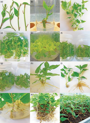
Figure 1. In vitro micropropagation protocol for A. rubricaulis: (a) New emerged annual sprout. (b) Shoot initiation from nodal segment bearing one bud. (c,d) Multiple shoots at the end of a multiplication cycle on LP medium. (e) Leaf chlorosis on N6 medium. (f) Comparison of shoot development under sucrose (left) or glucose (right) as carbon source treatment. (g) Comparison of shoot development under Kobe agar (left) or HP 696 (right). (h) Spontaneous rooting on MS surface medium. (i) Comparison of root development between an agar-solidified medium (left) and a transfer in a vermiculite medium (right): in vitro rooting on BdR 0.5 mg L−1 AIB. (j) Detail of the root system on agar medium. (k) Rooted plants on vermiculite medium. (l) Acclimatised plants in greenhouse.
Culture conditions
The explants were incubated in a growth room under a 16-h photoperiod provided by Philips cool-white fluorescent tubes (TL-D 58W840) at 40 μmol m−2 s−1 at 23 ± 2°C. Shoot multiplication and rooting were performed under the same conditions.
Shoot multiplication and growth rate: effect of medium composition
The initiated shoots were micropropagated in 500 mL glass jars covered by plastic screw caps, containing 135 mL of medium supplemented with 1.5 mg L−1 BA. The culture vessels were inoculated with 12 two-node cuttings coming from the previous multiplication cycle. Efficient multiplication has been achieved using a 4-to-6-week subculture period. The relative performance of the basal medium, carbon source and agar brand was compared. The six basal media evaluated were DKW (Driver and Kunyuki Citation1984), MS, N6, B5, WPM and LP. Glucose and sucrose were compared at 30 g L−1 each. Three gelling agents were tested: HP696, Kobe (Roth Sochiel, Lauterbourg, France) and Bacto (Becton Dickinson France, Le Pont de Claix, France) at 9.5, 8 and 9 g L−1, respectively, depending on the gel strength given by the supplier. The number of newly produced shoots from one initial explant, the highest shoot mean length, the multiplication index (total number of new explants bearing two nodes for the subsequent cycle) and the number of spontaneous rooting explants were recorded.
Rooting and acclimatisation: effect of medium and growth regulator
Shoots longer than 2.5 cm, derived from stock proliferating cultures, were used as explants for rooting experiments. The shoots were grown in two commonly used media known for their good root initiation rate on woody plants; BdR (Bourrain and Navatel Citation2000) and M4R (Nemeth Citation1981), containing IBA, IAA (Duchefa Biochemie B.V., Haarlem, The Netherlands) or NAA (Duchefa Biochemie B.V., Haarlem, The Netherlands) at 0.5 or 1 mg L−1 and supplemented with 30 g L−1 sucrose. After 7 days in the dark at 23 ± 2°C, the induced shoots either were directly transferred to a 16-h photoperiod without changing the medium or were transferred onto a PGR-free medium comprising 1/4 MS macro salts with 30 g L−1 glucose. The medium was solidified with 0.8% (w/v) HP696 agar and supplemented with vermiculite (Vermex M; Efisol, Strasbourg, France) in a 135:170 (v/v) mix of medium:vermiculite. Plants were removed from the aseptic in vitro conditions after a minimum of 4 weeks and rinsed with tap water to remove any remaining medium from the root system. The percentage of rooted plants and root quality was recorded. The rooted plants were then potted individually in plastic pots (9 cm × 9 cm × 9.5 cm) filled with a 70:30 (v/v) mix of white and brown peat containing 30 kg m−3 of clay with 1 kg m−3 balanced fertiliser (12-12-17 + trace elements) (AP10; Dumona, L’Isle d’Abeau, France). The plants were placed under greenhouse conditions at almost 100% relative humidity in a plastic tunnel with natural daylight. The relative humidity was provided by an adiabatic water atomisation air humidifier (Defensor® ABS3; Walter Meier, Pfäffikon, Switzerland). To condition the plants to ex vitro environmental conditions, the relative humidity was decreased gradually over 15 days by opening the plastic tunnel to allow air exchange and to remove excess condensed water. The plantlets were irrigated as required and fertilised once a week with a balanced commercial liquid fertiliser (Liquoplant 2.5-2.5-7 + 1.4 MgO + trace elements; Plantin, Courthezon, France). After 45 days under ex vitro conditions, survival percentage was recorded.
Statistical analysis
The data shown on shoot multiplication stage were the mean of 2 experiments using 24 explants each. The experiment on rooting was performed on a minimum of 20 homogeneous plants. Standard errors (SEM) of the arithmetic means were calculated for each treatment. Statistical analysis of the multiplication quantitative data was carried out using the ANOVA test. The Newman–Keuls test was applied at p = .05 probability level to find significant differences between the means. The statistical analyses were performed with Statbox 7.1 (FBC Software, Issy Les Moulineaux, France) for Windows.
Results
Explant sterilisation and in vitro establishment
Using greenhouse explants coming from newly emerged shoots, buds started sprouting as early as the second week. The surface sterilisation was very easy with the two Actinidia species studied. No contamination was observed on A. rubricaulis and only 2% on A. melanandra. Due to some explants that did not break, the final percentage of establishment success was 89% and 88%, respectively. Both species present hairless stems which make them easy to surface sterilise. Bristly-hair clones of A. chinensis grown in the same conditions as A. rubricaulis and A. melanandra gave 7% contamination using the same disinfection protocol (Bourrain Citation2015). More contamination also occurred when A. chinensis explants were harvested in autumn rather than spring with newly overwintered emerged shoots (Bourrain, unpublished data). The propagation of sterile cultures is possible using sodium hypochlorite tablets at an optimum balanced concentration/application time: 0.6% available chlorine for 25 min in these study conditions. The use of commercial bleach tablets as a sterilising agent is an easy and cheap solution that does not burn the plant tissue in contrast to the use of a liquid solution of concentrated sodium hypochlorite (previous data not shown). Within 2–4 weeks, the explants developed new sterile shoots ((b)). To avoid the proliferation of bacterial and fungal contaminations when the culture is established, and so have the possibility of propagating sterile material, the choice and the origin of explants were of extreme importance.
Shoot multiplication and growth rate: effect of medium composition
The two Actinidia species studied exhibited the same behaviour in all experiments (). Among the three criteria measured, the number of newly produced shoots from one initial explant did not vary with the six basal media tested. An average of 2–2.8 new shoots were formed from one explant with A. rubricaulis and 2.5–3.4 with A. melanandra, regardless of the basal salt used. Shoot length and the multiplication index (number of two-node cuttings obtained from one explant, for the subsequent cycle, which is correlated to the length of the shoot) were significantly different between the different basal media. LP medium ((c,d)) gave the best proliferation response, followed by B5 (). Leaf surface area (data not shown) and shoot length were better on LP than on B5. The leaves were narrower and more sharply serrated on B5. The multiplication index on LP medium was 6.1 and 6.4 for A. rubricaulis and A. melanandra, respectively, with a maximum shoot length of 55.7 and 51.6 mm. This means that more than six new explants can be obtained for the subsequent cycle from one initial explant. N6 and MS were the worst basal salts for shoot proliferation of both species, with N6 medium even resulting in greenish-yellow coloured shoots ((e)). Regardless of medium, the proliferation rate was mainly due to the elongation of the shoots rather than the formation of new shoots.
Table 1. Effect of proliferating medium composition.
Table 2. Effect of carbon source in proliferating medium.
Table 3. Effect of agar brand.
Significant differences were observed between glucose and sucrose with DKW basal salt ((f)), with sucrose giving better results than glucose for the number of newly formed shoots, the shoot length and the multiplication index ().
Agar is one of the most frequently used gelling agents. Despite their many disadvantages, agar-solidified media are very often used in commercial laboratories. Whatever the choice of inert support, gelling agent brand usually has a significant influence on the potential and quality of micropropagation (Debergh Citation1983); this is why three agar brands were evaluated in our study. As expected, significant differences were observed. The multiplication index was 3.6, 2.0 and 1.6, respectively, for Kobe, Bacto and HP696 with A. rubricaulis and 5.2, 3.2 and 2.2 with A. melanandra. Kobe agar brand gave by far the best results compared to the two other brands ((g); ). The multiplication index was nearly double for the Kobe agar brand. Unlike the basal salts, agar brand also had a significant effect on the number of new shoots formed from one initial explant. The best response was observed in Kobe agar medium with a new shoot number of 2.6 for A. rubricaulis and 3.7 for A. melanandra, against only 1.7 and 2.5 in Bacto agar medium and even less in HP696.
Spontaneous rooting has been observed during the multiplication stage on the surface medium in all treatments, even without any auxin in the medium ((h)). However, the basal salts showed a significant difference with regard to root formation. MS and N6 media gave more than two roots per plant, LP, WPM and DKW gave around one root and B5 was the medium that led to the formation of the minimum number of roots, not all the plants rooted on B5.
Rooting and acclimatisation: effect of medium and growth regulator
Rooting percentage was always 100%, regardless of the basal salt (BdR or M4R), the auxin type (IBA, IAA, NAA) and concentration (0.5 or 1 mg L−1), and the transfer or not onto a PGR-free medium. However, the quality of the roots was affected by the structure of the medium ((i)). In gelled medium, the root length was less than 10 mm, unlike in 135:170 (v/v) mix of agar medium:vermiculite, where the root length reached 50 mm. Also in gelled medium, only very small roots were formed around a callus in the medium, with some longer roots formed directly on the medium surface. Aerial roots were also noticed on the basal part of the shoot itself ((j)). Agar solid media alone are known to form anoxic conditions, which may be the reason for A. rubricaulis and A. melanandra roots having limited growth in the medium but better growth outside the medium. The vermiculite allowed the enhancement of the root system as well as the production of secondary roots ((k)), which make the acclimatisation stage easier. Over 99% of the vermiculite-rooted plants were successfully acclimatised and survived in greenhouse conditions, even those with shorter roots ((l)). The two Actinidia species studied were not susceptible to rapid desiccation after removal from in vitro culture leading to simple management during the acclimatisation stage.
Discussion and conclusion
In vitro establishment is a key factor for the process of micropropagation. The percentage of uncontaminated explants is an important economic criterion. A. rubricaulis and A. melanandra are easy to sterilise using 0.6% available chlorine for 25 min. Shen et al. (Citation1990) failed to sterilise A. chinensis in the presence of sodium hypochlorite (1% available chlorine, 5–15 min), but they succeeded when using explants from new annual growth, excised in spring, and treated with 0.1% HgCl2 for 3–8 min. They also reported better responses with soft-hair clones than with bristly-hair ones, and showed that explant type, origin and sampling season have a great influence on the surface sterilisation. With field-derived material, Debenham et al. (Citation2016) found that contamination was a significant issue. Disinfection was ineffective for 31% of the explants for A. chinensis and 53% for A. polygama. They also observed contamination a few weeks after disinfection and believed that many genotypes harboured endogenous contaminants. We do not observe contamination in A. rubricaulis and A. melanandra within 2 years after establishment. However, Monette (Citation1986), using non-axenic shoot tips, succeeded in producing plants in the presence of two survival bacterial contaminants that did not cause necrosis of the cultures.
Although DKW and WPM media are known for good results with multiplication of woody species (eg Juglans, Prunus and Malus), this was not the case for Actinidia spp. In our study, DKW and WPM media performances concerning shoot multiplication and growth rate were average, and LP medium and B5 gave better results. Our results are in agreement with those reported for A. chinensis using LP (Piagnani et al. Citation1986) or B5 (Barbieri and Morini Citation1988) media, but unlike those using N6 medium (Lin et al. Citation1994). N6 medium gave the worst results for A. rubricaulis and A. melanandra. Debenham et al. (Citation2016) observed no obvious differences in plant performance when A. chinensis and A. polygama were grown on MS, LP, B5 or WPM. In their study, LP and B5 media gave paler and smaller leaves, without detrimental impact on growth. In our trials, the leaves are narrower and more sharply serrated with B5 than with other basal formulations.
Akbas et al. (Citation2007) examined the effects of carbon source (sucrose, maltose and dextrose at 3%) on A. deliciosa. Sucrose was the optimal carbohydrate on shoot production in their study. Glucose is the carbon source frequently used in our laboratory on different species (eg Prunus, Malus, Pyrus and Fragaria), with very good results. So, it seemed relevant to test this carbon source on A. rubricaulis and A. melanandra in comparison to the traditional sucrose. The inferior result indicates that, unlike for other species, glucose is not an appropriate carbon source for Actinidia spp. micropropagation,
Spontaneous rooting was reported by Moncalean et al. (Citation2001) in A. deliciosa during the multiplication stage. In liquid medium, the spontaneous rooting percentage decreased in parallel with the increase in the BA incubation period, for example, only 20% of spontaneous rooting was observed with explants grown in a BA-free medium or when the incubation time was less than 8 h in the presence of BA. Our results showed 100% spontaneous rooting in the presence of 1.5 mg L−1 BA regardless of the basal salt.
Nasib et al. (Citation2008) observed in A. deliciosa that the root induction was highly affected by the length of shoots and that an appropriate length was a pre-requisite for efficient root formation. In that study, the length of the shoots at the end of a subculture was between 30 and 40 mm. In our experiments, the use of a length of over only 25 mm was proved to be sufficient for the two Actinidia species. Shoots of less than 25 mm size also root (data not shown). A. rubricaulis and A. melanandra are very easy-to-root species.
Several authors have indicated the formation of hyperhydric shoots in Actinidia spp. during micropropagation. Hyperhydricity has been proposed to be the correct term to designate the hyperhydric malformations frequently affecting herbaceous and woody shoots during their in vitro vegetative propagation. This physiological disorder was also previously known under the terms of vitrification, vitrescence, glassiness and translucency (Debergh et al. Citation1992). A. chinensis cultured in liquid medium showed a slightly vitrescent appearance compared to those produced in agar-gel medium without presenting problems at the rooting and acclimatisation stages (Monette Citation1986). Feito et al. (Citation1994) observed 57% of vitrified shoots in liquid media and only 5% on solidified media. Moncalean et al. (Citation1999) reduced the percentage of hyperhydric shoots using cellulose plugs as an inert support in A. deliciosa liquid culture. Marino and Bertazza (Citation1990) reported that 1 or 2 mg L−1 of BA sometimes caused vitrification in a 7 g L−1 Bacto agar-solidified medium which did not affect proliferation, only the older leaves became vitrified. Piagnani et al. (Citation1986) observed that a high level of BA (4.5 mg L−1) frequently caused vitrification. 1.5 mg L−1 BA used in our experiment did not cause the formation of hyperhydric shoots. Furthermore, no hyperhydricity was observed using any of the basal salts or carbon sources. In this study, the different media gave some limited vitrescence on A. chinensis (data not shown), however, A. rubricaulis and A. melanandra did not seem to be susceptible to this physiological disorder.
The procedure reported in this paper for the first time on A. rubricaulis and the second time on A. melanandra shows the possibility for rapidly producing, in quantity, true-to-type, homogenous, healthy and high-quality plants whatever the time of the year. In vitro micropropagation of these two Actinidia species is a far more efficient method of propagation than by conventional cuttings. This enhancement is of importance for the kiwifruit sector as it results in an improvement in the evaluation of the sensitivity of the commercial kiwifruit varieties to Psa.
Acknowledgements
The author sincerely thanks Sylvie Parkinson for the revision of the manuscript as well as INRA Grande Ferrade (Bordeaux, France) for providing plants from which cuttings were realised in order to produce the starting material for the in vitro establishment. Sylvain Teissonnière is particularly thanked for his picture on acclimatised plants. This research was supported by funds from the CASDAR 2012–2015, France.
Disclosure statement
No potential conflict of interest was reported by the author.
References
- Akbas F, Isikalan C, Namli S. 2009. Callus induction and plant regeneration from different explants of Actinidia deliciosa. Appl Biochem Biotechnol. 158:470–475. doi: 10.1007/s12010-008-8401-2
- Akbas F, Isikalan C, Namli S, Basaran D. 2007. Micropropagation of kiwifruit (Actinidia deliciosa). Int J Agric Biol. 3:489–493.
- Barbieri C, Morini S. 1988. Shoot regeneration from callus cultures of Actinidia chinensis (Cv. Hayward). Acta Hortic. 227:470–472. doi: 10.17660/ActaHortic.1988.227.96
- Bourrain L. 2015. Culture in vitro du kiwi (Actinidia chinensis) – introduction par mise en culture de bourgeons axillaire. Infos CTIFL. 308:37–41.
- Bourrain L, Navatel JC. 2000. Production de plants de châtaignier de la variété Marigoule, Castanea crenata x Castanea sativa, par multiplication in vitro. In: Verger M, editor. Troisième rencontre du groupe de la Sainte Catherine. Orléans (France): Cirad-Inra; p. 38–47.
- Chu C. 1978. The N6 medium and its applications to anther culture of cereal crops. In: Proceedings of Symposium on Plant Tissue Culture. Beijing: Science Press; p. 43–50.
- Debenham MC, Seelye JF, Mullan AC. 2016. An in vitro repository for clonal kiwifruit. Acta Hortic. 1113:93–98. doi: 10.17660/ActaHortic.2016.1113.13
- Debergh P. 1983. Effects of agar brand and concentration on the tissue culture medium. Physiol Plant. 59:270–276. doi: 10.1111/j.1399-3054.1983.tb00770.x
- Debergh P, Aitken-Christie J, Cohen D, Grout B, von Arnold S, Zimmerman R, Ziv M. 1992. Reconsideration of the term vitrification as used in micropropagation. Plant Cell Tiss Org Cult. 30:135–140. doi: 10.1007/BF00034307
- Dimassi-Theriou K, Bosabalidis AM. 1997. Effects of light, magnesium and sucrose on leaf anatomy, photosynthesis, starch and total sugar accumulation, in kiwifruit cultured in vitro. Plant Cell Tiss Org Cult. 47:127–134. doi: 10.1007/BF02318948
- Driver JD, Kunyuki AH. 1984. In vitro propagation of paradox walnut rootstock. Hortsci. 19:507–509.
- Feito I, Rodriguez A, Centeno ML, Sanchez-Tamès R, Fernandez B. 1994. Effect of the physical nature of the culture medium on the metabolism of benzyladenine and endogenous cytokinins in Actinidia deliciosa tissues cultured in vitro. Physiol Plant. 91:449–453. doi: 10.1111/j.1399-3054.1994.tb02973.x
- Ferguson AR, Seal AG. 2008. Kiwifruit. In: Hancock JF, editor. Temperate fruit crop breeding. New York: Springer; p. 235–264.
- Gamborg OL, Miller RA, Ojima K. 1968. Nutrient requirements of suspension cultures of soybean root cells. Exp Cell Res. 50:151–158. doi: 10.1016/0014-4827(68)90403-5
- Harada H. 1975. In vitro organ culture of Actinidia chinensis PL. as a technique for vegetative multiplication. J Hort Sci. 50:81–83.
- Kim M, Kim SC, Moon DY, Song KJ. 2007. Rapid shoot propagation from micro-cross sections of kiwifruit (Actinidia deliciosa cv. ‘Hayward’). J Plant Biol. 50(6):681–686. doi: 10.1007/BF03030613
- Kovac J. 1993. Micropropagation of Actinidia kolomikta. Plant Cell Tiss Org Cult. 35:301–303. doi: 10.1007/BF00037286
- Kumar S, Sharma DR. 2002. In vitro propagation of kiwi. J Hortic Sci Biotech. 77(5):503–508. doi: 10.1080/14620316.2002.11511530
- Lin QL, Chen ZQ, Wu JS. 1994. Propagation in vitro of some excellent clones of kiwi fruit. Journal of Fujian Agricultural University. 23(3):271–274.
- Lloyd G, McCown B. 1980. Commercially-feasible micropropagation of mountain laurel, Kalmia latifolia, by use of shoot-tip culture. Int Plant Prop Soc Proc. 30:421–427.
- Marino G, Bertazza G. 1990. Micropropagation of Actinidia deliciosa cvs. ‘Hayward’ and ‘Tomuri’. Sci Hortic. 45:65–74. doi: 10.1016/0304-4238(90)90069-Q
- Moncalean P, Canal MJ, Feito I, Rodriguez A, Fernandez B. 1999. Cytokinins and mineral nutrition in Actinidia deliciosa (Kiwi) shoots cultured in vitro. J Plant Physiol. 155:606–612. doi: 10.1016/S0176-1617(99)80061-3
- Moncalean P, Rodriguez A, Fernandez B. 2001. In vitro response of Actinidia deliciosa explants to different BA incubation periods. Plant Cell Tiss Org Cult. 67:257–266. doi: 10.1023/A:1012732429147
- Monette PL. 1986. Micropropagation of kiwifruit using non-axenic shoot tips. Plant Cell Tiss Org Cult. 6:73–82. doi: 10.1007/BF00037760
- Murashige T, Skoog F. 1962. A revised medium for rapid growth and bio assays with tobacco tissue cultures. Physiol Plant. 15:473–497. doi: 10.1111/j.1399-3054.1962.tb08052.x
- Nasib A, Ali K, Khan S. 2008. An optimized and improved method for the in vitro propagation of kiwifruit (Actinidia deliciosa) using coconut water. Pak J Bot. 40(6):2355–2360.
- Nemeth G. 1981. Adventitious root induction by substituted 2-chloro-3-phenyl-propionitriles in apple rootstocks cultivated in vitro. Sci Hortic. 14:253–259. doi: 10.1016/0304-4238(81)90020-0
- Pedroso MC, Oliveira MM, Pais MS. 1992. Micropropagation and simultaneous rooting of Actinidia deliciosa var. deliciosa ‘Hayward’. Hortsci. 27(5):443–445.
- Piagnani C, Eccher T, Castelli S. 1986. Micropropagation of Actinidia chinensis: effects of growth regulators on proliferation rate. Acta Hortic. 79:887–890. doi: 10.17660/ActaHortic.1986.179.158
- Prado MJ, Herrera MT, Vasquez RA, Romo S, Gonzalez MV. 2005. Micropropagation of two selected male kiwifruit and analysis of genetic variation with AFLP markers. Hortsci. 40(3):740–746.
- Quoirin M, Lepoivre P. 1977. Etude de milieux adaptés aux cultures in vitro de Prunus. Acta Hortic. 78:437–442. doi: 10.17660/ActaHortic.1977.78.54
- Revilla MA, Rey MA, Gonzalez-Rio F, Gonzalez MV, Diaz-Sala C, Rodriguez R. 1992. Micropropagation of kiwi (Actinidia spp.). In: Bajaj YPS, editor. Biotechnology in agriculture and forestry, high-tech and micropropagation II. Vol 18, Berlin: Springer-Verlag; p. 399–423.
- Rey M, Fernandez T, Gonzalez V, Rodriguez R. 1992. Kiwifruit micropropagation through callus shoot-bud induction. In Vitro Cell Dev Biol. 28:148–152. doi: 10.1007/BF02823064
- Sharma DR, Shirkot P. 2004. Biotechnological interventions for genetic amelioration of Actinidia deliciosa var. deliciosa (kiwifruit) plant. IJBT. 3:249–257.
- Shen XS, Wan JZ, Luo WY. 1990. Propagation in vitro of Chinese gooseberry (Actinidia chinensis) through the development of axillary buds. Sci Hortic. 42:45–54. doi: 10.1016/0304-4238(90)90146-6
- Standardi A. 1981. Micropropagazione dell’Actinidia chinensis Planch. mediante coltura ‘in vitro’ di apici meristematici. Frutticoltura. 43:23–27.
- Takahashi W, Sugawara F, Yamamoto N, Bando E, Matsushita J, Tanaka O. 2004. Plant regeneration in Actinidia polygama Miq. by leaf, stem, and petiole culture with zeatin, and from stem-derived calli on low-sucrose medium. J For Res. 9:85–88. doi: 10.1007/s10310-003-0053-z
- Viss PR, Brooks EM, Driver JA. 1991. A simplified method for the control of bacterial contamination in woody plant tissue culture. In Vitro Cell Dev Biol Plant. 27:42. doi: 10.1007/BF02632060
- Wang T, Gleave AP. 2012. Applications of biotechnology in Kiwifruit (Actinidia). In: Agbo EC, editor. Innovations in biotechnology, ISBN: 978-953-51-0096-6, InTech, DOI:10.5772/28673 . http://www.intechopen.com/books/innovations-in-biotechnology/applications-of-biotechnology-in-kiwifruit-actinidia-.
- Wiyaporn S, Subhadrabandhu S, Sahavacharin O. 1990. In vitro vegetative multiplication of kiwi plant. Acta Hortic. 279:447–460. doi: 10.17660/ActaHortic.1990.279.49
- Wu YJ, Xie M, Long QJ. 2011. In vitro organogenesis and plant regeneration from leaves of Actinidia eriantha Benth. cv White (kiwifruit). New Zeal J Crop Hort. 39(4):231–240. doi: 10.1080/01140671.2011.582876
- Xu X, Yao X, Chen H. 2003. Application of modern biotechnology on kiwifruit. Acta Hortic. 610:525–531. doi: 10.17660/ActaHortic.2003.610.70