Abstract
Background: The study of human anatomy is a core component of health science programs. However large student enrolments and the content-packed curricula associated with these programs have made it difficult for students to have regular access to cadaver laboratories.
Methods: Adobe Flash MX™ was used with cadaver digital photographs and textbook-derived illustrations to develop interactive anatomy images that were made available to undergraduate health science students enrolled in first-year combined anatomy and physiology (ANP) courses at the University of Ottawa. Colour coding was used to direct student attention, facilitate name-structure association, improve visualization of structure contours, assist students in the construction of anatomical pathways, and to reinforce functional or anatomical groupings. The ability of two-dimensional media to support the visualization of three-dimensional structure was extended by developing the fade-through image (students use a sliding bar to move through tissues) as well as the rotating image in which entire organs such as the skull were photographed at eight angles of rotation. Finally, students were provided with interactive exercises that they could repeatedly try to obtain immediate feedback regarding their learning progress.
Results: Survey data revealed that the learning and self-testing tools were used widely and that students found them relevant and supportive of their self-learning. Interestingly, student summative examination outcomes did not differ between those students who had access to the online tools and a corresponding student group from the previous academic year who did not.
Conclusion: Interactive learning tools can be tailored to meet program-specific learning objectives as a cost-effective means of facilitating the study of human anatomy. Virtual interactive anatomy exercises provide learning opportunities for students outside the lecture room that are of especial value to visual and kinesthetic learners.
Introduction
Undergraduate university students, the youngest of adult learners, are expected to assume significant responsibility for their educational success as they explore new concepts in large class settings. Nevertheless, this responsibility must also be shared by course instructors whose mandate is to provide an organized and engaging educational environment that addresses the diverse learning needs of large student populations. The study of human anatomy is a core component of health science programs including medicine, nursing, human kinetics, physiotherapy and occupational therapy (Granger et al. Citation2006). However, the level of anatomical knowledge and structural detail that is required for each body system is very much discipline-dependent and anatomy instruction must be tailored to meet the specific needs of each health science discipline (Terrell Citation2006).
The lecture room, the cadaver laboratory and the virtual laboratory are all appropriate venues for anatomy instruction. While lectures afford the opportunity for the instructor to present all learners with a package of well-organized information developed around a series of learning objectives, the potential exists, unfortunately, for a good deal of this learning to be passive as students listen to the instructor while they look at projected slides showing labeled images of body anatomy. Interactive laboratory sessions are helpful in that they provide some opportunity for students to manipulate body structures so that they can appreciate their three-dimensional form and visualize their interrelationships with surrounding tissues and organs. However, anatomy laboratory sessions require significant investments of time, space and resources (Rizzolo & Stewart Citation2006), are usually of limited duration, and often involve group sessions in which a student's view and/or ability to manipulate the cadaver is restricted. Furthermore, a challenge facing a number of health science programs, including those offered at the University of Ottawa, is that many of these programs have such large student enrolments and/or such information-dense curricula that it is simply not possible to offer weekly anatomy laboratories as part of the core curriculum (Granger et al. Citation2006; Rizzolo & Stewart Citation2006). Hence, the challenge exists for anatomy instructors to develop cost-effective, student-friendly learning resources that target program-specific anatomical learning objectives and are able to either replace or extend the learning environment of the cadaver laboratory (Clark Citation1994; Granger et al. Citation2006).
The learning characteristics of adult students have been extensively studied and numerous theories have been proposed. For example, in his widely-read theory of andragogy, Knowles described adult students as self-directed learners whose knowledge acquisition occurs best when it is exploratory and task-oriented, rather than via rote memorization (Knowles et al. Citation1984; Pratt Citation1993). Simply defined, learning involves the reorganization and transfer of new information from the limited confines of working memory to the limitless repository of long term memory (Kirschner Citation2002; Kirschner et al. Citation2006). Working memory, or what we can consider as conscious memory, is characterized by storage durations (5–20 sec if not readdressed) as well as content capacities (3–7 new elements at a time, depending on information complexity) that are both very limited (Miller Citation1956; Sweller et al. Citation1998; Kirschner Citation2002; Kirschner et al. Citation2006; Terrell Citation2006). Long term memory, on the other hand, encompasses our repository of accumulated knowledge and, therefore, has a capacity that is theoretically boundless (Terrell Citation2006). Indeed, the challenge facing each learner is to organize new information, as it is added to this repository, so that it can be successively retrieved and applied in a timely fashion when needed (Sweller et al. Citation1998; Kirschner Citation2002, Kirschner et al. Citation2006; Terrell Citation2006). With that in mind, a challenge facing anatomy educators is to effectively guide learning so as to maximize the efficiency with which new knowledge is encoded in long-term memory (Kirschner et al. Citation2006).
This paper describes the development and implementation of program-tailored online anatomy learning and self-testing tools for undergraduate Faculty of Health Science students enrolled in first-year combined anatomy and physiology (ANP) courses at the University of Ottawa. These tools were made available to students via supplementary WebCT™-based course web sites. When designing these online exercises, care was taken to apply instructional design principles such as cognitive load theory (Moreno & Mayer Citation2000; Kirschner Citation2002, Heo & Chow Citation2005; Terrell Citation2006) and Gagné's Nine Events of Instruction (Gagné et al. Citation1992) so that extraneous cognitive load (defined as the ineffective cognitive load that reduces ease of student learning), would be minimized and the ability of these tools to capture student attention and promote interactive, effective learning could be maximized. As an important first step, and as recommended by Robert Gagné's second (inform learners of objectives) and fifth (provide guidance) events of instruction (Gagné et al. Citation1992), the online exercises were embedded within a framework of carefully organized course learning objectives so that students would try each activity in order and at the most appropriate stage in their learning. Students began with worked examples (Heo & Chow Citation2005; Kirschner et al. Citation2006) of textbook diagrams and cadaver images in which all relevant information was made available to them, step by step. Then, and in compliance with Robert Gagné's sixth event of instruction (provide opportunities for practice; Gagné et al. Citation1992), students were provided with many opportunities to practice applying their new knowledge via unlimited access to the self-testing exercises. These exercises resembled very closely the types of questions students were subsequently asked on summative examinations and assessed student learning at two rather basic levels of competence (anatomy-based knowledge is largely factual with minimal opportunities for higher forms of knowledge application such as synthesis or analysis), but still at increasing levels of difficulty (van Merrienboer & Sweller Citation2005). They began with the easier drag-and-drop labeling activities that were then followed by the more challenging type-in-labeling exercises.
Formative assessment of the anatomy tools was conducted using student surveys. Furthermore, student performance on summative examinations was compared between students who had access to the online learning tools and a comparable student population previous academic year) that did not have access to the supplementary course web sites.
Materials and methods
Materials
Adobe Flash MX™ was used to create a number of different types of interactive online images, as described below.
Interactive self-learning images
The roll-over image
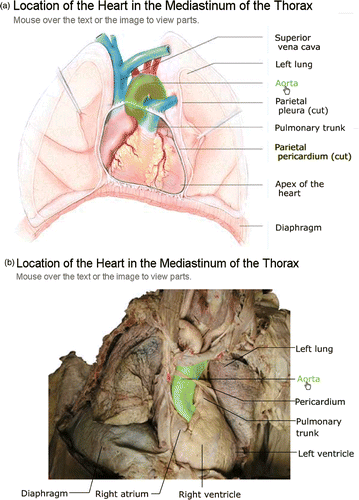
The basic roll-over image uses colour coding to allow the learner to visually link an anatomical structure with its corresponding label. Whenever possible, unlabeled textbook illustrations (Marieb Citation2001) were paired with cadaver images (dissections and photography courtesy of S. Goodwin, U. Ottawa) photographed at the same orientation so as to facilitate comparison of structure location and morphology between the textbook diagram and the in situ photograph (). To create interactivity, and as directed by the course learning objectives, a finite number of labels was added to each set of paired images in the form of mouse-operated buttons. Next, each of the labeled anatomical structures was outlined and filled with a bright and contrasting colour, keeping the fill partially transparent so that the visibility of the underlying structure was maintained at the same time that its shape was being clearly delineated and its proximity to nearby body components revealed.
Figure 1. Matching (a) textbook (From Marieb EN: Human Anatomy & Physiology, San Francisco, 2001, Benjamin Cummings, used by permission of Pearson Education, Inc.) and (b) cadaver roll-over images of the location of the heart in the mediastinum of the thorax showing the aorta. A comparison of (a) and (b) shows that the ‘empty spaces’ that are frequently included between structures for clarity in textbook drawings do not actually exist in the body and that structures are often less perfect in shape when seen in situ.
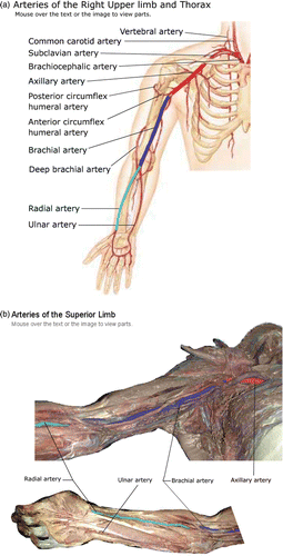
Within the roll-over images, the selective use of color and the ability to add additional highlighted components to an initial starting structure allows student attention to be directed along an anatomical pathway, such as the pathway of arterial blood flow down the abdominal aorta and into the left and right common iliac arteries (). Similarly, the pathway of arterial blood flow through the axillary, brachial and radial arteries as it flows down the upper limb toward the wrist can be followed as each pathway component is highlighted in succession (). While this pathway appears artificially simple when the vessels are shown in relative isolation in a textbook drawing (), students are made aware that the anatomy of the upper limb is actually considerably more complex and that these arteries must travel in between and around nerves, muscles and veins to reach their destinations ().
Figure 2. Cadaver image dissected to reveal the major abdominal arteries. (a) Roll-over highlight of abdominal aorta. (b) Roll-over highlight with the abdominal aorta still emphasized but now adding in two branches, the common iliac arteries.
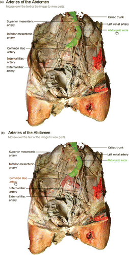
Figure 3. Corresponding (a) textbook (From Marieb EN: Human Anatomy & Physiology, San Francisco, 2001, Benjamin Cummings, used by permission of Pearson Education, Inc.) and (b) cadaver roll-over images of the right upper limb and thorax highlighted to allow the arterial pathway of blood flow to the wrist to be followed.
Variations on the roll-over image
The simple roll-over image does not address the pedagogical issue that three-dimensional anatomy is being studied using two-dimensional learning tools. Hence, variations such as the multi-angle rotation and the fade-through image (Macromedia Citation2006) were developed to encourage students to make associations between structures linked by location and/or function.
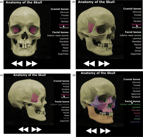
Multi-angle rotation. For the multi-angle interactive image, cadaver specimens were mounted on a movable stage and photographed at eight equally-spaced angles of rotation between 0° and 360°). Each image was then placed within a different frame in the video function of Flash so that the user will be able to use arrow buttons to navigate completely around the body structure in either direction (). As shown with this interactive rotational image of the skull (), students can also highlight a single structure (e.g. the sphenoid bone) and follow that structure through one complete revolution ().
Grouping. For some roll-over images, structure labels were organized so as to group related anatomical components. For example, the group label ‘facial bones’ was added to the image of the skull in such a way that when the mouse is placed over this group label, the labels for all of the individual facial bones as well as the bones themselves within the skull are highlighted at the same time (). Furthermore, by combining the grouping function with rotational ability, students are able to follow these bones as a complete structural unit when the skull is rotated and appreciate their physical relationships with one another from eight different points of view.
Figure 4. Rotating roll-over image of the skull with the sphenoid bone highlighted so that it can be followed as the skull is manipulated to be viewed at (a) 0°, (b) 45°, and (c) 90° of rotation. (d) The grouping function has been activated to allow all of the facial bones to be viewed simultaneously at 45° of rotation.
The fade-through image
The fade-through image allows students to appreciate the relationship between superficial and more deeply situated structures within an organ or body region. A minimum of two photographs of the same structure at different body depths were required for the construction of each fade-through interactive image. Using Flash, the photographs were placed directly on top of each other and a slider-bar was created that was linked directly to the transparency property of the top image. This gives the learner the ability to adjust the transparency of the top image so as to produce a range of image views extending from visualization of exclusively the top image, degrees of simultaneous relative visibility of both images (to identify physical relationships), to exclusive visibility of only the bottom image. While shows just a single screen capture of the structure of the heart taken at 72% transparency of the top image, the slide bar tool can be adjusted in very small increments between 100% and 0% transparency of the upper image so that the students can finely control the rate at which they see a more superficial view being replaced a deeper one. Furthermore, by moving the slide bar back and forth, students can make repeated and detailed comparisons to come to a greater understanding of the three-dimensional structure of this organ and the potentials for functional interaction between its components.
Figure 5. Fade-through textbook (From Marieb EN: Human Anatomy & Physiology, San Francisco, 2001, Benjamin Cummings, used by permission of Pearson Education, Inc.) image of the heart with a sliding bar allowing the transparency of the more superficial of the image components to be adjusted between 0 and 100%.
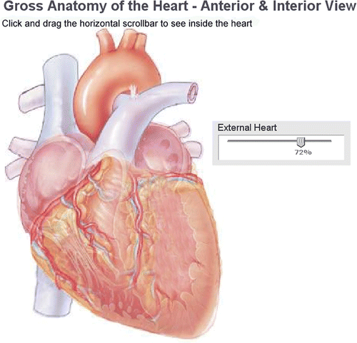
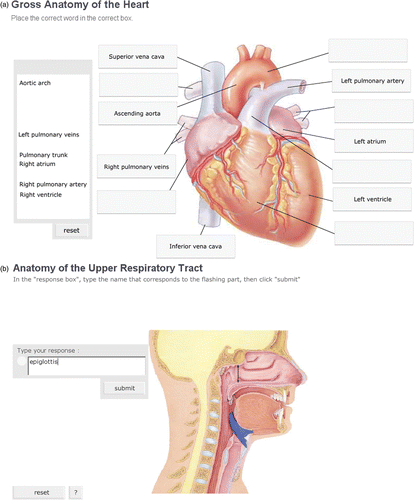
Figure 6. Reusable learning applets. (a) Partially completed drag-and-drop image of the heart. (b) Self-testing image of the upper respiratory tract in which the epiglottis has been highlighted and its corresponding label correctly entered. (reprinted by permission of Pearson Education, Inc.).
Interactive self-testing images
Drag-and-drop
Drag-and-drop labeling exercises were created to allow self-testing at the most basic level of knowledge, the simple recognition of a structure name when viewing that structure (Anderson & Krathwohl Citation2001). For these self-testing exercises, roll-over images were modified so that the label lines linked to empty boxes rather than structure names, and an alphabetical list of labels was provided to the left of the diagram. If a label was dragged and dropped into the correct box, it remained in place. If the label was placed incorrectly, it would automatically return to its original place within the label list, allowing the student unlimited opportunities to try to associate it with the appropriate structure.
Type-In labeling
Recognition of a structure name represents the simplest form of learning. However, the applied use of new knowledge in the workplace requires that a student know and can use structure names in context rather than simply recognizing them from a list. To promote the ability to recall and correctly spell structure names, type-in labeling applets were developed that required the user to key the name of a structure into an empty text box when that structure was highlighted on the computer screen. Students have three chances to type in the correct name before it appears in the box. Once the entire exercise has been successfully completed, the student is presented with a fully and accurately labeled diagram for review.
Evaluation methods
Formative evaluation
These learning and self-testing tools were provided online to 275 undergraduate University of Ottawa health science students throughout the term during which they were studying the anatomy and physiology of the endocrine, cardiovascular and respiratory systems. Students could use each tool as often as desired throughout the duration of the course. The anatomy tools were combined with interactive physiology self-tests to form a WebCT-based course web site that supplemented the learning accomplished in the lecture hall. At the end of the course, students were asked to complete an anonymous questionnaire evaluating the anatomy-based learning and self-testing components of the course web site.
This survey was composed of ten close-ended questions that allowed students to provide feedback pertaining to their ease and extent of use of each of the anatomy exploratory and self-testing exercises as well as their assessment of the ability of these learning tools to promote their learning and comprehension of course content. For each type of tool students were also provided with the option to not provide feedback because they had chosen not to make use of that particular tool. The final part of the survey contained open-ended questions that asked students to identify the strengths and weaknesses of the learning tools, so that any shortcomings could be addressed.
Summative evaluation
Although difficult to rigorously assess, the ultimate test of a learning tool is its ability to enhance student knowledge retention and transfer. Student outcomes on summative examinations were compared between the first group of 275 students to use the interactive web sites (LT Group) and a comparable student population (241 students) who took this course in the absence of supplemental learning tools during the previous academic year (No-LT Group). Summative examinations results were analysed by the t-test assuming unequal variances (Microsoft Excel XP) while student groupings by letter grade were evaluated using the Chi-square test (SPSS 8.0).
Results
In contrast to students studying medicine at the University of Ottawa, students enrolled in most other health-science-related programs do not have access to cadaver laboratories. All of their anatomy learning must be accomplished through lecture and online supplementary course web sites.
Student feedback
A response rate of 49% (136/275 students) was obtained for the survey evaluating the online learning and self-testing tools that were provided to undergraduate Faculty of Health Science students studying anatomy and physiology. Approximately two-thirds of respondents reported that they used the interactive learning images (64%) as well as the drag-and-drop (66%) and type-in labeling self-testing exercises (68%) and that they found these instructional tools to be helpful as they prepared for summative examinations. More than 90% of respondents indicated that the interactive exercises were relevant to course content, the supplied directions to students were clear, and the activities themselves were user-friendly.
Web site features that were identified by students as particularly useful included the anatomy learning tools themselves, the ability to test their knowledge online, and the existence of unlimited access to the interactive exercises. On the other hand, an important concern raised by students was a lack of flexibility associated with the type-in labeling self-tests. For a number of the labels, students noted that more than one correct answer should have been recognized, due to the existence of redundant terminology, optional use of capital letters or hyphens and occasional confusion as to whether the structure name should be provided in the singular or the plural. They suggested that the answer key should be expanded to accommodate all possible alternate responses.
Student learning outcomes
The two classes that were compared (LT vs No–LT) had similar student compositions in that approximately 44% of each class consisted of undergraduate nursing students while the remaining 56% of students were enrolled in the human kinetics program. Interestingly, the mean final grade obtained by students did not differ (p > 0.05) between the LT (69.4%, n = 275) and the No-LT groups (69.5%, n = 241). The two student populations were also compared with regard to the per cent of students attaining letter grades in the A, B, C, D and below D (failure) ranges. In agreement with the similar class averages, there was also no significant difference (p > 0.05) in the distribution of marks throughout this grading scale ().
Table 1. Comparison of class distribution of final grades for students who had access to online learning tools (LT Group) or did not have access to these tools (No-LT Group) throughout the academic term.*
Discussion
In this paper we describe the use Adobe Flash MX 2004™ to create interactive images that undergraduate health science students can use to compare the appearance of organ system components between textbook diagrams and cadaver images, explore three-dimensional body anatomy, develop a deeper understanding of structural and functional grouping of anatomical components, and obtain feedback on their progress in learning. When inserted into course web sites as instructive modules, these interactive images provide opportunities for self-directed study that can reinforce the learning that students have begun when viewing body structures presented as static images during lectures and/or manipulating body components in the anatomy laboratory.
Cognitive load theory provides a number of strategies which can be applied to the design of instructional tools that will maximize student use of the limited confines of working memory when learning (Moreno & Mayer Citation2000; Kirschner Citation2002, Heo & Chow Citation2005; Terrell Citation2006). Where applicable, certain of these strategies were applied to the development of the online activities described in the current paper. For example, specific colour coding applied to paired structures and labels within the worked examples provided an effective discriminative tool that not only enhanced contrast and clearly delineated structure boundaries, but also helped students to follow pathways (e.g. circulatory pathways) within a given area of the body. Colour coding also made use of economical pre-attentive information processing to facilitate information organization while minimizing cognitive effort (Heo & Chow Citation2005). Furthermore, the placement of structure labels as closely as possible to the structures being identified in each of the images reduced extraneous cognitive load by applying the spatial and temporal contiguity principles (Moreno & Mayer Citation2000). To further expand the limited boundaries of working memory, students were encouraged to mentally package related pieces of new information (e.g. all of the facial bones) as small chunks or schemata (Chi et al. Citation1982; Kirschner Citation2002) through use of the Flash-associated grouping function. Finally, by proceeding from worked examples through drag-and-drop and then type-in labeling exercises, students were taken through a graded series of applications that gradually asked them to supply more and more of the required information (van Merrienboer & Sweller Citation2005).
What is the value in devoting so much time and energy to the development of these learning and self-testing tools when interactive anatomy software [e.g. anatomy.tv (Primal Pictures), Anatomy & Physiology Revealed (McGraw-Hill), W3D-VBS (Temkin et al. Citation2006) and the Visible Human Project (Spitzer & Scherzinger Citation2006)] already exists in the educational marketplace? The commercially-available programs are indeed excellent educational tools that pride themselves on providing very high levels of anatomical detail. However, while they do provide levels of instruction that are suitable for medical students, these are levels that are often far in excess of what is required for most other health science programs. For example, nursing and human kinetics students would be faced with a surfeit of information through which they must search for those particular pieces that they need to know for summative examinations. An important benefit of the approach described in this paper is that effective learning is facilitated by customizing anatomical images in order to address only the learning needs of a particular program. This minimizes the distraction of extraneous cognitive load (Kirschner Citation2002) and allows students to concentrate on only those anatomical details for which they are responsible.
Related to the notion of information overload is the controversy surrounding the provision of multiple views of anatomical structures (Garg et al. Citation1999; Levinson et al. Citation2007). Certainly some of the anatomy software described in the previous paragraph can overwhelm students because not only are multiple angles of rotation provided for each body structure, but these rotations are permitted along both the horizontal and vertical axes and may involve a complete organ or just a component of that organ that has been taken out of context and is being manipulated in isolation. With regard to the current self-learning tools, rotation was used for only selected body components such as the skull and the heart because these are body components that one would view as entire discrete structures. The number of rotations was kept low (no more than eight) and the rotations were always along only the horizontal axis. The purpose was to mimic as closely as possible a student's ability, when in the lab, to turn over a structure such as the heart to see what it looks like from the other side. In contrast to other studies investigating the influence of multiple views on learning (Levinson et al. Citation2007), the multiple views did not provide new material for formative student assessment. Rather, they served as supplementary learning tools so that a student could rotate a body part, for example, to see how a blood vessel or a bone continues around the periphery of that structure. Students were informed that their summative examination questions would be derived from only their textbook diagrams.
When given access to these online tools, the majority of students did use them and they reported that they found the learning and self-testing tools to be user-friendly, relevant and helpful. However, there remained approximately one-third of students who did not use the online tools. One strategy that could be used to encourage students to develop a personal schedule of regular practice application would be to routinely assign (or at least remind them of) specific exercises that should be done when they are leaving at the end of a lecture. The concerns that students raised regarding the inflexibility of the type-in labeling exercises are indeed valid and, in an effort to minimize any confusion associated with these self-assessments, the type-in labeling exercises will be modified to permit alternative correct responses. While it was not possible to demonstrate a beneficial influence of the online anatomy exercises on student learning outcomes, there are likely a number of factors that contribute to this result. An initial confounding factor was an inability to link individual student use of the learning and self-testing exercises with student performance on summative exams. Within the confines of WebCT, it was simply not possible to track individual student use of each of the learning and self-testing exercises. Furthermore, student survey data was collected anonymously so as to protect student privacy. Hence it was not possible to determine if more favourable student outcomes might have been preferentially associated with those students who chose to make use of these additional learning and self-assessment opportunities compared to those who did not. Finally, given that a proportion of the summative exam questions are changed every year, the two student populations did not write midterm and final exams that were identical in their composition.
A more important value of these online learning tools may be their ability to appeal to learning styles that are often not addressed very strongly in the lecture room. Undergraduate classrooms are composed of heterogeneous populations of learners and these anatomy learning tools do address some of this heterogeneity. Fleming and Mills (Citation1992) and Fleming & Bonwell (Citation2001) described four types of learning preferences: visual, aural, read/write and kinesthetic and developed a simple online questionnaire (Fleming Citation2007) that students can use to recognize their primary learning style(s). Survey data collected from medical and health science students at the University of Ottawa have shown them to display a broad range of learning preferences and, as has been reported for other health science students, to frequently use two, three or even four learning approaches simultaneously (multimodal learner) when tackling a new situation (Baykan & Naçar Citation2007; Slater et al. Citation2007; Carnegie Citation2008). Interestingly, kinesthetic (hands-on) learning was a popular choice for many of these students (Carnegie Citation2008). By allowing students to not only visualize anatomical structures online, but also to manipulate them, much as they would if they were in the laboratory, and to carry out labeling exercises that provide feedback on their progress in learning, these anatomy tools have very definite instructional value for kinesthetic learners. But they have value for other learning styles as well. The extensive use of images and colour coding is helpful for visual learners while the type-in labeling activities address certain strengths of the read-write learner (Fleming & Bonwell Citation2001).
Accessibility to learning experiences is also becoming an increasingly important issue in undergraduate education. Many health science programs (including those offered at the University of Ottawa), have large undergraduate enrolments, anatomy laboratory space is limited, and there are insufficient resources to be able to afford the annual salaries for the prosectors and lab demonstrators that are needed to produce weekly laboratory sessions (Granger et al. Citation2006; Rizzolo & Stewart Citation2006). Virtual anatomy laboratory modules provide alternatives to the infrastructure-demanding cadaver laboratories by offering limitless online opportunities for students to view, manipulate and practice their identification of anatomical components. There is, of course, a cost associated with the initial development of these online exercises and cost is an important consideration when developing new instructional materials (Clark Citation1994). The tools described in this paper were developed during the summer by a health sciences student (POB) who had previously taken this anatomy and physiology course and was already familiar with Adobe Flash MX™ software. With minimal guidance, he was able to develop interactive tools that targeted the course learning objectives and some others that gave special attention to those anatomical structures that were visually challenging for students. Body anatomy does not really change. Hence once the initial investment in a student stipend has been made, one has now acquired a small library of reusable learning objects that can be incorporated into a variety of anatomy and physiology courses year after year.
In summary, we have described the development of a number of interactive learning and self-assessment exercises using Flash that undergraduate students studying anatomy at the University of Ottawa did use to promote their self-directed learning and to allow them to self-test their progress in learning. These tools can be custom-developed to meet program-specific learning objectives associated with various health sciences disciplines and allow the boundaries of the traditional anatomy laboratory to be extended in a cost-effective manner so that students can have unlimited access to virtual anatomy laboratories in which they can repeatedly explore, manipulate and evaluate their understanding of body structure.
Acknowledgements
The authors gratefully acknowledge the provision by Ms. Shannon Goodwin of careful and clean dissections as well as high quality photographs of human cadavers. The authors also thank Dr. Henri Lescault for his valued expertise regarding the accuracy of structure boundary identification throughout the preparation of these learning tools. The authors gratefully acknowledge the copyright permission granted by Pearson Education for the use of selected diagrams from E.N. Marieb: Human Anatomy and Physiology, 5th Edition, 2001 in the development of these online learning and self-testing tools. Finally, the authors would like to thank the Centre for e-Learning for their valuable assistance with the establishment of the course web site. This research was supported in part by a Teaching and Learning Grant (J. Carnegie) from the Centre for University Teaching, University of Ottawa.
Declaration of interest: The authors report no conflicts of interest. The authors alone are responsible for the content and writing of the paper.
References
- Anderson LW, Krathwohl DR. A Taxonomy for Learning, Teaching and Assessing: A Revision of Bloom's Taxonomy of Educational Objectives. Longman, New York, NY 2001
- Baykan Z, Naçar M. Learning styles of first-year medical students attending Erciyes University in Kayseri, Turkey. Adv Physiol Educ 2007; 31: 158–160
- Carnegie J. Know your audience: linking effective physiology instruction with student learning preferences. HAPS Educator 2008; 12(2)25–27
- Chi M, Glaser R, Rees E. Expertise in Problem Solving. Handbook of Learning and Cognitive Processes. Introduction of Concepts and Issues. Erlbaum, Hillsdale, NJ 1982; 1
- Clark R. Media will never influence learning. Educ Technol Res Devel 1994; 42: 21–29
- Deubel P. An investigation of behaviorist and cognitive approaches to instructional multimedia design. J Educ Multimed Hypermed 2003; 12(1)63–90
- Fleming N. VARK: A guide to learning styles 2007, Available at: http://www.vark-learn.com (retrieved May 5, 2007)
- Fleming N, Bonwell C. VARK: How Do I Learn Best? A Student's Guide to Improved Learning. Fleming and Bonwell, ChristchurchNZ 2001
- Fleming ND, Mills C. Not another inventory, rather a catalyst for reflection. Improv Acad 1992; 11: 137–142
- Gagné RM, Briggs LJ, Wager WW. Principles of Instructional Design,4th. Harcourt Brace Jovanovich, Fort Worth, TX 1992
- Garg A, Norman GR, Spero L, Maheshwari P. Do virtual computer models hinder anatomy learning?. Acad Med 1999; 74(Suppl 10)S87–S89
- Granger NA, Calleson DC, Henson OW, Juliano E, Wineski L, Mcdaniel MD, Burgoon JM. Use of web-based materials to enhance anatomy instruction in the health sciences. Anatom Record (Part B: New Anat) 2006; 289B: 121–127
- Heo M, Chow A. The impact of computer augmented online learning and assessment tool. Educ Technol Soci 2005; 8(1)113–125
- Kirschner PA. Cognitive load theory: Iimplications of cognitive load theory on the design of learning. Learn Instruct 2002; 1: 1–10
- Kirschner PA, Sweller J, Clark RE. Why minimal guidance during instruction does not work: An analysis of the failure of constructivist, discovery, problem-based, experiential, and inquiry-based teaching. Educ Psychol 2006; 4: 75–86
- Knowles MS & Associates. Andragogy in Action. Applying Modern Principles of Adult Education. Jossey Bass, San Francisco, CA 1984
- Levinson AJ, Weaver B, Garside S, Mcginn H, Norman GR. Virtual reality and brain anatomy: a randomized trial of e-learning instructional designs. Med Educ 2007; 41: 495–501
- Macromedia. (n.d.). Flash Professional 8, Available at: http://www.macromedia.com/software/flash/flashpro/ (retrieved April 15, 2006)
- Marieb E. Human Anatomy & Physiology,5th. Pearson Education, San Francisco, CA 2001
- Miller G. The magic number seven, plus or minus two: some limits of our capacity for processing information. Psychol Rev 1956; 63: 81–97
- Moreno R, Mayer RE. A learner-centered approach to multimedia explanations: deriving instructional design principles from cognitive theory 2000, Available at: http://imeg.wfu.edu/articles/2000/2/05/index.asp (retrieved May 25, 2005)
- Pratt DD. Andragogy after twenty-five years. Adult Learning Theory: An Update. pp 15–25, Meerriam S. Jossey Bass, San Francisco, CA 1993
- Rizzolo LJ, Stewart WB. Should we continue teaching anatomy by dissection when …?. Anatom Rec (Part B: New Anat) 2006; 289B: 215–218
- Slater JA, Lujan HL, Dicarlo SE. Does gender influence learning style preferences of first-year medical students?. Adv Physiol Educ 2007; 31: 336–342
- Spitzer VM, Scherzinger AL. Virtual anatomy: An anatomist's playground. Clin Anat 2006; 19: 192–203
- Sweller J, Van Merrienboer JJG, Paas FGWC. Cognitive architecture and instructional design. Educ Psychol Rev 1998; 10: 251–296
- Temkin B, Acosta E, Malvankar A, Vaidyanath S. An interactive three-dimensional virtual body structures system for anatomical training over the internet. Clin Anat 2006; 19: 267–274
- Terrell M. Anatomy of learning: Instructional design principles for the anatomical sciences. Anatom Record (Part B: New Anat) 2006; 289B: 252–260
- van Merrienboer JJG, Sweller J. Cognitive load theory and complex learning: Recent developments and future directions. Educ Psychol Rev 2005; 17: 147–177