Abstract
Background: Knowledge of the benefits of incorporating medical simulation into healthcare curricula is rapidly increasing. Though impeded by the high cost of complicated technology, medical simulation devices offer the ability to provide safe and controlled training environments, exposure to rare clinical scenarios, as well as unlimited training opportunities.
Methods: This report describes a novel, inexpensive method of broadcasting normal and abnormal auscultatory findings to a relatively normal appearing stethoscope for use in training of healthcare professionals.
Results: Using wireless transmitter broadcasting to a stethoscope fitted with a receiver apparatus, the student is able to perform a typical medical exam with auscultation of an unlimited variety of clinical sounds from anatomically appropriate sources while being observed from another room.
Conclusions: Implications of this low-cost device include limitless training possibilities worldwide and across disciplines. The simplicity and portability of this device increases potential for use in rapid training of recognition of clinical signs associated with chemical/biological warfare agents, mass casualty incidents and field military applications. This is the first device to simulate clinically relevant sounds in a realistic manner on standardized patients and mannequins. The benefits of such simulation in medical education ultimately serve to increase trainee confidence and consequently, improve patient care and safety.
Introduction
Throughout time, a variety of training methods have been used to teach medical students, residents, nurses and other health care professionals entering the medical field. Common methodologies have included classroom and bedside instruction, as well as medical simulation. All have effectively served as a means to impart the vital information necessary to care for their patients. Though use of medical simulation devices has been documented as early as 1737 (Moran Citation2007), simulation in medical education has been employed more frequently during the last decade. In addition to improving tested skills, the use of simulation provides a risk-free environment to practice high-stakes events as well as uncommon events in situations of known and controlled underlying causes, thus preventing harm to patients (Hammond Citation2004). Despite the many benefits to medical education, the financial component of innovative technology has impeded the integration of medical simulation into many educational curriculums.
Classroom teaching remains the mainstay of education for healthcare personnel. This method of teaching is touted for its ability to provide an enriching environment of human discussion and feedback between teachers and learners, thus allowing for immediate clarification of important concepts that may be difficult for students to learn in other venues (Finley et al. Citation1998). Classroom teaching, however, does not allow the student to learn at his or her convenience by being available at all times, nor can the student learn for the length of time most preferable (Finley et al. Citation1998).
Bedside teaching of medical personnel is another traditional teaching method. A patient population with high acuity and shorter hospital stays, as well as managed care restrictions has limited the learner's access to patients with different types of diseases (Issenberg et al. Citation2003). Lack of patient access can have a negative impact on the trainees’ education, as ‘expertise in any skill such as bedside cardiology results from deliberate practice that includes “informative feedback and opportunities for repetition and correction of errors”’ (Ericsson Citation1996; Issenberg et al. Citation2003).
Simulation, in general, has been used for many decades to train other disciplines, including pilots of various aircraft. Flight simulation was employed during World War II to train military pilots due to its cost effectiveness and time efficiency. Commercial pilots, as well as the National Aeronautics Space Association use simulation for training today (Grenvik et al. Citation2004). The same ideology has recently been transferred to the medical setting. Critical emphasis has been placed on integration of medical simulation into curricula for medical students and other health care providers during the last decade. Simulation Based Medical Education (SBME), a method of medical education which employs simulative aids, is integrated with other methods of teaching including lectures, bedside teaching, and problem-based learning with an overall emphasis on enhancing patient safety by learning from mistakes with increased practice to prevent errors in real life (Ziv et al. Citation2005).
Unique benefits of simulation in medicine include: providing a safe and controlled environment for the practice of risky procedures, unlimited practice with difficult procedures and situations that rarely appear in uncontrolled clinical settings, development and preparation of specific scenarios and rare events to provide training opportunities, quick feedback to trainee and instructor/evaluator, a system to repeat different types of scenarios, practicing and improving team skills and reducing instructor costs (Grenvik et al. Citation2004).
According to the report, ‘To Err Is Human: Building a Safer Health System’, in 1999, deaths due to medical error may be ranked between the fifth to eighth leading cause of death in the United States (Kohn et al. Citation2000; Grenvik et al. Citation2004). Preventable medical events cost our nation anywhere from $17 billion to $29 billion per year (Kohn et al. Citation2000; Grenvik et al. Citation2004). As such, the use of simulated technology also has the potential to help reduce costs through minimizing preventable mistakes by allowing residents and physicians to practice their skills in different situations before a similar event occurs in the clinical setting. Additionally, this practice may occur at their convenience and for as long or often as necessary.
Together with the economic benefit of using simulators, a panoply of advantages exist including the protection of patients from endangerment, increased possibilities for medical research, more advanced techniques in specialty areas and the incorporation of evidence-based training into medical education (Grenvik et al. Citation2004).
The use of simulators cannot replace training with actual patients. However, simulation training is needed to improve the skill, comfort and understanding of procedures for the learner despite decreased opportunity to see patients with diverse conditions. A recent study looked at pertinent cardiology skills acquired by osteopathic internists with the use of ‘Harvey’, the Cardiac Patient Simulator (CPS), and the UMedic multimedia computer system. The internists who used the CPS and UMedic in a 2 h workshop significantly improved identification of 10 cardiac auscultatory findings (compared to a pre-test of the same 10 cardiac auscultatory findings). Results of the study suggest significant improvement for internists’ cardiac auscultatory skill following the use of simulation (Issenberg Citation2003).
Unfortunately, the benefits of medical simulation in education are often overshadowed by high costs. One of the earliest sophisticated medical simulators, Sim One, was largely dismissed due to cost restrictions despite ‘promising early reports of its effectiveness in training’ (Bradley Citation2006). Due to the importance of simulation in medical training, the need exists to identify and/or develop novel, inexpensive methods of mimicking key clinical scenarios for the educational benefit of health care providers and patients alike. The objective of this report is to describe an inexpensive method of broadcasting abnormal auscultatory findings to a relatively normal appearing stethoscope.
Materials and methods
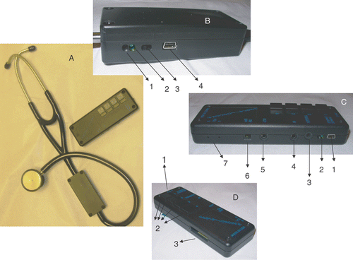
An inexpensive method of broadcasting normal and abnormal auscultatory findings to a relatively normal looking stethoscope has been developed (). To this point, it has not been possible to ‘play’ abnormal sounds realistically and in real-time on an otherwise normal standardized patient. This allows for the integration of human interaction and communication skills, and medical information gathering and processing, with realistic physical findings in a given scenario. Additionally, it is possible that learning these sounds in the context of the ‘whole’ patient could lead to better acquisition and retention of this knowledge. The wireless transmitter (A) sends medically appropriate teaching sounds to a training stethoscope (B). The device comes with 12 common sounds that may be used to perform 10 or more common clinical cases. Beyond this, any MP3 file of the instructor's choice may be used for this purpose, and will expand the potential number of possible cases. Sounds may be obtained by any number of stethoscope recording devices or from any sound files that have been converted to MP3 format. This allows any instructor to choose sounds of appropriate quality and specificity, although caution should be taken to obtain sounds that have been validated by at least one expert in the field. The scope of possible sounds may, therefore, include pulmonary, cardiac, abdominal and/or vascular. The pre-selected sound(s) are sent from the transmitter to the receiver in an undetected fashion such that they appear to be originating from the point of contact with the stethoscope head. The student is able to carry out a typical exam without any specific instruction regarding how to use the stethoscope portion of the device or the close supervision of faculty. Although the transmitter is limited to storing 12 sounds at one time, these are quickly and easily changed by plugging in another Secure Digital (SD) card with a different set of sounds. Additionally, an input jack (C) allows for other devices such as a PDA, laptop, cassette player, etc. to be used to provide the transmitted sounds instead of the MP3 files on the card.
Figure 1. The Ventriloscope®. (A) The Ventriloscope® device including transmitter and receiver. (B) The receiver: (1) Charging LED; (2) Power LED; (3) Power Switch; and (4) USB Charging Port. (C) The transmitter (view 1): (1) USB Charging Outlet; (2) LEDs; (3) Trigger Jack; (4) Input Jack; (5) Output Jack; (6) Power Switch; and (7) Volume Control. (D) The transmitter (view 2): (1) ‘ABC' Switch; (2) Sound Selection Buttons; and (3) Secure Digital Card Slot.
Sound quality for the sounds transmitted through the FCC and CE approved Ventriloscope® have been formally tested using a Fonix 6500 Hearing Aid Analyzer (Frye Electronics, Tigard OR) to detect frequency response and distortion for sounds ranging from 250 to 8000 Hz. Sounds were clear with minimal background noise. Additionally, digital sound (MP3) files are stable and do not deteriorate, therefore, as long as the sound recorded to the file is of a good quality, it will continue to be so with no background hissing, distortion or static.
Transmission occurs wirelessly to a relatively normal appearing stethoscope that does not conduct the patient's own physiological sounds. Contrary to other simulators with limited areas for auscultation, these broadcasted sounds are heard wherever the head of the stethoscope is placed making findings such as an ocular or thyroid bruit possible, as well as many others.
The most probable placement of the transmitter is with the trained patient who can then precisely control both the timing and type of transmitted sounds based on tactile feedback. The trained patient may control the transmitter with pre-loaded abnormal or normal sounds. Once the medical trainee places the stethoscope over the appropriate area of the standardized patient, the patient presses a button to allow transmission of one of 12 sounds wirelessly to the training stethoscope. The maximum range of this device is approximately 20 feet. Alternatively, the instructor may be in control of the transmitter through distant supervision. Control of the sounds from a distance is possible by hard-wiring the distant sound generator to the transmitter near the patient through the input jack. This allows the controls close to the patient to be overridden and the sounds originating from afar are transmitted. Using a live video feed to cue the distant instructor to the selection and timing of sounds allows an almost unlimited distance between instructor and student.
Since MP3 files are used, they may be quickly distributed by email to virtually anywhere in the world in an instant. This may have important implications in the event of a new infectious agent or chemical/biological warfare agent to allow front-line care providers to be quickly trained using simulation in recognition of these new conditions. Also, knowledge of converting a given recording into MP3 format using an audio recording and editing program such as ‘GoldWave’ is becoming more common, which lends even greater variability and flexibility to this device.
To add to the versatility of the Ventriloscope®, this system allows for the detection of appropriate sounds at differing locations, or for different patient positions. For example, the character or loudness of a cardiac murmur may be changed with a special maneuver of the patient by using a different sound played when the maneuver is performed. With the aid of a ‘trigger’ jack on the Ventriloscope®, vascular phenomena can be synchronized with the patient's own pulse. The ‘trigger’ jack accepts input from a separate external pulse sensor and subsequently automatically times the transmitted heart sounds with the patient's actual pulse. As an alternative, without the use of additional devices, the standardized patient may palpate his/her own pulse and press the buttons correlating with vascular sounds in synchrony. In this way, a student palpating the carotid pulse to time the murmur or other sound would hear the abnormal sound at the appropriate time in the cardiac cycle. The respiratory sounds may also be correlated with the patients own inspiration and/or expiration. Volume can be controlled from the transmitter, but the volume of the recorded sounds is also preserved, allowing for the transmission of sounds of various volumes from the same SD card. Additionally, different sounds can be played over the same area so that for a case of pneumonia, E to A changes, as well as whispered pectoriloquy, bronchophony and bronchial breath sounds could all be heard over the same location at different times depending on what the patient is instructed to do by the examining physician. This is generally not possible to do on a high fidelity mannequin. The transmitter may be located behind a one-way mirror, out of sight of the student during a teaching scenario, easily hidden within the room, or even concealed in the exam gown. The small size and low weight of this device allows for great portability and use in a number of locations without special equipment. It can also be operated in total darkness. These features may prove advantageous in the conducting of Mass Casualty Incident (MCI) drills and military applications, especially in field locations.
Although only one stethoscope can be used per transmitter, two or more sets of stethoscopes and transmitters can be operated in the same room at the same time. Two high tech chips in the transmitter and receiver automatically pair together and select a frequency not currently in use by any other transmitter. This prevents unwanted transmission of two or more sounds on the same frequency. Also, the chip randomly changes frequencies multiple times per second, allowing for greater security as well as a clearer signal. In many scenarios, several participants may be simultaneously assessing different aspects of the patient using a stethoscope. The current arrangement allows for each to hear completely appropriate sounds, despite listening over different areas of the patient or mannequin.
Through the use of an output jack, this device may be adapted for use in small or large group teaching settings when using patient simulators or trained patients via speakers. The output jack also allows for earphone use during training of the patients as well as recording for verification of sounds played during a testing situation. The ability to record the actual sounds played provides verification of a patient's performance, or can be used to verify that correct sounds were actually played during an examination if questions arise.
The Ventriloscope® has some technical limitations that are easily remedied. Though the device transmits well through the body, it may not transmit through large amounts of tissue. Transmission issues may be avoided by keeping the transmitter receiver pair visible to each other or at least minimizing the amount of tissue between the two. Additionally, each stethoscope must be controlled by one transmitter. For group teaching, speakers or other devices can be attached to the output jack. Finally, training of instructors or patients is required, however a 1 h training session will usually allow mock-up of a standard heart failure case.
Discussion
In order to effectively present future physicians, nurses, practicing physicians and other health care providers with the technology provided by medical simulators, cost becomes a considerable factor. Other similar simulators may retail for approximately $25,000 without any options or accessories. Highlighted features of the more expensive systems include wireless infrared sound systems, small group instruction, and a variety of sounds including heart, breath and bowel. These features may be accomplished using the device described here for approximately $4,000. The technology required to provide greater patient safety and reduce preventable and costly mistakes need not be expensive or complex in order to strengthen and build pertinent skills that are required for effective medical care. Additionally, advances and accessibility to technology make medical simulation much simpler and more affordable.
The benefits of such technology are emphasized by Issenberg, in reference to a training workshop for physicians-in-training and practicing physicians, ‘Our results indicate that a 2-hour workshop using simulation instruments such as Harvey and UMedic can result in a significant improvement in cardiovascular auscultation skills–…’. (Issenberg et al. Citation2003) Students participating in the training workshop had an average pre-test score of 37%, while the post-test score average soared to 81% in identifying 10 normal and abnormal cardiac auscultatory findings (Issenberg et al. Citation2003).
One limitation to using standardized patients to train medical students and residents to detect sound abnormalities is the inability to demonstrate realistic abnormal auscultatory findings. With this device, standardized patients can provide congruent auscultatory findings to match a verbal account of symptoms. The simulator described here provides an inexpensive, yet adequate method of broadcasting auditory findings to a conventional stethoscope with only slight modification. Students will gain experience and improve the skill required to provide quality care, without being limited by high costs and lack of standardized patients.
In order to provide the best medical care to patients, a variety of training methods must be used. This includes a combination of classroom, bedside and simulation training. Limitations of both classroom and bedside training can be offset by the use of simulation in which feedback is timely and a variety of diseases and scenarios may be experienced.
The high costs of some forms of medical simulation may be a thing of the past, as newer and more versions become available. With the appearance of newer innovations that reduce cost and thus increase accessibility, particularly in the area of auscultation, it is now possible to expand training to include these newer methods of training.
A simple system that allows for wireless transmission of abnormal and/or normal auditory findings from a transmitter has been presented here. Modification of a stethoscope to function as the receiver, while still appearing relatively normal, has been discussed. This apparatus has been found to be very useful in conjunction with the use of standardized patients, allowing for the integration of communication skills and professionalism with realistic physical findings. It is also beneficial when used in conjunction with patient simulators or in small or large group teaching settings. This innovative teaching method is simple, effective, inexpensive and versatile.
To our knowledge, this is the first simulator to allow abnormal auscultatory findings to be successfully simulated in a realistic way on a standardized patient. This product offers another option to medical simulation while providing the benefits of low cost, reproducibility of sounds, unlimited variability in auditory findings (including pulmonary, cardiac, abdominal and/or vascular sounds), product simplicity and the ability to teach small or large groups. The benefits of such education at all training levels will serve to increase trainee confidence and consequently, improve patient care and safety.
Declaration of interest: The authors report no conflicts of interest. The authors alone are responsible for the content and writing of the article.
Additional information
Notes on contributors
Amy Castilano
AMY CASTILANO, BS, drafted the manuscript through intellectual and editorial contribution.
Nairmeen Haller
NAIRMEEN A HALLER, PhD, contributions were intellectual and editorial.
Cheryl Goliath
CHERYL GOLIATH, Med, contributions were intellectual and editorial.
Paul Lecat
PAUL LECAT, MD, is the inventor of the Ventriloscope® with a patent pending. His contributions were intellectual and editorial.
References
- Bradley P. The history of simulation in medical education and possible future directions. Med Educ 2006; 40: 254–262
- Ericsson KA. The road to excellence: The acquisition of expert performance in the arts and sciences, sports, and games. Lawrence Erlbaum Associates, New Jersey 1996
- Finley J, Sharratt G, Nanton M, Chen R, Roy D, Paterson G. Auscultation of the heart: A trial of classroom teaching versus computer-based independent learning. Med Educ 1998; 32: 357–361
- Grenvik A, Schaefer Jr, DeVita M, Rogers P. New aspects on critical care medicine training. Curr Opin Crit Care 2004; 10: 233–237
- Hammond J. Simulation in critical care and trauma education and training. Curr Opin Crit Care 2004; 10: 325–329
- Issenberg S, Gordon M, Greber A. Bedside cardiology skills training for the osteopathic internist using simulation technology. J Am Osteopath Assoc 2003; 103: 603–607
- Kohn LT, Corrigan J, Donaldson MS. To err is human: Building a safer health system. National Academy Press, Washington, DC 2000, Institute of Medicine (U.S.) Committee on Quality of Health Care in America.
- Moran M. Jacques de Vaucanson: The father of simulation. J Endourol 2007; 21: 679–683
- Ziv A, Ben-David S, Ziv M. Simulation based medical education: An opportunity to learn from errors. Med Teach 2005; 27: 193–199